. 2015 Dec;14(12):1262-8.
doi: 10.1038/nmat4444. Epub 2015 Oct 12.

Cell-mediated fibre recruitment drives extracellular matrix mechanosensing in engineered fibrillar microenvironments

Affiliations

Cell-mediated fibre recruitment drives extracellular matrix mechanosensing in engineered fibrillar microenvironments

Brendon M Baker et al. Nat Mater. 2015 Dec.

Abstract

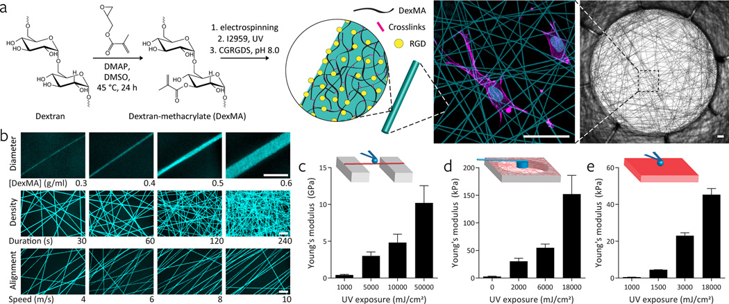
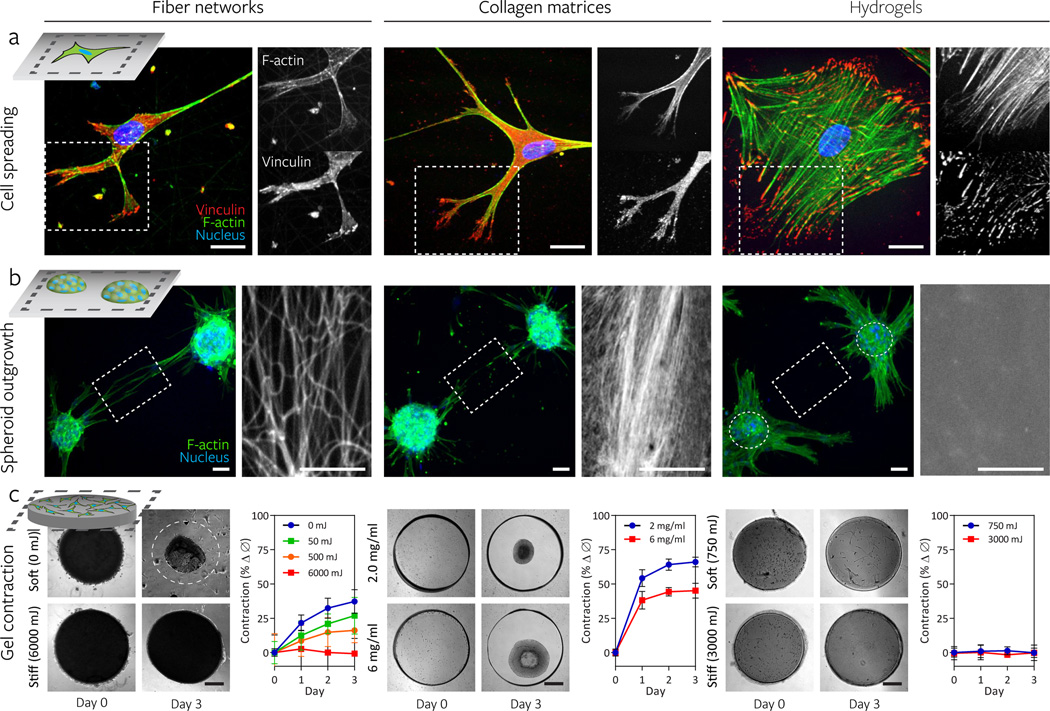
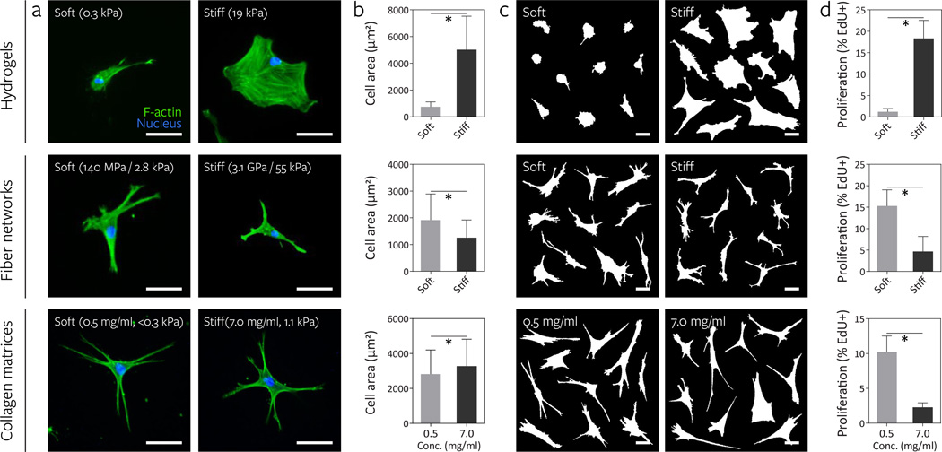
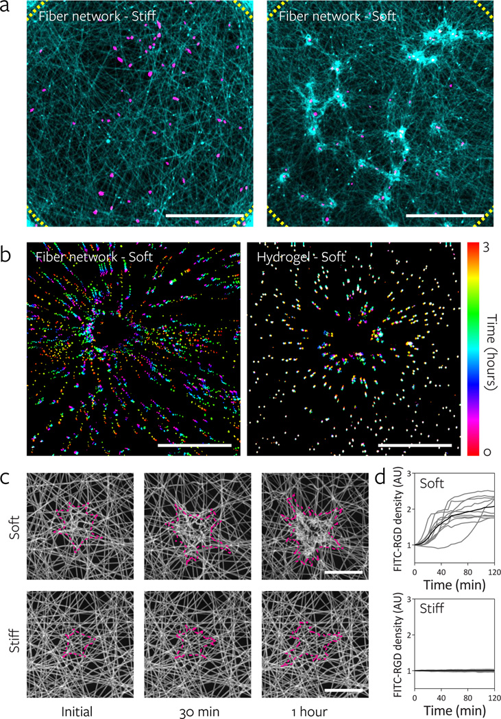
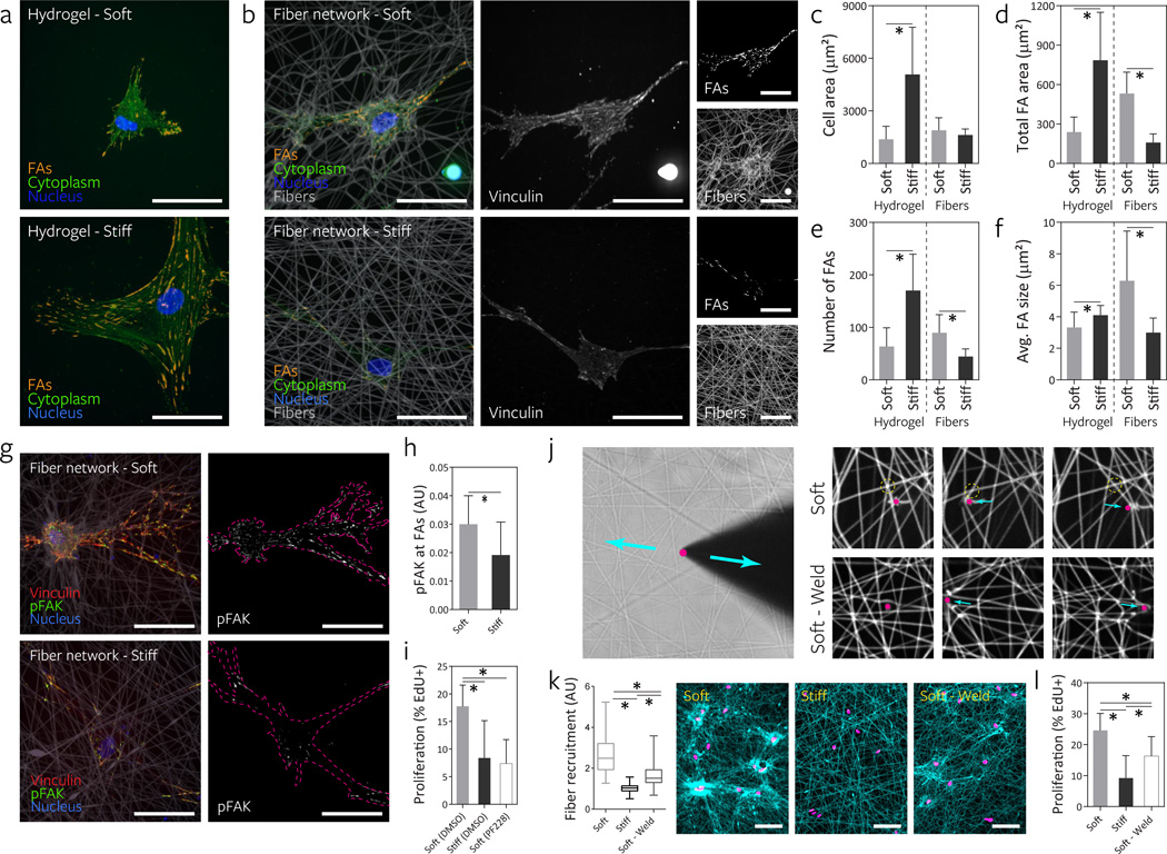
To investigate how cells sense stiffness in settings structurally similar to native extracellular matrices, we designed a synthetic fibrous material with tunable mechanics and user-defined architecture. In contrast to flat hydrogel surfaces, these fibrous materials recapitulated cell-matrix interactions observed with collagen matrices including stellate cell morphologies, cell-mediated realignment of fibres, and bulk contraction of the material. Increasing the stiffness of flat hydrogel surfaces induced mesenchymal stem cell spreading and proliferation; however, increasing fibre stiffness instead suppressed spreading and proliferation for certain network architectures. Lower fibre stiffness permitted active cellular forces to recruit nearby fibres, dynamically increasing ligand density at the cell surface and promoting the formation of focal adhesions and related signalling. These studies demonstrate a departure from the well-described relationship between material stiffness and spreading established with hydrogel surfaces, and introduce fibre recruitment as a previously undescribed mechanism by which cells probe and respond to mechanics in fibrillar matrices.


Figures

Figure 1
Figure 1. A novel approach to engineering fibrillar microenvironments with tunable mechanical and architectural features
a, Hierarchical overview of fabricating cell-adhesive suspended networks of dextran methacrylate (DexMA) fibers. Dextran is reacted with glycidyl methacrylate to generate DexMA. Following addition of a photoinitiator, DexMA is electrospun onto microfabricated substrates to define networks of suspended fibers. Networks are photo-crosslinked to tune fiber stiffness and the RGD is incorporated to enable cell attachment (scale bars, 100 µm). b, Through modulation of the electrospinning fabrication process, networks with varying fiber diameter (polymer solution concentration), density (fiber collection duration), and alignment (collection surface translation speed) can be generated, enabling the modeling of diverse fibrillar ECMs present in different tissue systems throughout the body (scale bars: 10 µm). c, Young’s modulus of individual fibers isolated over PDMS troughs and measured by three point bending AFM; n ≥ 12, mean ± s.d. d, Young’s modulus of DexMA fiber networks measured by cylindrical indentation with a calibrated cantilever; n ≥ 5, mean ± s.d. e, Young’s modulus of DexMA flat hydrogels determined by AFM spherical probe nanoindentation and Hertz contact mechanics; n ≥ 6, mean ± s.d.
Figure 2
Figure 2. Synthetic fiber networks induce similar topographical and mechanical interactions with cells as collagen matrices at multiple length scales
Comparison of cell spreading (a), spheroid outgrowth (b), and gel contraction (c) across hMSC-seeded DexMA fiber networks (left column), type I collagen matrices (middle column), and flat DexMA hydrogels (right column). a, Cytosolic and focal adhesion-localized vinculin (red), counter-stained for F-actin (green) and cell nuclei (blue) with phalloidin and Hoechst 33342, respectively. Dashed box designates region shown with F-actin or vinculin channel isolated (scale bars, 50 µm). b, Outgrowth of hMSC spheroids (100 cells/spheroid) stained for F-actin (green) and cell nuclei (blue). Rhodamine methacrylate-coupled DexMA fibers and hydrogel surface and Picrosirius Red-stained collagen are shown in the region spanning two spheroids, as indicated by the dashed box (scale bars, 50 µm). c, DexMA fibers and hydrogels and collagen were processed into thick circular slabs and seeded at high density with hMSCs. Initial and final states of soft (top) and stiff (bottom) constructs. Cell-mediated contraction was determined by normalizing construct diameter to the initial diameter; mean ± s.d., n ≥ 6 (scale bars, 500 µm).
Figure 3
Figure 3. Increasing fiber stiffness suppresses cell spreading and proliferation
The effect of altering material stiffness on hMSC spreading and proliferation was examined on DexMA hydrogels (top row, soft: 290 Pa, stiff: 19.1 kPa) and fiber networks (middle row, soft: 140 MPa fiber, 2.8 kPa network; stiff: 3.1 GPa fiber, 55 kPa network). Low (0.5 mg/mL, <0.3 kPa) and high (7.0 mg/mL, 1.1 kPa) concentration type I collagen matrices where bulk stiffness and adhesive ligand density increase in tandem were included for comparison (bottom row). a, Actin cytoskeletal organization of representative hMSCs 16 h after seeding, stained for F-actin (green) and cell nuclei (blue) (scale bars, 50 µm). b, Quantification of cell area; mean ± s.d., n ≥ 64 cells, * P < 0.05. c, Cell outlines of ten representative cells (scale bars, 50 µm). d, Proliferation of hMSCs over two days as determined by EdU incorporation; mean ± s.d., n ≥ 13 ROI with totals of 750–1500 cells analyzed, * P < 0.05.
Figure 4
Figure 4. Lower fiber and network stiffness enables cell-mediated reorganization of the material and clustering of adhesive ligands local to the cell
a, Reorganization of stiff and soft fiber networks 16 h after hMSC seeding. Fibers imaged by coupling with rhodamine methacrylate (cyan) and thresholded cell nuclei labeled with Hoechst 33342 (magenta). Dotted lines indicate the periphery of the suspended network (scale bars, 500 µm). b, Temporally color-coded overlays capturing the motion of beads embedded within soft fibers (fiber: 140 MPa, network: 2.8 kPa) and soft hydrogels (290 Pa) over a 3 h time course following hMSC seeding. c, Time-lapse images of FITC-RGD coupled fiber recruitment during the first two hours of hMSCs spreading on soft (top) and stiff (bottom) networks. Cell outlines shown in magenta (scale bars, 50 µm). d, Quantification of FITC-RGD fluorescence intensity in a 50 µm diameter circular region centered on the cell’s nucleus. Intensity was normalized to adjacent acellular areas; n = 10 cells.
Figure 5
Figure 5. Fibrillar ECM remodeling promotes focal adhesion (FA) formation and FAK phosphorylation to increase proliferation
a, FA formation of representative hMSCs seeded on DexMA hydrogels of low and high stiffness as visualized by cytosol extraction, vinculin immunostaining, and subsequent image analysis to identify FAs (orange). The cell’s cytoplasm (green) and nucleus (blue) are also shown (scale bars, 50 µm). b, FA formation of representative hMSCs on DexMA fiber networks of low and high fiber stiffness 16 h after seeding. Composite images (left) showing FAs (orange), cytoplasm (green), nuclei (blue) and fibers (grey). Single channel images of vinculin (middle) and fibers (right, bottom) as well as identified FAs (right, top) (scale bars, 50 µm). Cell area (c), total FA area (d), total number of FAs (e), and average FA size (f); mean ± s.d., n = 12 cells, * P < 0.05. g, Merged images of representative hMSCs 16 h after seeding co-stained for vinculin (red) and phospho-FAK (green). Single channel images of phospho-FAK with cells outlined in magenta (scale bars, 50 µm). h, Quantification of phospho-FAK localization to FAs determined by fluorescence intensity; mean ± s.d., n = 10 cells, * P < 0.05. i, Effect of FAK phosporylation inhibition on proliferation of hMSCs over two days, as determined by EdU incorporation; mean ± s.d., n ≥ 9 ROI with totals of 450–1500 cells analyzed, * P < 0.05. j, To test fiber-fiber connectivity, a diamond sharpened blade was placed adjacent to individual fibers and reciprocated via micromanipulator. Soft networks as fabricated in all previous studies possess limited connectivity as demonstrated by free sliding of fibers (top row) in contrast to “welded” networks with high fiber-fiber connectivity (bottom row). k, Remodeling of fiber networks 16 h after MSC seeding. Fiber recruitment in 50 µm diameter circular regions centered on the cell nucleus. Fibers imaged by coupling with rhodamine methacrylate (cyan) and thresholded cell nuclei labeled with Hoechst 33342 (magenta) (scale bars, 100 µm). Fluorescence intensity was normalized to adjacent acellular areas. l, Effect of altering fiber-fiber connectivity on proliferation of hMSCs over two days as determined by EdU incorporation; mean ± s.d., n ≥ 9 ROI with totals of 450–1500 cells analyzed, * P < 0.05.

References

    1. Discher DE, Janmey P, Wang Y. Tissue Cells Feel and Respond to the Stiffness of Their Substrate. Science. 2005;310:1139–1143. - PubMed
    1. Wozniak MA, Chen CS. Mechanotransduction in development: a growing role for contractility. Nat Rev Mol Cell Biol. 2009;10:34–43. - PMC - PubMed
    1. Jaalouk DE, Lammerding J. Mechanotransduction gone awry. Nat Rev Mol Cell Biol. 2009;10:63–73. - PMC - PubMed
    1. Pelham RJ, Wang Y. Cell locomotion and focal adhesions are regulated by substrate flexibility. PNAS. 1997;94:13661–13665. - PMC - PubMed
    1. Yeung T, et al. Effects of substrate stiffness on cell morphology, cytoskeletal structure, and adhesion. Cell Motil. Cytoskeleton. 2005;60:24–34. - PubMed

Publication types