. 2015 Oct 13:5:15042.
doi: 10.1038/srep15042.

Inhibition of Transforming Growth Factor-β Receptor signaling promotes culture expansion of undifferentiated human Endometrial Mesenchymal Stem/stromal Cells

Affiliations

Inhibition of Transforming Growth Factor-β Receptor signaling promotes culture expansion of undifferentiated human Endometrial Mesenchymal Stem/stromal Cells

Shanti Gurung et al. Sci Rep. .

Abstract

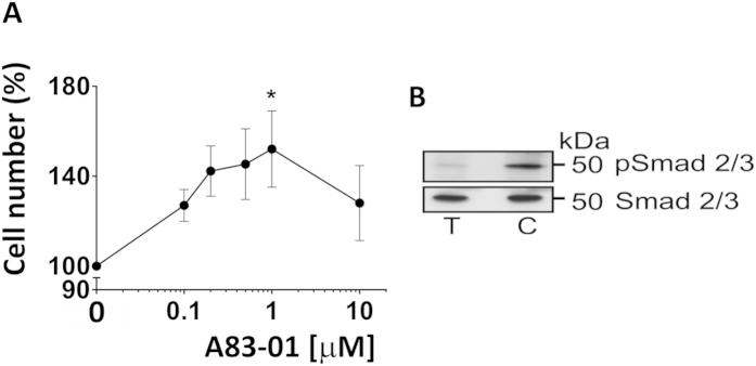
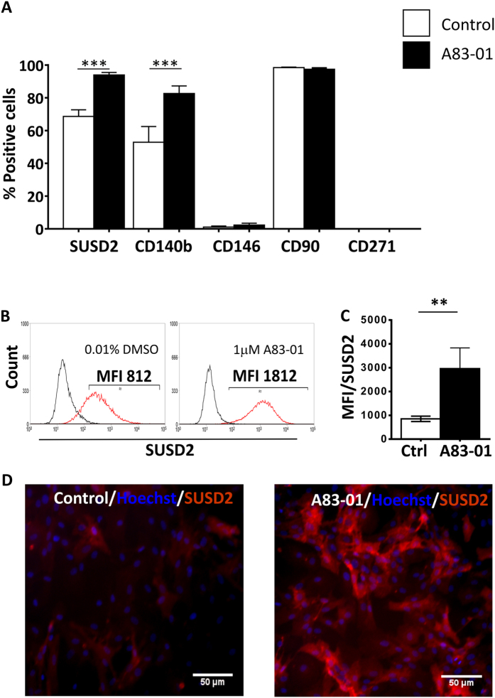
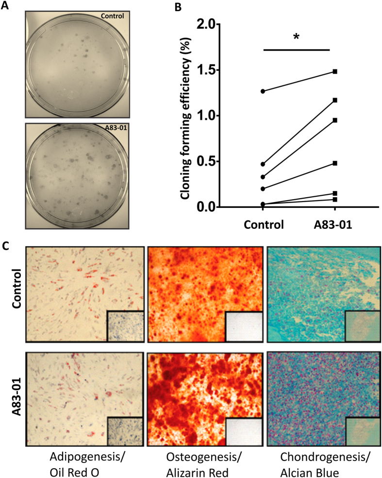
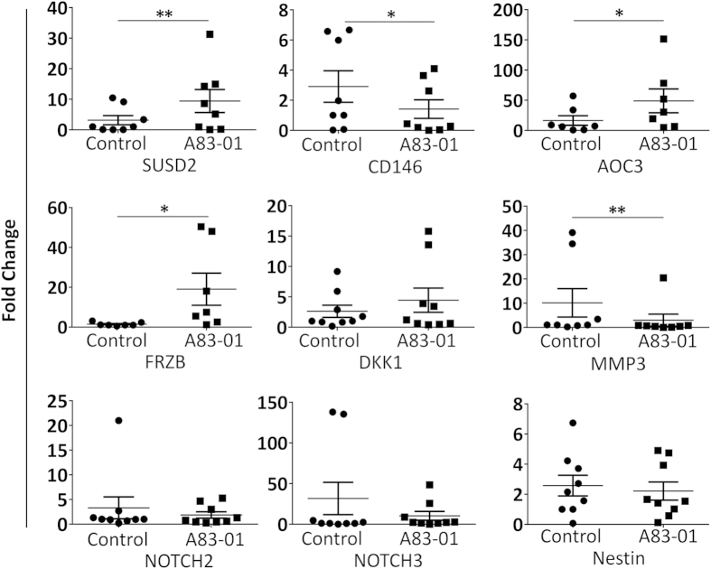
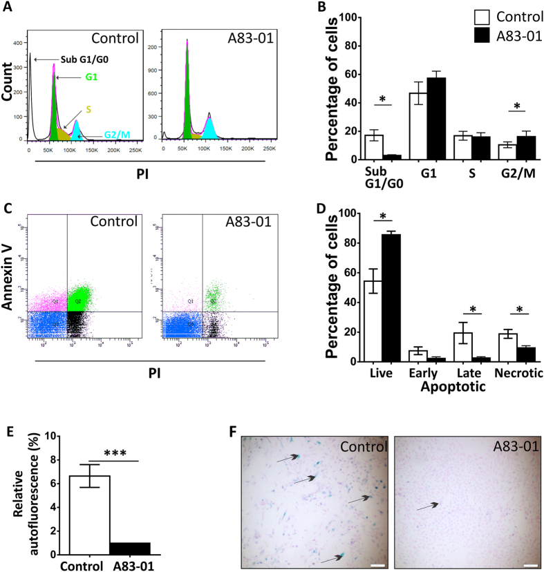
Human endometrial MSC (eMSC) are a novel source of MSC easily harvested from the highly regenerative uterine lining. We have developed protocols for eMSC isolation from single cell suspensions using magnetic bead-sorting using a perivascular marker antibody to SUSD2 and culture expansion in serum free medium (SFM). Similar to other MSC, eMSC spontaneously differentiate into fibroblasts during culture expansion decreasing their purity and efficacy. The aim of this study was to determine if A83-01, a TGF-β receptor inhibitor prevents eMSC differentiation in culture. SUSD2(+) eMSC were cultured in SFM with bFGF/EGF in 5% O2/5% CO2. At passage 6, eMSC were incubated with or without A83-01 for 7 days, then analysed for MSC properties. A83-01 dose dependently promoted SUSD2(+) eMSC proliferation and blocked apoptosis via the SMAD 2/3 pathway. Fewer A83-01 treated cells were autofluorescent or stained with β-galactosidase, indicating reduced senescence. A83-01-treated cells had higher cloning efficiency, differentiated into mesodermal lineages and expressed MSC phenotypic markers. These data suggest that A83-01 maintains SUSD2(+) eMSC stemness, promoting proliferation by blocking senescence and apoptosis in late passage cultures through binding to TGF-β receptors. Small molecules such as A83-01 may enable the expansion of undifferentiated MSC for use in tissue engineering and cell-based therapies.


Figures

Figure 1
Figure 1. Dose Response curve of A83-01 promotion of eMSC proliferation.
(A) Passage 3 eMSC incubated A83-01 in SFM in 5%O2/5%CO2 at 37 °C for 7 days was assessed by MTS cell viability assay. Means for triplicates were obtained for each sample at each concentration, then normalised to vehicle control DMSO (100%) and plotted as mean ± SEM of n = 6 patient samples. (B) Passage 6 eMSC lysates with or without 1μM A83-01 were immunoblotted with anti-SMAD 2/3 or anti-pSMAD 2/3 antibodies. A83-01 inhibited TGF-βR-induced phosphorylation of SMAD 2/3. (C = control, T =  treated).
Figure 2
Figure 2. Phenotype of P6 eMSC cultured with or without A83-01 in serum free medium in 5%O2.
(A) % Positive cells for MSC surface markers on passage 6 eMSC (n = 8) cultured in 1μM A83-01(black bars) or in 0.01% DMSO (white bars) for 7 days and assessed by single-colour flow cytometry. (B) Representative flow cytometric histograms of SUSD2+ eMSC treated with (black bar) and without (white bar) 1 μM A83-01 and MFI summarised in (C). (D) SUSD2 expression on control (left panel) and A83-01 treated (right panel) eMSC by immunofluorescence (red). Data are mean ± SEM of n = 8 different patient samples. **p < 0.01, ***p < 0.001.
Figure 3
Figure 3. Functional MSC properties of P6 eMSC cultured with or without A83-01 in serum free medium.
(A) Representative culture plates seeded at clonal density (50 cell/cm2). (B) Graph shows Colony Forming Efficiency of P6 eMSC pre-treated with 1μM A83-01 or 0.01% DMSO vehicle for 7 days in 5% O2 in SFM followed by clonal culture at 50 cells/cm2 in SFM for 4 weeks. (C) Multilineage mesodermal differentiation of 0.01% DMSO treated control and 1 μM A83-01 treated P6 eMSC showing adipogenic, osteogenic, and chondrogenic differentiation (controls were cultured in 1% serum media) for four weeks in 5%O2. Oil Red O was used to visualise cellular lipid vesicles for adipogenic differentiation, Alizarin Red to detect calcium mineralisation for osteogenic differentiation and Alcian Blue to detect acidic polysaccharides in the extracellular matrix for cartilage differentiation. Representative images from n = 3 samples. Data are mean ± SEM of n = 6 different samples *p < 0.05.
Figure 4
Figure 4. Quantitative RT-PCR analysis of MSC genes.
P6 eMSC cultures treated (black squares) or untreated control (black circles) with 1 μM A83-01 in 5%O2/5%CO2/90%N at 37 °C for 7 days. qRT-PCR analysis of SUSD2, CD146, AOC3, FRZB, MMP3, DKK1, NOTCH3, NOTCH2 and NESTIN. β-Actin or GAPDH were used to normalise the mRNA level, and fold change was calculated using 2−ΔΔCT. Data are mean ± SEM on n = 7 different tissue samples. *p < 0.05; **p < 0.01.
Figure 5
Figure 5. A83-01 blocks apoptosis and promotes eMSC proliferation.
P6 eMSC treated with 1 μM A83-01 or 0.01% DMSO cultured for 7 days in SFM in 5%O2/5%CO2/90%N were assessed by (A) Cell cycle analysis of PI stained cells. Shows representative PI staining on a linear axis of a flow cytometry plot (B) the percentage of cells in SubG1/G0, G1, S and G2/M stages of the cell cycle (black bar A83-01 treated, White bar control). Data are mean ± SEM, n = 7 patient samples; *p < 0.05. (C) Annexin-V and PI staining. Representative flow cytometric plots. The lower left quadrant of each panel shows the viable cells, upper left early apoptotic; upper right late apoptotic and lower right necrotic cells and (D) graph showing the percentage of live, apoptotic and necrotic cells. Data are mean ± SEM, n = 6 patient samples, *p < 0.05. (E) Relative autofluorescence by flow cytometry of unstained P6 cells treated (black bar) and untreated (white bar) with 1 μM A83-01. Data are mean ± SEM, n = 10, ***p < 0.005. (F) Representative images showing the staining of senescence-associated β-galactosidase (SA-β-gal) in cultured P6 eMSC treated with or without 1 μM A83-01.

References

    1. Crisan M. et al. Perivascular multipotent progenitor cells in human organs. Ann. N. Y. Acad. Sci. 1176, 118–123, 10.1111/j.1749-6632.2009.04967.x (2009). - DOI - PubMed
    1. Friedenstein A. J. et al. Precursors for fibroblasts in different populations of hematopoietic cells as detected by the in vitro colony assay method. Exp. Hematol. 2, 83–92 (1974). - PubMed
    1. Pittenger M. F. et al. Multilineage potential of adult human mesenchymal stem cells. Science 284, 143–147 (1999). - PubMed
    1. Dominici M. et al. Minimal criteria for defining multipotent mesenchymal stromal cells. The International Society for Cellular Therapy position statement. Cytotherapy 8, 315–317, 10.1080/14653240600855905 (2006). - DOI - PubMed
    1. Caplan A. I. & Bruder S. P. Mesenchymal stem cells: building blocks for molecular medicine in the 21st century. Trends in molecular medicine 7, 259–264 (2001). - PubMed

MeSH terms

Substances