

YK-4-279 effectively antagonizes EWS-FLI1 induced leukemia in a transgenic mouse model
- PMID: 26462019
- PMCID: PMC4741957
- DOI: 10.18632/oncotarget.5520
YK-4-279 effectively antagonizes EWS-FLI1 induced leukemia in a transgenic mouse model
Erratum in
-
Correction: YK-4-279 effectively antagonizes EWS-FLI1 induced leukemia in a transgenic mouse model.Oncotarget. 2024 Feb 22;15:143. doi: 10.18632/oncotarget.28524. Oncotarget. 2024. PMID: 38386842 Free PMC article. No abstract available.
Abstract
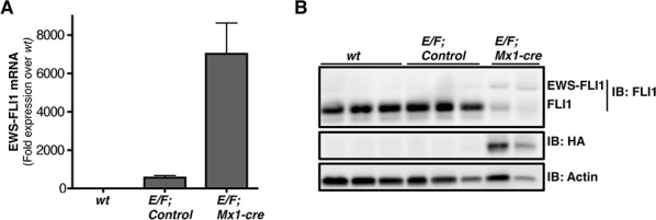
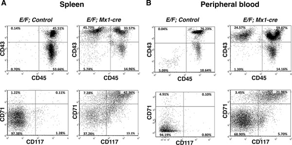
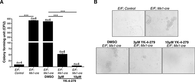
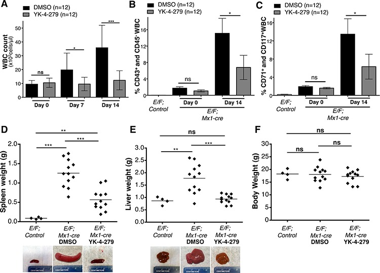
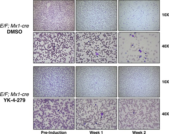
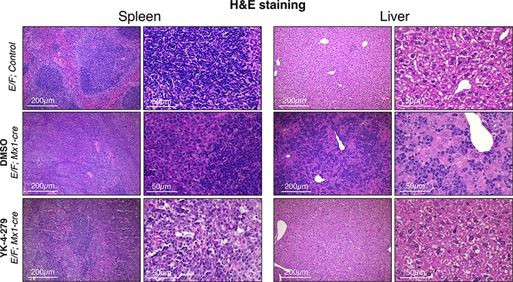
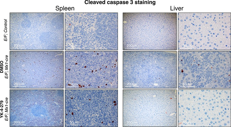
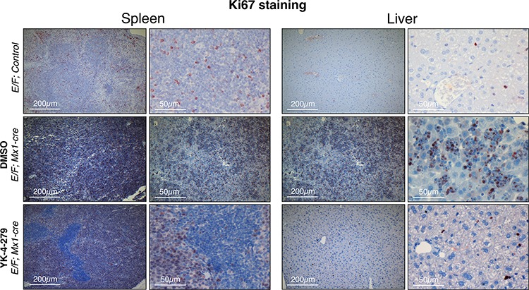
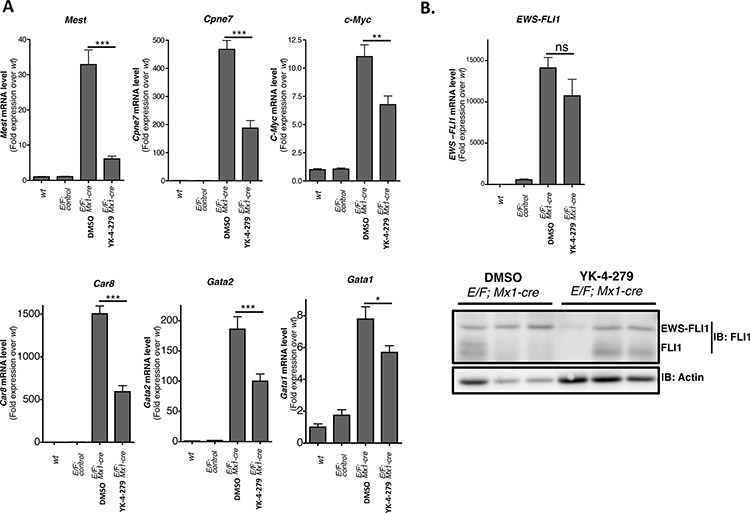
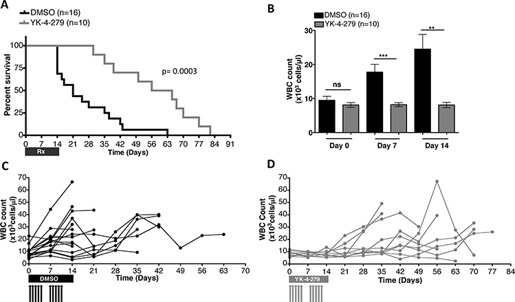
Ewing sarcoma is an aggressive tumor of bone and soft tissue affecting predominantly children and young adults. Tumor-specific chromosomal translocations create EWS-FLI1 and similar aberrant ETS fusion proteins that drive sarcoma development in patients. ETS family fusion proteins and over-expressed ETS proteins are also found in acute myeloid leukemia (AML) and acute lymphoblastic leukemia (ALL) patients. Transgenic expression of EWS-FLI1 in mice promotes high penetrance erythroid leukemia with dense hepatic and splenic infiltrations. We identified a small molecule, YK-4-279, that directly binds to EWS-FLI1 and inhibits its oncogenic activity in Ewing sarcoma cell lines and xenograft mouse models. Herein, we tested in vivo therapeutic efficacy and potential side effects of YK-4-279 in the transgenic mouse model with EWS-FLI1 induced leukemia. A two-week course of treatment with YK-4-279 significantly reduced white blood cell count, nucleated erythroblasts in the peripheral blood, splenomegaly, and hepatomegaly of erythroleukemic mice. YK-4-279 inhibited EWS-FLI1 target gene expression in neoplastic cells. Treated animals showed significantly better overall survival compared to control mice that rapidly succumbed to leukemia. YK-4-279 treated mice did not show overt toxicity in liver, spleen, or bone marrow. In conclusion, this in vivo study highlights the efficacy of YK-4-279 to treat EWS-FLI1 expressing neoplasms and support its therapeutic potential for patients with Ewing sarcoma and other ETS-driven malignancies.
Keywords: ETS fusion proteins; EWS-FLI1; YK-4-279; erythoid leukemia; ewing sarcoma.
Conflict of interest statement
United States Patent and Trademark Office awarded the patent for YK-4-279 to Georgetown University; inventors include A.Ü. and J.T. A license agreement has been executed between Georgetown University and Tokalas, Inc. for the patent. J.T and A.Ü. are shareholders of Tokalas, Inc.
Figures

Similar articles
-
Inhibition of the oncogenic fusion protein EWS-FLI1 causes G2-M cell cycle arrest and enhanced vincristine sensitivity in Ewing's sarcoma.Sci Signal. 2017 Oct 3;10(499):eaam8429. doi: 10.1126/scisignal.aam8429. Sci Signal. 2017. PMID: 28974650 Free PMC article.
-
BET bromodomain inhibitors suppress EWS-FLI1-dependent transcription and the IGF1 autocrine mechanism in Ewing sarcoma.Oncotarget. 2016 Jul 12;7(28):43504-43517. doi: 10.18632/oncotarget.9762. Oncotarget. 2016. PMID: 27259270 Free PMC article.
-
Pharmacokinetic modeling optimizes inhibition of the 'undruggable' EWS-FLI1 transcription factor in Ewing Sarcoma.Oncotarget. 2014 Jan 30;5(2):338-50. doi: 10.18632/oncotarget.1495. Oncotarget. 2014. PMID: 24481407 Free PMC article.
-
Therapeutic opportunities in Ewing sarcoma: EWS-FLI inhibition via LSD1 targeting.Oncotarget. 2016 Apr 5;7(14):17616-30. doi: 10.18632/oncotarget.7124. Oncotarget. 2016. PMID: 26848860 Free PMC article. Review.
-
Blocking the road, stopping the engine or killing the driver? Advances in targeting EWS/FLI-1 fusion in Ewing sarcoma as novel therapy.Expert Opin Ther Targets. 2014 Nov;18(11):1315-28. doi: 10.1517/14728222.2014.947963. Epub 2014 Aug 27. Expert Opin Ther Targets. 2014. PMID: 25162919 Review.
Cited by
-
Tumor Cell Dormancy: Threat or Opportunity in the Fight against Cancer.Cancers (Basel). 2019 Aug 19;11(8):1207. doi: 10.3390/cancers11081207. Cancers (Basel). 2019. PMID: 31430951 Free PMC article. Review.
-
RGD delivery of truncated coagulase to tumor vasculature affords local thrombotic activity to induce infarction of tumors in mice.Sci Rep. 2017 Aug 15;7(1):8126. doi: 10.1038/s41598-017-05326-9. Sci Rep. 2017. PMID: 28811469 Free PMC article.
-
EWS-FLI1 and RNA helicase A interaction inhibitor YK-4-279 inhibits growth of neuroblastoma.Oncotarget. 2017 Oct 19;8(55):94780-94792. doi: 10.18632/oncotarget.21933. eCollection 2017 Nov 7. Oncotarget. 2017. PMID: 29212266 Free PMC article.
-
Transcription/Replication Conflicts in Tumorigenesis and Their Potential Role as Novel Therapeutic Targets in Multiple Myeloma.Cancers (Basel). 2021 Jul 27;13(15):3755. doi: 10.3390/cancers13153755. Cancers (Basel). 2021. PMID: 34359660 Free PMC article. Review.
-
Targeting Transcription Factors for Cancer Treatment.Molecules. 2018 Jun 19;23(6):1479. doi: 10.3390/molecules23061479. Molecules. 2018. PMID: 29921764 Free PMC article. Review.
References
-
- Delattre O, Zucman J, Melot T, Garau XS, Zucker JM, Lenoir GM, Ambros PF, Sheer D, Turc-Carel C, Triche TJ, et al. The Ewing family of tumors—a subgroup of small-round-cell tumors defined by specific chimeric transcripts. N Engl J Med. 1994;331:294–299. - PubMed
-
- Aman P, Panagopoulos I, Lassen C, Fioretos T, Mencinger M, Toresson H, Hoglund M, Forster A, Rabbitts TH, Ron D, Mandahl N, Mitelman F. Expression patterns of the human sarcoma-associated genes FUS and EWS and the genomic structure of FUS. Genomics. 1996;37:1–8. - PubMed
-
- Bertolotti A, Melot T, Acker J, Vigneron M, Delattre O, Tora L. EWS, but not EWS-FLI-1, is associated with both TFIID and RNA polymerase II: interactions between two members of the TET family, EWS and hTAFII68, and subunits of TFIID and RNA polymerase II complexes. Mol Cell Biol. 1998;18:1489–1497. - PMC - PubMed
-
- Rossow KL, Janknecht R. The Ewing's sarcoma gene product functions as a transcriptional activator. Cancer Res. 2001;61:2690–2695. - PubMed
-
- Yang L, Chansky HA, Hickstein DD. EWS. Fli-1 fusion protein interacts with hyperphosphorylated RNA polymerase II and interferes with serine-arginine protein-mediated RNA splicing. J Biol Chem. 2000;275:37612–37618. - PubMed
Publication types
MeSH terms
Substances
Grants and funding
LinkOut - more resources
Full Text Sources
Other Literature Sources
