. 2015 Nov 3;6(34):36338-53.
doi: 10.18632/oncotarget.5537.

Mitochondrial ATF2 translocation contributes to apoptosis induction and BRAF inhibitor resistance in melanoma through the interaction of Bim with VDAC1

Affiliations

Mitochondrial ATF2 translocation contributes to apoptosis induction and BRAF inhibitor resistance in melanoma through the interaction of Bim with VDAC1

Zongwei Gao et al. Oncotarget. .

Abstract

Background: The mitochondrial accumulation of ATF2 is involved in tumor suppressor activities via cytochrome c release in melanoma cells. However, the signaling pathways that connect mitochondrial ATF2 accumulation and cytochrome c release are not well documented.

Methods: Several melanoma cell lines, B16F10, K1735M2, A375 and A375-R1, were treated with paclitaxel and vemurafenib to test the function of mitochondrial ATF2 and its connection to Bim and voltage-dependent anion channel 1 (VDAC1). Immunoprecipitation analysis was performed to investigate the functional interaction between the involved proteins. VDAC1 oligomerization was evaluated using an EGS-based crosslinking assay.

Results: The expression and migration of ATF2 to the mitochondria accounted for paclitaxel stimuli and acquired resistance to BRAF inhibitors. Mitochondrial ATF2 facilitated Bim stabilization through the inhibition of its degradation by the proteasome, thereby promoting cytochrome c release and inducing apoptosis in B16F10 and A375 cells. Studies using B16F10 and A375 cells genetically modified for ATF2 indicated that mitochondrial ATF2 was able to dissociate Bim from the Mcl-1/Bim complex to trigger VDAC1 oligomerization. Immunoprecipitation analysis revealed that Bim interacts with VDAC1, and this interaction was remarkably enhanced during apoptosis.

Conclusion: These results reveal that mitochondrial ATF2 is associated with the induction of apoptosis and BRAF inhibitor resistance through Bim activation, which might suggest potential novel therapies for the targeted induction of apoptosis in melanoma therapy.

Keywords: ATF2; Bim; Mcl-1; VDAC1; mitochondria.


Conflict of interest statement

CONFLICTS OF INTEREST

No potential conflicts of interest relevant to this article are reported.

Figures

Figure 1
Figure 1. ATF2-associated apoptosis in B16F10 and K1735M2 cells is mainly dependent on Bim
B16F10 cells (left panel) and K1735M2 cells (right panel) transfected with empty vector (EV) or ATF2T52A A. and scrambled or ATF2-specific shRNA B. were treated with paclitaxel (100 nM) for 24 h and subjected to apoptotic cell death analysis using Annexin V/PI staining. Columns represent the average values of three independent experiments; bars, s.e.m. *, p < 0.01 compared with the corresponding control groups (by one-way ANOVA). C. Immunoblotting analysis of the indicated proteins in the whole-cell lysates of B16F10 and K1735M2 cells that were treated with paclitaxel (100 nM) for the indicated times. The data are representative of three independent experiments, and the protein weights are indicated (in kDa). D. Immunoblotting analysis of the indicated proteins in the whole-cell lysates of B16F10 and K1735M2 cells stably transfected with scrambled or ATF2-specific shRNA that were treated with paclitaxel (100 nM) for 24 h. Representative figures of multiple experiments are shown. E. B16F10 cells were transfected with empty vector (EV), ATF2 (WT), ATF2T52A, or ATF2T52E, as well as ATF2 shRNA, and then treated with paclitaxel (100 nM, 12 h). Alternatively, prior to paclitaxel treatment, the mitochondrial accumulation of ATF2 was prevented with Leptomycin B (LMB, 40 ng/ml, 6 h). Cytosolic and mitochondrial fractions were subjected to western blot analysis with antibodies against Bim. β-actin and COX-IV were probed as loading controls. Representative figures of multiple experiments are shown. Lower panel: Quantitative data of relative intensity for E were calculated with respect to the corresponding loading control (n = 3). F. Confocal microscopy images of B16F10 cells transfected with scrambled or ATF2-specific shRNA that were treated with paclitaxel and stained with a Bim-specific antibody. The data are representative of three independent replicate coverslips per condition. Scale bar: 20 μm. Rr, Pearson's correlation coefficient; Mr, Mander's co-localization coefficient for red; Mg, Mander's co-localization coefficient for green.
Figure 2
Figure 2. Bim protein stabilization is regulated by ATF2
A. Bim mRNA levels in B16F10 cells transfected with ATF2-specific shRNA in the presence or absence of paclitaxel (100 nM) for 24 h (upper panel) and in A375 cells transfected with ATF2-specific shRNA in the presence or absence of vemurafenib (5 μM) for 24 h (lower panel). RNA levels are expressed relative to the levels detected in the 2 h untreated sample (assigned a value of 1; bars, s.e.m.). At least three independent experiments were performed. *p < 0.01 compared with the corresponding control groups (one-way ANOVA). B. Bim expression analysis in B16F10 cells (upper panel) and A375 cells (lower panel) was performed by a time course treatment of the transcriptional inhibitor actinomycin D (ActD, 2.5 μg/ml), followed by paclitaxel and vemurafenib treatment for 24 h, respectively. RNA levels are expressed relative to the levels detected in the 2 h untreated sample (assigned a value of 1; bars, s.e.m.). At least three independent experiments were performed. *p < 0.01, #p < 0.01 compared with the corresponding control groups (one-way ANOVA). C. Immunoblot for Bim in whole-cell lysates of B16F10 cells that were stably transfected with empty vector (EV) or ATF2T52A following cycloheximide (CHX, 50 μg/ml) incubation for the indicated times. The data are representative of three independent experiments. D. B16F10 cells transfected with ATF2-specific shRNA were treated with paclitaxel for 24 hr with or without the addition of the inhibitor PS341 (100 nM). Whole-cell lysates were subjected to western blot for Bim. Probing with antibody against β-actin was used as a loading control. Lower panel: Quantitative data of relative intensity for D and E. were calculated with respect to the sample at 0 h (assigned a value of 1).
Figure 3
Figure 3. Induction of Bim is critical for ATF2-associated apoptotic changes of the mitochondria and apoptosis
A. B16F10 cells stably transfected with ATF2-specific shRNA were treated with paclitaxel (100 nM) for 24 h. JC-1 flow cytometry analysis was performed, and representative flow cytometry plots of multiple experiments are shown. B. B16F10 cells stably transfected with scrambled or Bim-specific shRNA were treated with paclitaxel (100 nM) for 24 h. A western blot of the indicated proteins in whole-cell lysates is shown. β-actin was included as a loading control. C. B16F10 cells stably transfected with scrambled or Bim shRNA and Bad shRNA were treated with paclitaxel for 24 h. JC-1 flow cytometry analysis was performed, and representative flow cytometry plots are shown. B16F10 cells D. expressing scrambled or Bim shRNA were subjected to treatment with paclitaxel (100 nM) or ABT-737 (1 μM) for 24 h, and A375 cells E. expressing scrambled or Bim shRNA were subjected to vemurafenib (5 μM) treatment for 24 h. Apoptosis was then measured by Annexin V/PI staining (bars, s.e.m.) (n = 3). Columns represent the mean percentage of annexin V-positive cells from at least three independent experiments; bars, s.e.m. *p < 0.01 compared with the corresponding control groups (one-way ANOVA). F. B16F10 cells expressing scrambled or Bim shRNA were treated with paclitaxel (100 nM) for 24 h and subjected to western blotting for cytochrome c in the cytosolic and mitochondrial fractions. β-actin and COX-IV were used as loading controls. Representative figures of multiple experiments are shown.
Figure 4
Figure 4. Bim triggers mitochondrial VDAC upregulation and dimerization, Cytochrome c release and apoptosis
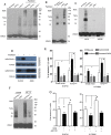
A. B16F10 cells stably transfected with empty vector (EV), ATF2T52A, or ATF2 shRNA were treated with paclitaxel (100 nM) for 24 h. The cells were then incubated with EGS (250 μM, 15 min), followed by SDS-PAGE and western blot analysis using an anti-VDAC1 antibody. The positions of the molecular weight protein standards are provided. Representative figures of multiple experiments are shown. B. B16F10 cells stably transfected with scrambled or Bim shRNA and Bad shRNA were treated with paclitaxel (100 nM) for 24 h. The cells were then subjected to crosslinking with EGS and western blot analysis using an anti-VDAC1 antibody. C. A375 cells stably transfected with scrambled or Bim shRNA and A375R cells were subjected to vemurafenib (5 μM) treatment for 24 h. The cells were then subjected to crosslinking with EGS and western blot analysis using an anti-VDAC1 antibody. D. B16F10 cells in the presence or absence of DIDS (100 μM, 1 hour) were treated with paclitaxel (100 nM) for 24 h. Western blot analysis of cytochrome c in the cytosolic and mitochondrial fractions was performed. β-actin and COX-IV were used as loading controls. Representative figures of multiple experiments are shown. E. B16F10 cells (left panel) and K1735M2 cells (right panel) stably transfected with empty vector (EV) or ATF2T52A in the presence or absence of DIDS were further treated with paclitaxel (100 nM) for 24 h and subjected to apoptosis analysis using Annexin V/PI staining. Columns represent the mean percentage of annexin V-positive cells from three independent experiments; bars, s.e.m. *, #p < 0.01 compared with the corresponding control groups (one-way ANOVA). F. A375 cells expressing scrambled or Bim-specific shRNA were treated with ABT737 (1 μM, 24 h) and subjected to crosslinking with EGS and western blot analysis using an anti-VDAC antibody. G. B16F10 and K1735M2 cells in the presence or absence of DIDS were treated with ABT737 and subjected to apoptotic cell death analysis using Annexin V/PI staining. Columns represent the mean percentage of annexin V-positive cells from three independent experiments; bars, s.e.m. *p < 0.01, #p < 0.01 compared with the control groups (one-way ANOVA).
Figure 5
Figure 5. Mitochondrial ATF2 does not directly bind to Bim following paclitaxel treatment
A. The association of Bim with ATF2 and Mcl-1 was detected by reciprocal immunoprecipitation assays. B16F10 cells (left panel) and K1735M2 cells (right panel) were treated with paclitaxel (100 nM) for 24 h. Bim was immunoprecipitated, and western blot analyses were performed for ATF2, Mcl-1 and Bim. Inputs for coimmunoprecipitation experiments were also subjected to western blot analysis. Representative figures of multiple experiments are shown. B. ATF2 was immunoprecipitated from B16F10 cells (left panel) and K1735M2 cells (right panel) treated with paclitaxel. Alternatively, ATF2-depleted supernatants were subjected to western blot analysis to determine the proportion of ATF2-bound and free Bim. Representative figures of multiple experiments are shown.
Figure 6
Figure 6. Mitochondrial ATF2 facilitates the association of Bim with VDAC1 following paclitaxel treatment
A. Bim was immunoprecipitated from paclitaxel-treated (100 nM, 24 h) cells (Left panel: B16F10 cells; Right panel: K1735M2 cells), and western blots were conducted to determine the association of Bim with VDAC1. Representative figures of multiple experiments are shown. B. Reciprocal immunoprecipitation-western blot analysis of paclitaxel-treated cells (Left panel: B16F10 cells; Right panel: K1735M2 cells) was performed for VDAC1, followed by western blot for Bim to determine VDAC1-bound Bim. Representative figures of multiple experiments are shown.
Figure 7
Figure 7. Mcl-1 is involved in the association of Bim with VDAC1
A. B16F10 cells (left panel), and K1735M2 cells (right panel) stably transfected with scrambled or Mcl-1 shRNA were treated with paclitaxel (100 nM) for 24 h, and Mcl-1 was immunoprecipitated, followed by immunoblotting for Bim to detect their interaction. Inputs for coimmunoprecipitation experiments were also subjected to western blot analysis. Representative figures of multiple experiments are shown. B. B16F10 cells (left panel) and K1735M2 cells (right panel) stably transfected with scrambled or Mcl-1 shRNA were treated with paclitaxel (100 nM) for 24 h, and VDAC1 was immunoprecipitated, followed by immunoblotting for Bim to detect their interaction. Representative figures of multiple experiments are shown. C. B16F10 cells (left panel), and K1735M2 cells (right panel) stably transfected with scrambled or Mcl-1 shRNA were treated with paclitaxel (100 nM) for 24 h and then subjected to apoptosis analysis using Annexin V/PI staining. Columns represent the mean percentage of annexin V-positive cells from three independent experiments; bars, s.e.m. *p < 0.01, #p < 0.01 compared with the paclitaxel + shMcl-1 group (one-way ANOVA).

Similar articles

Cited by

References

    1. Shah M, Bhoumik A, Goel V, Dewing A, Breitwieser W, Kluger H, Krajewski S, Krajewska M, Dehart J, Lau E, Kallenberg DM, Jeong H, Eroshkin A, et al. A role for ATF2 in regulating MITF and melanoma development. PLoS Genet. 2010;6:e1001258. - PMC - PubMed
    1. Roos WP, Quiros S, Krumm A, Merz S, Switzeny OJ, Christmann M, Loquai C, Kaina B. B-Raf inhibitor vemurafenib in combination with temozolomide and fotemustine in the killing response of malignant melanoma cells. Oncotarget. 2014;5:12607–12620. - PMC - PubMed
    1. Spagnolo F, Ghiorzo P, Queirolo P. Overcoming resistance to BRAF inhibition in BRAF-mutated metastatic melanoma. Oncotarget. 2014;5:10206–10221. - PMC - PubMed
    1. Lau E, Kluger H, Varsano T, Lee K, Scheffler I, Rimm DL, Ideker T, Ronai ZA. PKCε promotes oncogenic functions of ATF2 in the nucleus while blocking its apoptotic function at mitochondria. Cell. 2012;148:543–555. - PMC - PubMed
    1. Bhoumik A, Fichtman B, Derossi C, Breitwieser W, Kluger HM, Davis S, Subtil A, Meltzer P, Krajewski S, Jones N, Ronai Z. Suppressor role of activating transcription factor 2 (ATF2) in skin cancer. Proc Natl Acad Sci U S A. 2008;105:1674–1679. - PMC - PubMed

Publication types

MeSH terms