. 2015 Oct 12:3:12.
doi: 10.1186/s40170-015-0137-1. eCollection 2015.

Targeting glutamine metabolism sensitizes pancreatic cancer to PARP-driven metabolic catastrophe induced by ß-lapachone

Affiliations

Targeting glutamine metabolism sensitizes pancreatic cancer to PARP-driven metabolic catastrophe induced by ß-lapachone

Gaurab Chakrabarti et al. Cancer Metab. .

Abstract

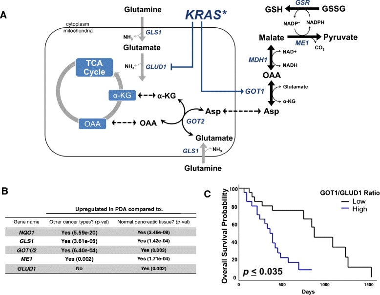
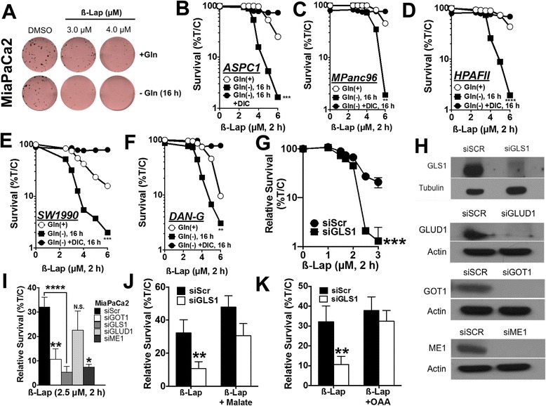
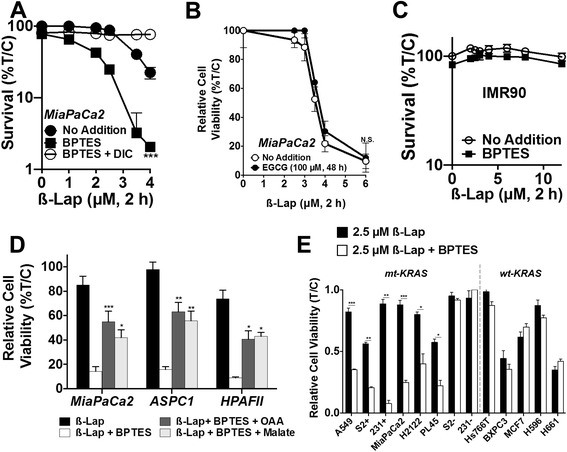
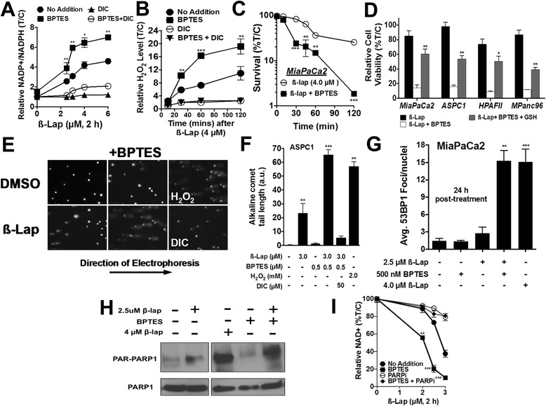
Background: Pancreatic ductal adenocarcinomas (PDA) activate a glutamine-dependent pathway of cytosolic nicotinamide adenine dinucleotide phosphate (NADPH) production to maintain redox homeostasis and support proliferation. Enzymes involved in this pathway (GLS1 (mitochondrial glutaminase 1), GOT1 (cytoplasmic glutamate oxaloacetate transaminase 1), and GOT2 (mitochondrial glutamate oxaloacetate transaminase 2)) are highly upregulated in PDA, and among these, inhibitors of GLS1 were recently deployed in clinical trials to target anabolic glutamine metabolism. However, single-agent inhibition of this pathway is cytostatic and unlikely to provide durable benefit in controlling advanced disease.

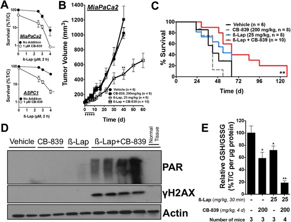
Results: Here, we report that reducing NADPH pools by genetically or pharmacologically (bis-2-(5-phenylacetamido-1,2,4-thiadiazol-2-yl)ethyl sulfide (BPTES) or CB-839) inhibiting glutamine metabolism in mutant Kirsten rat sarcoma viral oncogene homolog (KRAS) PDA sensitizes cell lines and tumors to ß-lapachone (ß-lap, clinical form ARQ761). ß-Lap is an NADPH:quinone oxidoreductase (NQO1)-bioactivatable drug that leads to NADPH depletion through high levels of reactive oxygen species (ROS) from the futile redox cycling of the drug and subsequently nicotinamide adenine dinucleotide (NAD)+ depletion through poly(ADP ribose) polymerase (PARP) hyperactivation. NQO1 expression is highly activated by mutant KRAS signaling. As such, ß-lap treatment concurrent with inhibition of glutamine metabolism in mutant KRAS, NQO1 overexpressing PDA leads to massive redox imbalance, extensive DNA damage, rapid PARP-mediated NAD+ consumption, and PDA cell death-features not observed in NQO1-low, wild-type KRAS expressing cells.

Conclusions: This treatment strategy illustrates proof of principle that simultaneously decreasing glutamine metabolism-dependent tumor anti-oxidant defenses and inducing supra-physiological ROS formation are tumoricidal and that this rationally designed combination strategy lowers the required doses of both agents in vitro and in vivo. The non-overlapping specificities of GLS1 inhibitors and ß-lap for PDA tumors afford high tumor selectivity, while sparing normal tissue.

Keywords: Glutamine metabolism; Metabolic cancer therapy; NQO1-bioactivated drugs; Transamination.


Figures

Fig. 1
Fig. 1
Glutamine metabolism genes are upregulated in PDA. a Glutamine utilization pathway in PDA. Asp aspartate, GSR glutathione-disulfide reductase. b NQO1 and glutamine metabolism enzymes assessed in patient tumor tissue in PDA versus 17 other cancer types (central columns, “other” cancers include solid and hematological malignancies) and PDA versus normal pancreatic tissue (right most column). Data were obtained from Oncomine (www.oncomine.org). c Kaplan–Meier survival curve corrected for multiple comparisons of 45 PDA patients grouped according to high versus low GOT1:GLUD1 expression assessed using the PROGgene webtool (http://watson.compbio.iupui.edu/chirayu/proggene/database/?url=proggene)
Fig. 2
Fig. 2
Inhibiting glutamine metabolism sensitizes PDA to ß-lap. a Colony formation assay of MiaPaCa2 cells depleted of glutamine for 16 h, followed by treatment with ß-lap for 2 h. p < 0.01 at 3 μM and p < 0.0001 at 4 μM. bf Glutamine deprivation experiments with a dose range of ß-lap in ASPC1, MPanc96, HPAFII, SW1990, and DAN-G PDA cell lines. g ß-lap dose response in MiaPaCa2 cells with knockdown of GLS1. h Western blots for GLS1, ME1, GOT1, and GLUD1 upon knockdown in MiaPaCa2 cell lines for 48 h using siRNA. i Relative survival of GOT1, GLS1, GLUD1, and ME1 MiaPaCa2 knockdown cells treated with 2.5 μM ß-lap for 2 h. j, k GLS1 was knocked down in MiaPaCa2, and either 3 mM oxaloacetate (OAA) or 3 mM dimethyl malate was added for 24 h followed by a 2-h treatment with 2.5 μM ß-lap. Relative survival represents means of CellTiter-Glo survival assay 48 h after treatment plotted as percentage treated/control (T/C), ±SE from sextuplicate samples. All results were compared using Student’s t tests. *p < 0.05; **p < 0.01; ***p < 0.001; ****p < 0.0001
Fig. 3
Fig. 3
GLS1 inhibition by BPTES sensitizes PDA to ß-lap in an NQO1-dependent manner. a Clonogenic survival assay of MiaPaCa2 pre-treated ± 500 nM BPTES for 48 h followed by the addition of ß-lap ±50 μM for 2 h. Data represent survival means ± SE from quadruplicate samples. b MiaPaCa2 cells treated with 100 μM of the GLUD1 inhibitor, EGCG, for 48 h followed by 2-h ß-lap dose response. Relative cell viability represents mean of CellTiter-Glo survival assay 48 h after ß-lap treatment plotted as percentage treated/control (T/C) ± SE from sextuplicate samples. c Clonogenic survival assay of normal lung fibroblast cell line, IMR90, pre-treated with ±500 nM BPTES for 48 h followed by 2 h of ß-lap treatment. Data represent survival means ± SE from quadruplicate samples. d MiaPaCa2, ASPC1, and HPAFII PDA cell lines were pre-treated with ± 500 nM BPTES for 48 h, either 3 mM oxaloacetate (OAA) or 3 mM dimethyl malate for the last 24 h, and 2.5 μM ß-lap for 2 h. Relative cell viability represents means of CellTiter-Glo survival assay 48 h after treatment plotted as percentage (T/C) ± SE from sextuplicate samples. e Various cancer cell lines pre-treated with ± 500 nM BPTES (sub-growth inhibitory) for 48 h followed by the addition of 2.5 μM ß-lap for 2 h. Mutant (mt) KRAS lines: A549 non-small cell lung (NSCL), PL45 PDA, NQO1 expressing S2-013 (S2+) PDA, NQO1 expressing MDA-MB-231 (231+) triple-negative breast, H2122 NSCL, NQO1-deficient S2-013 (S2−) PDA, and NQO1 deficient MDA-MB-231 (231−) triple-negative breast cancer cells. Wild-type (wt) KRAS lines: Hs766T PDA, BxPC3 PDA, MCF7 breast, as well as NQO1+ H596 NSCL and H661 NSCL cancer cell lines
Fig. 4
Fig. 4
GLS1 inhibition decreases anti-oxidant defenses and increases susceptibility to ß-lap-induced DNA damage. a Relative NADP+ to NADPH ratio in MiaPaCa2 cells pre-treated with BPTES for 48 h (500 nM) and then treated with ß-lap for 2 h. NADP+ and NADPH levels were measured immediately after 2 h treatment. b Relative extracellular H2O2 was measured through luminescence assay from the media of 4 μM ß-lap ± DIC, ±BPTES-treated MiaPaCa2 over a time-frame of 120 min, ±SE from sextuplicate samples. c Clonogenic survival of MiaPaCa2 pre-treated ± 500 nM BPTES for 48 h followed by the addition of ß-lap for various incubation times. Data represent survival means ± SE from quadruplicate samples. d MiaPaCa2, ASPC1, HPAFII, and MPanc96 pre-treated with ±500 nM BPTES and ±GSH reduced ethyl ester for 48 h followed by the addition of ß-lap for 2 h. e, f Alkaline comet assay of ASPC1 PDA cell lines pre-treated with ±500 nM BPTES followed by 2 h of ß-lap. g Average 53BP1 foci 24 h post treatment in MiaPaCa2. h Western blot for PAR formation with indicated treatment after 15 min of ß-lap exposure. i Relative NAD+ levels ±BPTES with various doses of ß-lap after 2 h of treatment. All results were compared using Student’s t tests. *p < 0.05; **p < 0.01; ***p < 0.001
Fig. 5
Fig. 5
GLS1 inhibition sensitizes pancreatic cancer to ß-lap in vivo. a Clonogenic survival of MiaPaCa2 and ASPC1 cells pre-treated with 1 μM CB-839 for 48 h followed by 2 h of ß-lap dose response. b Subcutaneous tumors grown from MiaPaCa2 cells in nude mice were allowed to reach a volume of 100 mm3, after which, the mice were treated every other day with vehicle (HPßCD, n = 6), sub-efficacious dose of CB-839 (oral gavage, 200 mg/kg, twice daily for 10 days, n = 8), sub-efficacious dose of ß-lap (IV, 25 mg/kg, n = 8) or sub-efficacious doses of CB-839 and ß-lap (n = 10) for a total of five doses (arrows). Tumor growth was monitored until tumors reached 1000 mm3. Error bars, SEM. c Survival of tumor-bearing mice represented as a Kaplan–Meier plot. The mice were sacrificed when tumors reached 1000 mm3. Statistically analyzed with the log-rank test for trend. d Western blot of PAR and γH2AX from a set of tumors harvested 30 min after treatment with ß-lap ± CB-839 (4-day treatment), n = 3 tumors per group, same doses as above. e Relative GSH/GSSG ratio in treated groups normalized to microgram of tumor protein. Unless stated otherwise, all results were compared using Student’s t tests. *p < 0.05; **p < 0.01; ***p < 0.001

References

    1. Hidalgo M. Pancreatic cancer. N Engl J Med. 2010;362(17):1605–1617. doi: 10.1056/NEJMra0901557. - DOI - PubMed
    1. Bryant KL, Mancias JD, Kimmelman AC, Der CJ. KRAS: feeding pancreatic cancer proliferation. Trends Biochem Sci. 2014;39(2):91–100. doi: 10.1016/j.tibs.2013.12.004. - DOI - PMC - PubMed
    1. Cox AD, Fesik SW, Kimmelman AC, Luo J, Der CJ. Drugging the undruggable RAS: mission possible? Nat Rev Drug Discov. 2014;13(11):828–851. doi: 10.1038/nrd4389. - DOI - PMC - PubMed
    1. Kong B, Qia C, Erkan M, Kleeff J, Michalski CW. Overview on how oncogenic Kras promotes pancreatic carcinogenesis by inducing low intracellular ROS levels. Front Physiol. 2013;4:246. doi: 10.3389/fphys.2013.00246. - DOI - PMC - PubMed
    1. Lyssiotis CA, Son J, Cantley LC, Kimmelman AC. Pancreatic cancers rely on a novel glutamine metabolism pathway to maintain redox balance. Cell Cycle. 2013;12(13):1987–1988. doi: 10.4161/cc.25307. - DOI - PMC - PubMed