. 2016 Mar;183(3):469-79.
doi: 10.1111/cei.12730. Epub 2015 Dec 8.

NET amyloidogenic backbone in human activated neutrophils

Affiliations

NET amyloidogenic backbone in human activated neutrophils

L Pulze et al. Clin Exp Immunol. 2016 Mar.

Abstract

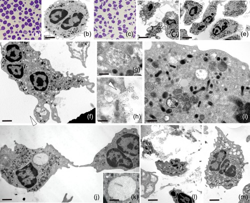
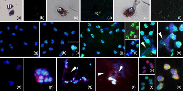
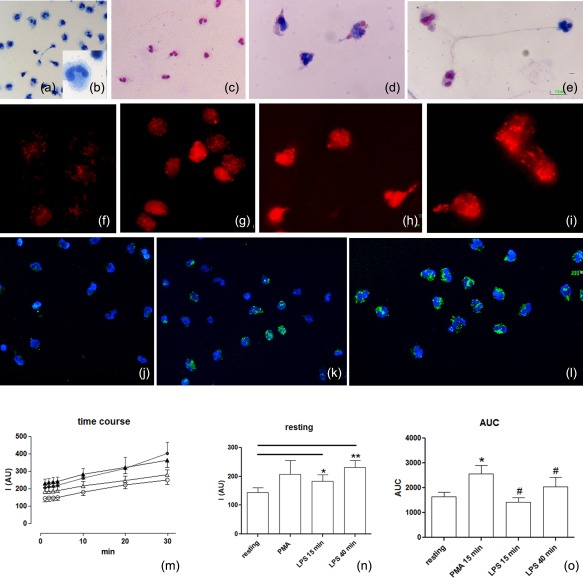
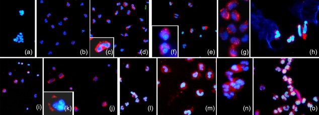
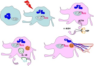
Activated human neutrophils produce a fibrillar DNA network [neutrophil extracellular traps (NETs)] for entrapping and killing bacteria, fungi, protozoa and viruses. Our results suggest that the neutrophil extracellular traps show a resistant amyloidogenic backbone utilized for addressing reputed proteins and DNA against the non-self. The formation of amyloid fibrils in neutrophils is regulated by the imbalance of reactive oxygen species (ROS) in the cytoplasm. The intensity and source of the ROS signal is determinant for promoting stress-associated responses such as amyloidogenesis and closely related events: autophagy, exosome release, activation of the adrenocorticotrophin hormone/α-melanocyte-stimulating hormone (ACTH/α-MSH) loop and synthesis of specific cytokines. These interconnected responses in human activated neutrophils, that have been evaluated from a morphofunctional and quantitative viewpoint, represent primitive, but potent, innate defence mechanisms. In invertebrates, circulating phagocytic immune cells, when activated, show responses similar to those described previously for activated human neutrophils. Invertebrate cells within endoplasmic reticulum cisternae produce a fibrillar material which is then assembled into an amyloidogenic scaffold utilized to convey melanin close to the invader. These findings, in consideration to the critical role played by NET in the development of several pathologies, could explain the structural resistance of these scaffolds and could provide the basis for developing new diagnostic and therapeutic approaches in immunomediated diseases in which the innate branch of the immune system has a pivotal role.

Keywords: ACTH axis; ROS evaluation; amyloidogenesis; exosomes; neutrophil extracellular trap.


Figures

Figure 1
Figure 1
Unstimulated and stimulated human neutrophils. Thin and semithin sections of resting (a,b) and activated neutrophils (c–m). The unstimulated cells (a,b) are roundish, but after stimulation (c) with both phorbol myristate acetate (PMA) (d) and lipopolysaccharide (LPS) (e–m) they lose their globoid shape. Starting from 15 min after stimulation, neutrophils show a transformed phenotype characterized by irregular profile, due to the presence of protrusions and pseudopodia (d,e). In a time lapse between 15 and 40 min from LPS activation (e–m), a series of events are detectable due to the presence of multi‐vesicular bodies (f,g), released exosomes (f,h) and autophagosomes containing portions of the cytosol (i). Activated cells show large dilated reticulum cisternae (j,k) (arrowheads) filled with spatially organized fibrillar material (k) or empty vacuoles (l,m) and material among cells (m). Scale bars. (a) 6 μm; (b) 5 μm; (d) 9 μm; (e) 3·5 μm; (f) 4·5 μm; (g) 1·9 μm.
Figure 2
Figure 2
Amyloidogenesis in lipopolysaccharide (LPS)‐stimulated neutrophils. (a–n) Detection of amyloid fibril presence with Congo red (CR) and thioflavin S (ThS) stainings. (a–f) CR staining: unstimulated neutrophils (a,b) in comparison with LPS‐stimulated cells (c–f) at the starting point of activation. The CR positive cells show the typical apple green birifrangence evidencing the spotted presence of amyloid structures in the cytoplasm. Nuclei are counterstained with haematoxylin (n). (g–n) Identification of amyloid fibrils with ThS: unstimulated neutrophils (g) in comparison with stimulated cells at 15 min (h–j) and 40 min (k–n) from LPS administration. Within cells (h–j) and extracellularly (k–n), amyloid fibrils are localized by ThS bright fluorescence. Nuclei and DNA material in neutrophil extracellular traps (NETs) are stained with 4′,6′‐diamino‐2‐phenylindole (DAPI) and marked in brilliant blue. Both single staining, for ThS and DAPI (k,l) and the two overlaid signals are proposed to better identify the presence of amyloid and DNA materials. Note the co‐localization of blue (DAPI) and green (ThS) in NETs released on stimulation (arrowheads). The semi‐quantitative analysis of the concomitant amyloid fibril presence (ThS) and DNA (DAPI) of NETs showed a ratio of 0·78 ± 0·03. (o–u) (PMEL17) immunolocalization: immunofluorescence staining showing, in comparison to control (o), the increased expression of Pmel17 that is present in the cytoplasm of stimulated neutrophils (p–r) and in the NETs (q,r). DAPI (blue) staining for DNA shows distinct cores indicating nuclei, as well as diffuse patterns evidencing extracellular DNA in NETs (arrowheads). (s–u) Double‐labelling of neutrophils with ThS (green) and antibody against Pmel17 (red). The co‐localization of two signals (merge in yellow, u) is quite good. Nuclei (blue) are stained with DAPI.
Figure 3
Figure 3
Evaluation of intracellular reactive oxygen species (ROS). (a–e) Light microscopy: resting (a,b) and stimulated cells (c–e) are stained using the May–Grünwald–Giemsa technique. The differential staining of neutrophils depends on cytoplasmic pH (the pink mark indicates acid pH while the blue mark shows an alkaline pH). (f–o) ROS evaluation was obtained using fluorescence dyes: the MitoSOX red used as a selective indicator of mitochondrial superoxide and 2′,7′‐dichlorodihydrofluorescein diacetate (H2DCFDA) to detect the overall degree of cytoplasmic ROS. Most ROS (red signal) were generated in mitochondria (f–i) starting from the resting condition (f) up to 15 min (g) and 40 min (h,i) after lipopolysaccharide (LPS) stimulation. The increase of the overall degree of intracellular ROS (j–l), using H2DCFDA, is localized by fluorescence microscopy: (j) cells in resting condition (k,l) 15 and 40 min after LPS stimulation, respectively. In neutrophils, from the resting condition to neutrophil extracellular trap (NET) formation, the concentration of cellular ROS levels increased following appropriate [LPS and phorbol myristate acetate (PMA)] stimulation on a rapid time‐scale. (m) Spectrofluorimetric evaluation of the time–course of ROS generation in human neutrophils under resting conditions (empty circle), prestimulation for 15 min with PMA (filled circles) for 15 min (empty triangles) or 40 min (filled triangles) with LPS. (n) Delta variations of values measured in neutrophils under the different conditions. (o) Area under the curve (AUC) of fluorescence measured for detection of neutrophil ROS generations during the 30 min of detection. Data are presented as mean ± standard error of at least three to five separate experiments. *P < 0·05 versus resting PMN; #P < 0·05 versus PMA.
Figure 4
Figure 4
Characterization of lipopolysaccharide (LPS)‐stimulated neutrophils. (a–o) Immunocytochemical characterizations. Interleukin (IL)‐18 (a–d), adrenocorticotrophin hormone (ACTH) (e–h), alpha melanocyte‐stimulating hormone (α‐MSH) (i–j) and neutral endopeptidase (NEP) (l–o) expressions show high levels of positivity in activated neutrophils (b–d, f–h, j–k, m–o), while in resting cells the expression of IL‐18 (a), ACTH (e), α‐MSH (i) and NEP (l) is basal.
Figure 5
Figure 5
Schematic overview explaining the possible behaviour of lipopolysaccharide (LPS)‐activated neutrophils. LPS activation of neutrophils results in an increase of reactive oxygen species (ROS) levels. Concomitant cross‐talk between immune and neuroendocrine systems induces an activation of stress‐sensoring circuits to produce adrenocorticotrophin hormone (ACTH), neutral endopeptidase (NEP), alpha melanocyte‐stimulating hormone (α‐MSH) and release of interleukin (IL)‐18. In the cell‐linked events, such as production of exosomes, autophagocytosis and amyloidogenesis take place. The reticulum cisternae become an amyloid fibril reservoir. Subsequently, the exocytosed amyloid lattice co‐operates to form the backbone structure of NET. n = nucleus.

References

    1. Borregaard N, Cowland J. Granules of the human neutrophilic polymorphonuclear leukocyte. Blood 1997; 89:3503–21. - PubMed
    1. Brinkmann V, Zychlinsky A. Beneficial suicide: why neutrophils die to make NETs. Nat Rev Microbiol 2007; 5:577–82. - PubMed
    1. Papayannopoulos V, Zychlinsky A. NETs: a new strategy for using old weapons. Trends Immunol 2009; 30:513–21. - PubMed
    1. Mantovani A, Cassatella MA, Costantini C, Jaillon S. Neutrophils in the activation and regulation of innate and adaptative immunity. Nat Rev Immunol 2011; 11:519–31. - PubMed
    1. Remijsen Q, Kuijpers TW, Wirawan E, Lippens S, Vandenabeele P, Vanden Berghe T. Dying for a cause: NETosis, mechanisms behind an antimicrobial cell death modality. Cell Death Differ 2011; 18:581–8. - PMC - PubMed

Publication types