. 2016 Jan:85:60-72.
doi: 10.1016/j.nbd.2015.10.004. Epub 2015 Oct 14.

Cognitive and behavior deficits in sickle cell mice are associated with profound neuropathologic changes in hippocampus and cerebellum

Affiliations

Cognitive and behavior deficits in sickle cell mice are associated with profound neuropathologic changes in hippocampus and cerebellum

Li Wang et al. Neurobiol Dis. 2016 Jan.

Abstract

Strokes are perhaps the most serious complications of sickle cell disease (SCD) and by the fifth decade occur in approximately 25% of patients. While most patients do not develop strokes, mounting evidence indicates that even without brain abnormalities on imaging studies, SCD patients can present profound neurocognitive dysfunction. We sought to evaluate the neurocognitive behavior profile of humanized SCD mice (Townes, BERK) and to identify hematologic and neuropathologic abnormalities associated with the behavioral alterations observed in these mice. Heterozygous and homozygous Townes mice displayed severe cognitive deficits shown by significant delays in spatial learning compared to controls. Homozygous Townes also had increased depression- and anxiety-like behaviors as well as reduced performance on voluntary wheel running compared to controls. Behavior deficits observed in Townes were also seen in BERKs. Interestingly, most deficits in homozygotes were observed in older mice and were associated with worsening anemia. Further, neuropathologic abnormalities including the presence of large bands of dark/pyknotic (shrunken) neurons in CA1 and CA3 fields of hippocampus and evidence of neuronal dropout in cerebellum were present in homozygotes but not control Townes. These observations suggest that cognitive and behavioral deficits in SCD mice mirror those described in SCD patients and that aging, anemia, and profound neuropathologic changes in hippocampus and cerebellum are possible biologic correlates of those deficits. These findings support using SCD mice for studies of cognitive deficits in SCD and point to vulnerable brain areas with susceptibility to neuronal injury in SCD and to mechanisms that potentially underlie those deficits.

Keywords: Anemia; Anxiety; Cerebellum; Depression; Hippocampus; Learning; Memory; Neuronal injury; Pain.


Conflict of interest statement

Declaration of Conflicting Interests

The authors declare that there is no conflict of interest.

Figures

Figure 1
Figure 1
Cognitive learning and memory in Townes SCD mice. Homozygous and heterozygous Townes mice had cognitive deficits as they took significantly longer to reach criterion for spatial learning compared to control mice (A). Kaplan-Meier survival analysis of times to reaching criterion for spatial learning also indicates the presence of cognitive deficits in homozygous and heterozygous Townes mice as both took significantly longer time to reach criterion of spatial learning compared to control mice (both P<0.001, B). During the water T-maze spatial learning test, homozygous mice took significantly longer times to reach the platform on the second day of testing compared to control mice (C). SCD genotype had no effect on reversal learning as homozygous and heterozygous Townes reached criterion for reversal learning at similar times compared to control mice (both, P≥0.2, Figure 1D and 1E). During the reversal learning test, homozygous mice took longer times to reach the platform on the second and third days of testing compared to control mice (F). P values reflect post hoc analysis comparing the groups indicated by the brackets. Fifty-eight mice underwent the water T-maze test, N=17–24 per genotype including balanced numbers of age-matched male and females. * reflects p<0.05, † reflects p<0.01, and ‡, p<0.001.
Figure 2
Figure 2
Depression-like behavior in Townes SCD mice. Homozygous Townes mice spent significantly greater time immobile compared to controls and heterozygous Townes suggesting that homozygous animals have significant depression-like behavior compared with controls and heterozygous mice (A). Homozygous and heterozygous Townes mice spent significantly less time climbing during the forced swim test compared to control mice (B). Homozygous mice spent less time swimming compared to heterozygous mice (C). P values reflect post hoc analysis comparing the groups indicated by the brackets. Seventy-eight mice underwent the forced swim test, N=23–29 per genotype including balanced numbers of age-matched male and females. * reflects p<0.05, † reflects p<0.01, and ‡, p<0.001.
Figure 3
Figure 3
Anxiety-like behavior in Townes SCD mice. Homozygous mice spent a significantly greater percentage of time in closed arms and lower percentage of time in the center and open arms of the elevated plus maze compared with controls and heterozygous, thus suggesting that homozygous Townes mice displayed significant anxiety-like behavior compared with controls and heterozygous (A and B). In addition, homozygous mice had significantly lower number of entries onto the closed arms (C) but similar number of entries to the closed arms compared to heterozygous and controls Townes mice (D). P values reflect post hoc analysis comparing the groups indicated by the brackets. One hundred and two mice underwent the elevated plus maze test, N=26–48 per genotype including balanced numbers of age-matched male and females. * reflects p<0.05, † reflects p<0.01, and ‡, p<0.001.
Figure 4
Figure 4
Effect of genotype, age, and sex on voluntary wheel running distances in Townes SCD mice. Controlling for genotype, young and older females ran significantly longer distances compared with male mice (p<0.001, A, B, C, and D). The effect of genotype on distances ran on the voluntary wheel varied according to mouse age (P≤0.008 for age*genotype interactions). At young age (A and B), Townes controls, heterozygous and homozygous mice ran similar daily distances on the voluntary wheel (P>0.5). In contrast, at older age, female homozygous and heterozygous Townes mice overall (C), ran significantly shorter distances on the voluntary wheel compared to control mice (p<0.001 for homozygotes and p=0.005 heterozygotes compared to controls respectively). Among older Townes, male homozygous (D) mice overall, also ran significantly shorter distances compared to controls (p=0.012). Symbols reflect P values from post hoc analysis by day comparing homozygous and heterozygous with control. * reflects p<0.05, † reflects p<0.01, and ‡, p<0.001. Eighty-two mice participated in the voluntary wheel running test, N=12–15 per genotype including balanced numbers of age-matched male and females.
Figure 5
Figure 5
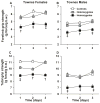
Effect of genotype and sex of Townes SCD mice on muscle strength. Female Townes mice (A and C), regardless of genotype, had greater forelimb and total grip strength compared to male (B and D) mice over all three days tested (P<0.001). Homozygous mice (male and female) had significantly lower forelimb and total grip strength compared to control Townes (all, p<0.001) and to heterozygous Townes (all, p≤0.008). Over the three days tested, heterozygous Townes had similar forelimb and total grip strength compared to control mice (all, P≥0.198). Symbols reflect P values from post hoc analysis by day comparing homozygous with control. * reflects p<0.05, † reflects p<0.01, and ‡, p<0.001. Forty-two mice participated in the grip force test, N=13–16 per genotype including balanced numbers of age-matched male and females. g force/ g b.w. indicates g force/ g of body weight.
Figure 6
Figure 6
Effect of age, sex, and genotype of Townes SCD mice on hematologic parameters. Overall, homozygous Townes mice had significantly lower hemoglobin, hematocrit, and red blood cell counts compared to control Townes (A, B, and C). In addition, among homozygous Townes, older mice had significantly lower hemoglobin (p<0.001) and hematocrit (p=0.004) compared to younger homozygous mice. In contrast, among control and heterozygous Townes, young and older mice had similar hemoglobin and hematocrit (all, p>0.49). Homozygous Townes mice also had significantly higher white blood cell counts compared to control and heterozygous mice (D). Among homozygous mice, males had higher white blood cell count compared to females (p<0.001). Among female, but not male mice, heterozygous Townes had higher platelet counts compared to controls and homozygous mice (E). Older homozygous male mice had higher platelet counts compared to male control mice. Symbols reflect P values from post hoc analysis comparing homozygous with control mice for each sex and age group. * reflects p<0.05, † reflects p<0.01, and ‡, p<0.001. Seventy-one mice participated in this experiment, N=9–13 per genotype including balanced numbers of age-matched male and females.
Figure 7
Figure 7
Neuropathology in Townes SCD mice. Representative Hematoxylin and Eosin (H&E) stained slides for each genotype of Townes mice (N=10–13 per genotype). Unlike control Townes, old homozygous mice had long bands of dark/pyknotic (shrunken) neurons seen mostly in the CA 1 and CA 3 fields of the hippocampus. In heterozygous Townes mice, similar dark/pyknotic neurons were present in CA1 and CA3 fields of the hippocampus in significantly lower numbers, than in that seen in homozygotes, and those dark/pyknotic neurons were interspersed with morphologically normal neurons. In young homozygotes, the degree of hippocampus injury was milder than that observed in old homozygous mice. In the cerebellum of old homozygous mice, there were decreases in neuronal density (neuronal drop-out) compared to control Townes mice. All hippocampus panels are shown in original magnification x40 and CA1, CA3, and cerebellum panels are shown in original magnification x200.
Figure 8
Figure 8
Iron tissue levels in cortex, hippocampus and cerebellum in Townes mice. Cerebellum iron tissue levels were significantly higher than those in cortex and hippocampus in controls, heterozygous and homozygous (all p<0.0015). In hippocampus, heterozygous and homozygous Townes combined had significantly lower total iron tissue levels compared to control mice (p=0.03). Fourteen mice were enrolled N=4–5 per genotype. * reflects p<0.05

Similar articles

Cited by

References

    1. Albani SH, et al. Behavior in the elevated plus maze is differentially affected by testing conditions in rats under and over three weeks of age. Front Behav Neurosci. 2015;9:31. - PMC - PubMed
    1. Andreotti C, et al. The Association of Cytokine Levels With Cognitive Function in Children With Sickle Cell Disease and Normal MRI Studies of the Brain. J Child Neurol 2014 - PMC - PubMed
    1. Armstrong FD, et al. Developmental function in toddlers with sickle cell anemia. Pediatrics. 2013;131:e406–14. - PMC - PubMed
    1. Barkus C. Genetic mouse models of depression. Curr Top Behav Neurosci. 2013;14:55–78. - PubMed
    1. Benton TD, et al. Psychiatric diagnosis in adolescents with sickle cell disease: a preliminary report. Curr Psychiatry Rep. 2011;13:111–5. - PubMed

Publication types

MeSH terms