. 2015 Oct 14:5:15024.
doi: 10.1038/srep15024.

How astrocyte networks may contribute to cerebral metabolite clearance

Affiliations

How astrocyte networks may contribute to cerebral metabolite clearance

Mahdi Asgari et al. Sci Rep. .

Abstract

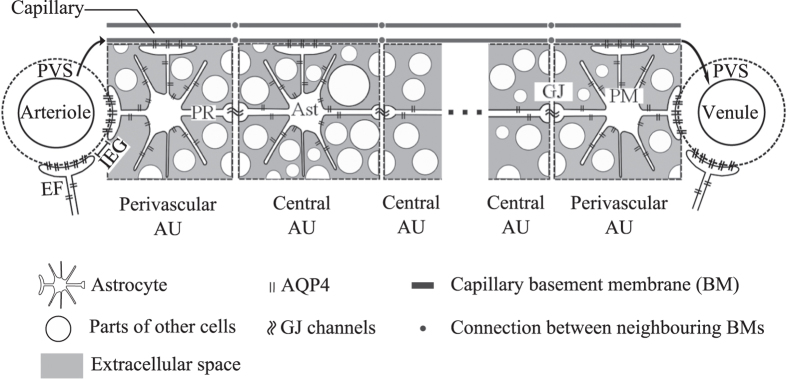
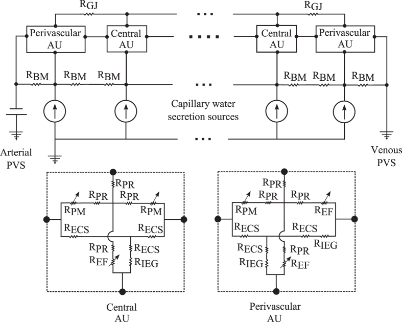
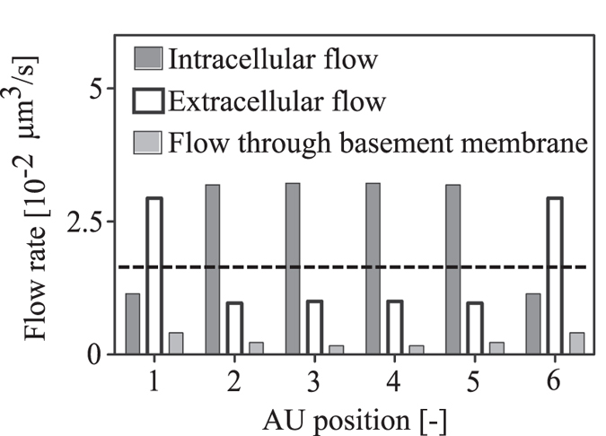
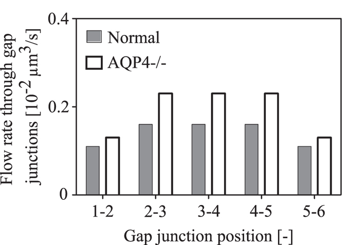
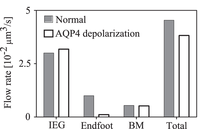
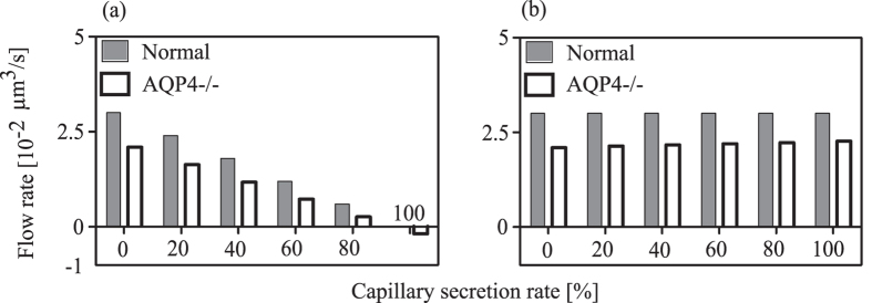
The brain possesses an intricate network of interconnected fluid pathways that are vital to the maintenance of its homeostasis. With diffusion being the main mode of solute transport in cerebral tissue, it is not clear how bulk flow through these pathways is involved in the removal of metabolites. In this computational study, we show that networks of astrocytes may contribute to the passage of solutes between tissue and paravascular spaces (PVS) by serving as low resistance pathways to bulk water flow. The astrocyte networks are connected through aquaporin-4 (AQP4) water channels with a parallel, extracellular route carrying metabolites. Inhibition of the intracellular route by deletion of AQP4 causes a reduction of bulk flow between tissue and PVS, leading to reduced metabolite clearance into the venous PVS or, as observed in animal studies, a reduction of tracer influx from arterial PVS into the brain tissue.


Figures

Figure 1
Figure 1. Sketch of the model domain consisting of an astrocyte network between arterial and venous paravascular spaces (PVS).
The network consists of several astrocyte units (AU) that each include one astrocyte (Ast) expressing aquaporin-4 (AQP4) water channels, extracellular space (ECS) and parts of the intracellular space of other cells including neurons, oligodendrocytes and microglia. Note that the schematic is not drawn to scale and does not represent the true ratio between intra- and extracellular space. The perivascular AUs are in direct contact with their neighbouring PVS via the respective astrocyte’s endfoot (EF). All astrocytes connect via endfeet to the basement membrane (BM) of the capillary associated with their AU. Neighbouring BMs connect to each other, and the perivascular BMs are additionally connected to the corresponding PVS (indicated by arrows in the figure). AQP4 covering each of these endfeet connect the capillary BMs or the PVS to the intra-astrocyte space. The intra-astrocyte and extracellular spaces within a given AU are in constant exchange of water through AQP4 water channels expressed in the astrocyte plasma membrane (PM). Gap junctions (GJ) connect the intracellular spaces of two adjacent astrocytes. PR: Astrocyte process. IEG: Inter-endfeet-gap.
Figure 2
Figure 2. Electrical analogue model of cerebral water transport between arterial and venous paravascular spaces (PVS).
Definitions of the abbreviations referring to the physical model domain are given in Fig. 1. The voltage source represents the driving pressure difference between arterial and venous paravascular spaces that are connected by resistances (R) representing the resistance to fluid flow of capillary basement membrane (BM) segments and astrocyte units (AU). Each astrocyte unit (AU) includes resistances of both intracellular (cell processes, PR) and extracellular (ECS) pathways which are linked by membrane resistances, namely those of the astrocyte endfoot membrane (EF) and the remainder of the astrocyte plasma membrane (PM). Since these membrane resistances are dependent on the AQP4 expression level, they are indicated as variable resistances (arrows). Gap junction (GJ) resistances connect the intracellular spaces of two neighbouring astrocytes.
Figure 3
Figure 3. Flow rate distribution between intracellular, extracellular and capillary basement membrane pathways under normal conditions.
AU Positions 1 and 6 refer the arterial and venous perivascular astrocyte units, respectively. The remaining positions refer to the central AUs. The dashed line indicates hypothetical extracellular flow rate (not including flow through basement membrane) in absence of the parallel, interconnected intracellular pathway.
Figure 4
Figure 4. Water flow rate from arterial PVS into the parenchyma as a function of the number of astrocytes between adjacent PVS.
6 astrocytes in this space correspond to the baseline, whereas the lowest shown number of 2 only includes the perivascular astrocytes. In configurations with less than 6 astrocytes, the resulting gaps are assumed to be filled with generic cells that do not express AQP4.
Figure 5
Figure 5. Effect of AQP4 deletion on water flow rate from PVS to the parenchyma through endfoot AQP4 channels, inter-endfeet-gaps (IEG), capillary basement membrane (BM) and in total.
(a) Baseline configuration with 6 astrocytes. (b) Configuration with only the two perivascular astrocytes with the remaining space filled with other cells not expressing AQP4. (c) Percentage change in flow rates from PVS to tissue after AQP4 deletion as a function of the number of AUs included in the network.
Figure 6
Figure 6. The effect of AQP4 deletion on water flow rate through the direct GJ connections between neighbouring astrocytes in the network.
Gap junction positions 1–2 and 5–6 refer to the connection between the arterial perivascular astrocyte and the first central astrocyte, and between the last central astrocyte and venous perivascular astrocyte, respectively.
Figure 7
Figure 7. Effect of AQP4 depolarization on water flow rate from PVS to the parenchyma through endfoot AQP4 channels, inter-endfeet-gaps (IEG), capillary basement membrane (BM) and in total.
Dark bars refer to the baseline state with AQP4 water channels polarized on the endfoot, whereas light bars refer to the depolarized state.
Figure 8
Figure 8. Effect of capillary water secretion rate on the water flow rate through inter-endfeet gaps connecting the perivascular AUs to the (a) arterial and (b) venous PVS, respectively.
The left most pair of bars in both panels show values for zero water secretion, whereas the right most pairs bars give the values for the case where all of the water inflow into the domain stems from capillary secretion. The white bars represent the AQP4 deletion state, in which next to the removal of the corresponding pathway in the AUs, a reduction of the capillary water secretion by 31% is imposed.

References

    1. Kurtcuoglu V. et al. Computational investigation of subject-specific cerebrospinal fluid flow in the third ventricle and aqueduct of Sylvius. J Biomech 40, 1235–1245 (2007). - PubMed
    1. Arbel-Ornath M. et al. Interstitial fluid drainage is impaired in ischemic stroke and Alzheimer’s disease mouse models. Acta Neuropathol 126, 353–364 (2013). - PMC - PubMed
    1. Carare R. O. et al. Solutes, but not cells, drain from the brain parenchyma along basement membranes of capillaries and arteries: significance for cerebral amyloid angiopathy and neuroimmunology. Neuropathol Appl Neurobiol 34, 131–144 (2008). - PubMed
    1. Cserr H. & Patlak C. Secretion and bulk flow of interstitial fluid. In: Physiology and pharmacology of the blood-brain barrier (ed^(eds). Springer (1992).
    1. Sykova E. & Nicholson C. Diffusion in brain extracellular space. Physiol Rev 88, 1277–1340 (2008). - PMC - PubMed

Publication types