

Fluorescence imaging enabled poly(lactide-co-glycolide)
- PMID: 26463014
- PMCID: PMC4681614
- DOI: 10.1016/j.actbio.2015.10.010
Fluorescence imaging enabled poly(lactide-co-glycolide)
Abstract
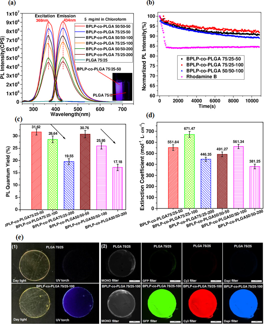
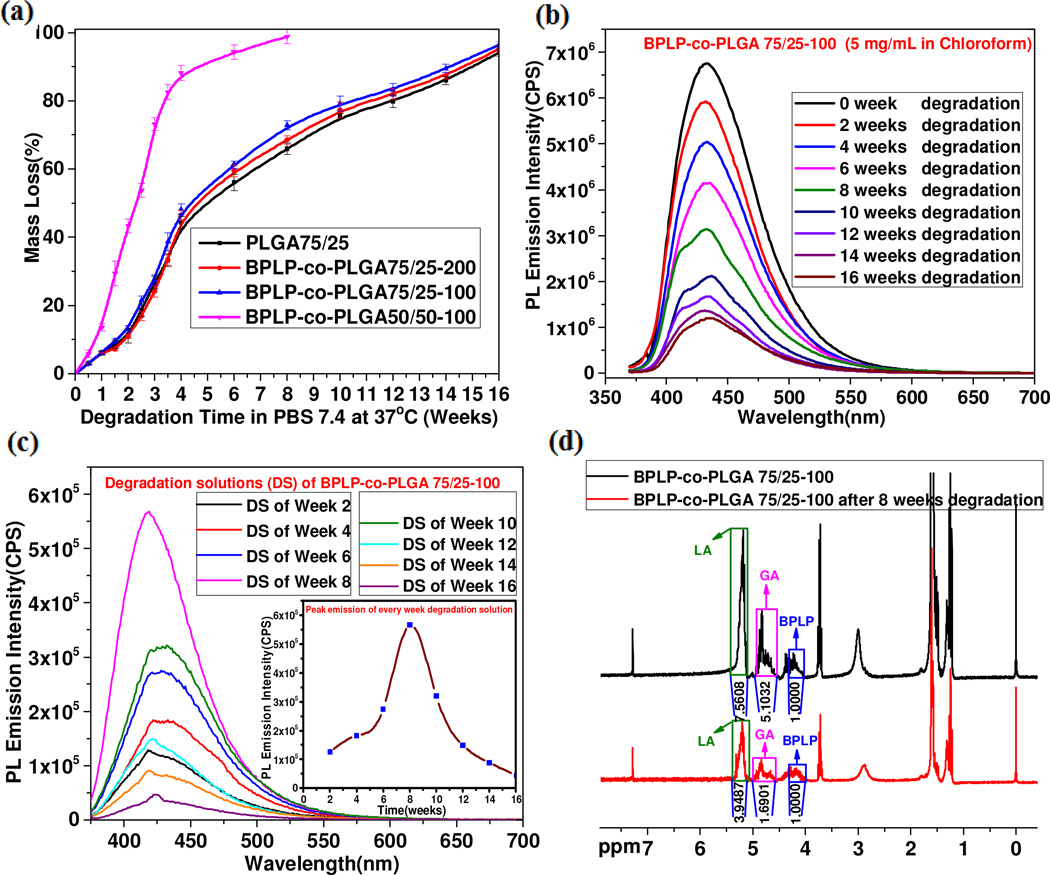
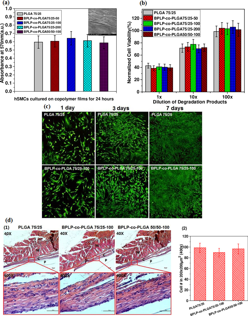
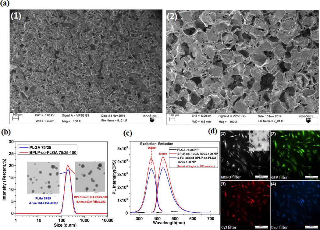
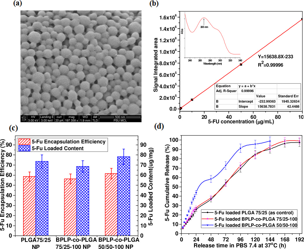
Fluorescent biomaterials have attracted significant research efforts in the past decades. Herein, we report a new series of biodegradable, fluorescence imaging-enabled copolymers, biodegradable photoluminescent poly(lactide-co-glycolide) (BPLP-co-PLGA). Photoluminescence characterization shows that BPLP-co-PLGA solutions, films and nanoparticles all exhibit strong, tunable and stable photoluminescence. By adjusting the molar ratios of L-lactide (LA)/glycolide (GA) and (LA+GA)/BPLP, full degradation of BPLP-co-PLGA can be achieved in 8-16 weeks. The fluorescence decay behavior of BPLP-co-PLGA can be used for non-invasive monitoring of material degradation. In vitro cytotoxicity and in vivo foreign body response evaluations demonstrate that BPLP-co-PLGA exhibits similar biocompatibility to poly(lactide-co-glycolide) (PLGA). The imaging-enabled BPLP-co-PLGA was fabricated into porous scaffolds whose degradation can be monitored through non-invasive imaging and nanoparticles that show theranostic potential demonstrated by fluorescent cellular labeling, imaging and sustained 5-fluorouracil delivery. The development of inherently fluorescent PLGA copolymers is expected to impact the use of already widely accepted PLGA polymers for applications where fluorescent properties are highly desired but limited by the conventional use of cytotoxic quantum dots and photobleaching organic dyes.
Statement of significance: This manuscript describes a novel strategy of conferring intrinsic photoluminescence to the widely used biodegradable polymers, poly(lactide-co-glycolide) without introducing any cytotoxic quantum dots or photo-bleaching organic dyes, which may greatly expand the applications of these polymers in where fluorescent properties are highly desired. Given the already significant impact generated by the use of PLGA and alike, this work contributes to fluorescence chemistry and new functional biomaterial design and will potentially generate significant impact on many fields of applications such as tissue engineering, molecular imaging and labeling, and drug delivery.
Keywords: Biodegradable; Bioimaging; Drug delivery; PLGA; Photoluminescence; Tissue engineering.
Copyright © 2015 Acta Materialia Inc. Published by Elsevier Ltd. All rights reserved.
Figures
Similar articles
-
Biological compatibility, thermal and in vitro simulated degradation for poly(p-dioxanone)/poly(lactide-co-glycolide)/poly(ethylene succinate-co-glycolide).J Biomed Mater Res B Appl Biomater. 2021 Nov;109(11):1817-1835. doi: 10.1002/jbm.b.34842. Epub 2021 Apr 24. J Biomed Mater Res B Appl Biomater. 2021. PMID: 33894107
-
Fluorescent labeling of degradable poly(lactide-co-glycolide) for cellular nanoparticles tracking in living cells.Int J Artif Organs. 2011 Feb;34(2):152-60. doi: 10.5301/ijao.2011.6420. Int J Artif Organs. 2011. PMID: 21374567
-
Sustained release of bee venom peptide from biodegradable thermosensitive PLGA-PEG-PLGA triblock copolymer-based hydrogels in vitro.Pharmazie. 2006 Mar;61(3):199-202. Pharmazie. 2006. PMID: 16599259
-
The manufacturing techniques of various drug loaded biodegradable poly(lactide-co-glycolide) (PLGA) devices.Biomaterials. 2000 Dec;21(23):2475-90. doi: 10.1016/s0142-9612(00)00115-0. Biomaterials. 2000. PMID: 11055295 Review.
-
Formulating poly(lactide-co-glycolide) particles for plasmid DNA delivery.J Pharm Sci. 2008 Jul;97(7):2448-61. doi: 10.1002/jps.21215. J Pharm Sci. 2008. PMID: 17918737 Review.
Cited by
-
Different PEG-PLGA Matrices Influence In Vivo Optical/Photoacoustic Imaging Performance and Biodistribution of NIR-Emitting π-Conjugated Polymer Contrast Agents.Adv Healthc Mater. 2021 Feb;10(4):e2001089. doi: 10.1002/adhm.202001089. Epub 2020 Aug 31. Adv Healthc Mater. 2021. PMID: 32864903 Free PMC article.
-
High-affinity mutant Interleukin-13 targeted CAR T cells enhance delivery of clickable biodegradable fluorescent nanoparticles to glioblastoma.Bioact Mater. 2020 May 7;5(3):624-635. doi: 10.1016/j.bioactmat.2020.04.011. eCollection 2020 Sep. Bioact Mater. 2020. PMID: 32405577 Free PMC article.
-
Nanoparticle eluting-angioplasty balloons to treat cardiovascular diseases.Int J Pharm. 2019 Jan 10;554:212-223. doi: 10.1016/j.ijpharm.2018.11.011. Epub 2018 Nov 5. Int J Pharm. 2019. PMID: 30408532 Free PMC article.
-
Polymeric biomaterials for biophotonic applications.Bioact Mater. 2018 Aug 6;3(4):434-445. doi: 10.1016/j.bioactmat.2018.07.001. eCollection 2018 Dec. Bioact Mater. 2018. PMID: 30151431 Free PMC article. Review.
-
Engineering multifunctional bioactive citrate-based biomaterials for tissue engineering.Bioact Mater. 2022 May 7;19:511-537. doi: 10.1016/j.bioactmat.2022.04.027. eCollection 2023 Jan. Bioact Mater. 2022. PMID: 35600971 Free PMC article. Review.
References
-
- Maia FR, Bidarra SJ, Granja PL, Barrias CC. Functionalization of biomaterials with small osteoinductive moieties. Acta Biomater. 2013;9:8773–8789. - PubMed
-
- Nair LS, Laurencin CT. Biodegradable polymers as biomaterials. Prog Polym Sci. 2007;32:762–798.
-
- Liu Q, Jiang L, Shi R, Zhang L. Synthesis, preparation, in vitro degradation, and application of novel degradable bioelastomers—A review. Prog Polym Sci. 2012;37:715–765.
Publication types
MeSH terms
Substances
Grants and funding
LinkOut - more resources
Full Text Sources
Other Literature Sources
Research Materials
Miscellaneous
