. 2015 Oct 13:15:209.
doi: 10.1186/s12866-015-0544-z.

Mutations in the bacterial cell division protein FtsZ highlight the role of GTP binding and longitudinal subunit interactions in assembly and function

Affiliations

Mutations in the bacterial cell division protein FtsZ highlight the role of GTP binding and longitudinal subunit interactions in assembly and function

Heidi A Arjes et al. BMC Microbiol. .

Abstract

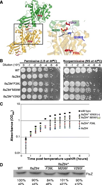
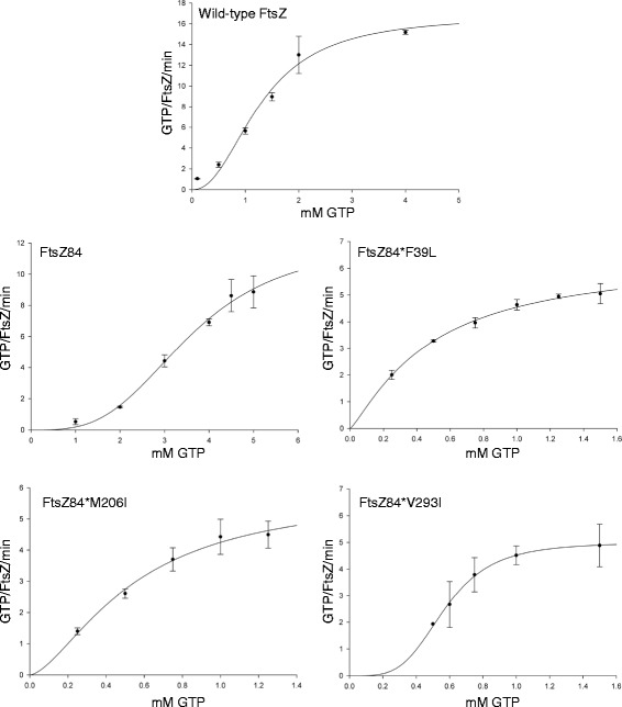
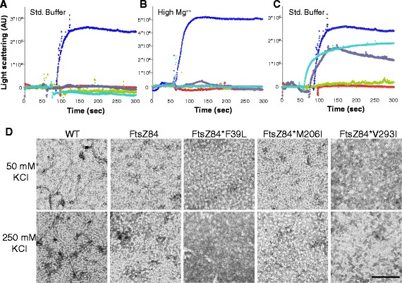
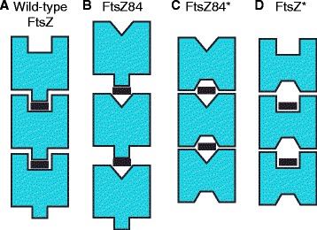
Background: Assembly of the tubulin-like GTPase, FtsZ, at the future division site initiates the process of bacterial cytokinesis. The FtsZ ring serves as a platform for assembly of the division machinery and constricts at the leading edge of the invaginating septum during cytokinesis. In vitro, FtsZ assembles in a GTP-dependent manner, forming straight filaments that curve upon GTP hydrolysis. FtsZ binds but cannot hydrolyze GTP as a monomer. Instead, the active site for GTP hydrolysis is formed at the monomer-monomer interface upon dimerization. While the dynamics of GTP hydrolysis and assembly have been extensively studied in vitro, significantly less is known about the role of GTP binding and hydrolysis in vivo. ftsZ84, a GTPase defective allele of Escherichia coli ftsZ, provides a striking example of the disconnect between in vivo and in vitro FtsZ assembly.

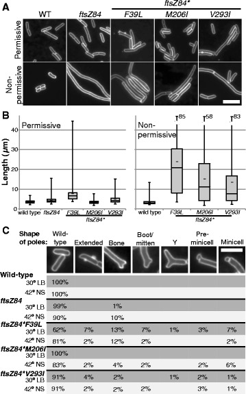
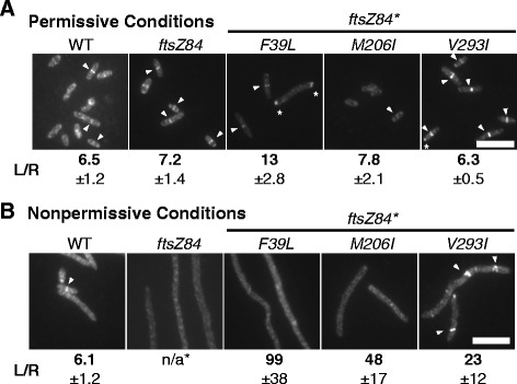
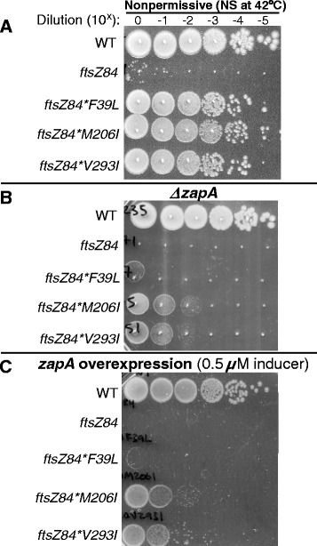
Results: Although ftsZ84 mutants are defective for FtsZ ring formation and division under nonpermissive conditions, they are near wild type for ring formation and division under permissive conditions. In vitro, however, purified FtsZ84 is defective in GTP binding, hydrolysis and assembly under standard reaction conditions. To clarify the nature of the FtsZ84 assembly defect, we isolated and characterized three intragenic suppressors of ftsZ84. All three suppressor mutations increased the apparent affinity of FtsZ84 for GTP, consistent with improved subunit-subunit interactions along the longitudinal interface. Although kinetic analysis indicates that the suppressor mutations increase the affinity of FtsZ84 for GTP, all three exhibit reduced rates of GTP hydrolysis and fail to support assembly in vitro.

Conclusion: Together, our data suggest that FtsZ, and potentially other enzymes whose assembly is similarly regulated, can compensate for defects in catalysis through increases in substrate binding and subunit-subunit interactions. In addition, these results highlight the dichotomy between commonly used in vitro assembly conditions and FtsZ ring formation in the complex intracellular milieu.


Figures

Fig. 1
Fig. 1
Secondary mutations restore temperature resistance to ftsZ84 cells without increasing FtsZ concentration. a Location of the FtsZ84 (G105S) point mutation and the three intragenic suppressors of that mutation mapped onto an FtsZ dimer from Staphylococcus aureus. G10S5S (blue) is the original ftsZ84 point mutation. GDP is shown in gray. The three intragenic suppressors F39L, M206I and V293I are highlighted in the inset. The crystal structure is modified from the S. aureus FtsZ dimer structure (PDB ID: 3WGN) [52]. b Plating efficiency assays of wild-type, ftsZ84, and the three intragenic ftsZ84 suppressor strains. Tenfold dilutions of cells cultured in permissive conditions were plated under permissive and nonpermissive conditions. This experiment was repeated 3 times with identical results. One representative experiment is shown. c Growth as measured by absorbance (OD600) of wild-type, ftsZ84, and the three intragenic ftsZ84 suppressor strains under nonpermissive conditions. Error bars represent standard deviation of 3 independent experiments. d Quantitative immunoblot indicates that intracellular FtsZ concentrations are wild type in ftsZ84 and ftsZ84* mutants under nonpermissive conditions. After ~3 mass doubling periods in LB no salt, cells were sampled at equivalent OD’s to ensure the same amount of protein was loaded per lane and protein bands were normalized to total protein (Ponceau staining) as a loading and transfer control. ImageJ software was used to quantify band intensity. The average and standard deviation of 3 independent experiments are shown below the blot
Fig. 2
Fig. 2
ftsZ84* suppressor mutants exhibit variable cell lengths and polar shapes under permissive and nonpermissive conditions. a Representative images of cell membrane staining of live wild-type cells, ftsZ84 and ftsZ84* strains under permissive and nonpermissive conditions. Bar = 5 μm. b Box and whisker plots of cell length distributions in permissive or nonpermissive conditions. Cells were grown in permissive conditions to an OD600 of 0.2-0.4 and sampled. For the nonpermissive conditions, cells were then backdiluted to an OD600 of 0.05 and grown to a sampling OD600 of 0.2 to 0.4. The box indicates the middle 50 % of values, the line indicates the median, the short bar indicates the mean, and the whisker bars represent the span of data in the lowest quartile (below the box) and the highest quartile (above the box). Values for ftsZ84*F39L and ftsZ84*V293I are conservative as many filaments extended past the field of view and could not be quantified. Note the upper values for the ftsZ84* mutants have been truncated; the number indicates the highest value measured. For permissive conditions, n = 146 (wild type), 132 (ftsZ84), 138 (ftsZ84*F39L), 139 (ftsZ84*M206I) and 110 (ftsZ84*V293I). For nonpermissive conditions, n = 102 (wild type), 42 (ftsZ84*F39L), 83 (ftsZ84*M206I), and 129 (ftsZ84*V293I). c ftsZ84* suppressor mutants exhibit branching, abnormal polar morphologies, and minicell formation. The fraction of cells exhibiting each morphology was determined by analyzing ~100-150 cells, the only exceptions being ftsZ84, ftsZ84*F39L, and ftsZ84*M206I under nonpermissive conditions where fewer cells were examined, respectively, as only whole cells were scored and it was rare to visualize an entire cell in the frame in these samples. For permissive conditions, n = 146 (wild type), 132 (ftsZ84), 138 (ftsZ84*F39L), 139 (ftsZ84*M206I), and 110 (ftsZ84*V293I). For nonpermissive conditions, n = 102 (wild type), 10 (ftsZ84), 42 (ftsZ84*F39L), 83 (ftsZ84*M206I) and 129 (ftsZ84*V293I). Bar = 3 μm
Fig. 3
Fig. 3
ftsZ84* suppressor mutations restore FtsZ ring formation to varying degrees under nonpermissive conditions. Representative images of cells grown under (a) permissive conditions or (b) nonpermissive conditions and labeled for cell wall and FtsZ. Arrowheads indicate FtsZ rings. Bar = 5 μm. The length per ring (L/R) ratio was calculated by dividing the total length of all cells by the number of FtsZ rings counted. Average and standard deviation of 3 biological replicates is shown below each image
Fig. 4
Fig. 4
Steady state kinetic analysis of FtsZ, FtsZ84, and FtsZ84* proteins. GTPase assays were carried out as described in materials and methods (50 mM MES, pH 6.5, 50 mM KCl, 2.5 mM MgCl2, 1 mM EGTA). FtsZ concentration was kept constant at 5 μM and GTP concentration were varied from 0.1 μM to 5 μM (different ranges were used depending on the FtsZ mutant analyzed). The best fit for our data was obtained using a Hill-modified Michaelis-Menten equation using Sigma Plot software. Km and Vmax values are listed in Table 3
Fig. 5
Fig. 5
ftsZ84* suppressor mutations do not restore assembly in vitro. a-b 90° angle light scattering data from FtsZ (blue), FtsZ84 (red), FtsZ84*F39L (green), FtsZ84*M206I (purple), and FtsZ84*V293I (aqua) a 50 mM MES, pH 6.5, 50 mM KCl, 2.5 mM MgCl 2, 1 mM EGTA or b 50 mM MES, pH 6.5, 50 mM KCl, 10 mM MgCl 2, 1 mM EGTA (methods). After establishing baseline, assembly was initiated by the addition of 1 mM GTP. c 90° angle light scattering data from suppressor mutations alone (in the absence of ftsZ84(G105S)). FtsZ (blue), FtsZ84 (red), FtsZ*F39L (green), FtsZ*M206I (purple), and FtsZ*V293I (aqua) Reaction conditions same as in (a). d Electron micrographs of FtsZ assembled in 50 mM KCl or 250 mM KCl (50 mM MES, pH 6.5, 50 mM KCl, 2.5 mM MgCl2, 1 mM EGTA) Bar = 100 nm
Fig. 6
Fig. 6
zapA deletions reduce ftsZ84* viability under nonpermissive conditions. a Plating efficiency of WT, ftsZ84, and ftsZ84* strains when grown in permissive conditions and plated onto nonpermissive conditions. b Plating efficiency assays of zapA deletions in wild-type, ftsZ84, and ftsZ84* suppressor strains. Tenfold dilutions of cells cultured in permissive conditions were plated under nonpermissive conditions. c zapA overexpression from a sodium salicylate inducible zapA plasmid (pKG110-zapA) does not restore viability to ftsZ84 and makes ftsZ84* suppressor strains more sensitive when plated under nonpermissive conditions with 0.5 μM of sodium salicylate. Identical results were obtained when using the IPTG inducible zapA vector (pQE80-H6-zapA) in ftsZ84 (see Additional file 1). b and c These experiments were performed at least 3 times with equivalent results
Fig. 7
Fig. 7
FtsZ84*M206I and FtsZ84*V293I mutations compensate FtsZ84 to increase GTPase activity and longitudinal subunit interactions. a Wild-type FtsZ efficiently binds to and hydrolyzes GTP (black rectangle). This promotes subunit interactions and polymerization. b FtsZ84 contains a mutation in the GTP binding site that reduces GTP binding and hydrolysis, thereby weakening FtsZ dimerization and subunit-subunit assembly. c FtsZ84*M206I and FtsZ84*V293I mutations compensate for FtsZ84’s GTP binding defect and help to restore subunit-subunit interactions. d Introducing FtsZ84*M206I and FtsZ84*V293I into wild-type FtsZ reduces GTPase activity and assembly

References

    1. Adams DW, Errington J. Bacterial cell division: assembly, maintenance and disassembly of the Z ring. Nat Rev Microbiol. 2009;7:642–653. doi: 10.1038/nrmicro2198. - DOI - PubMed
    1. Erickson HP, Anderson DE, Osawa M. FtsZ in bacterial cytokinesis: cytoskeleton and force generator all in one. Microbe. 2010;74:504–528. - PMC - PubMed
    1. Den Blaauwen T, Buddelmeijer N, Aarsman ME, Hameete CM, Nanninga N. Timing of FtsZ assembly in Escherichia coli. J Bacteriol. 1999;181:5167–5175. - PMC - PubMed
    1. Weart RB, Levin PA. Growth rate-dependent regulation of medial FtsZ ring formation. J Bacteriol. 2003;185:2826–2834. doi: 10.1128/JB.185.9.2826-2834.2003. - DOI - PMC - PubMed
    1. Errington J, Daniel RA, Scheffers D-J. Cytokinesis in bacteria. Microbe. 2003;67:52–65. - PMC - PubMed

Publication types

MeSH terms

LinkOut - more resources