. 2016 Jan 7;127(1):139-48.
doi: 10.1182/blood-2015-06-654194. Epub 2015 Oct 13.

Glutathione peroxidase 4 prevents necroptosis in mouse erythroid precursors

Affiliations

Glutathione peroxidase 4 prevents necroptosis in mouse erythroid precursors

Özge Canli et al. Blood. .

Abstract

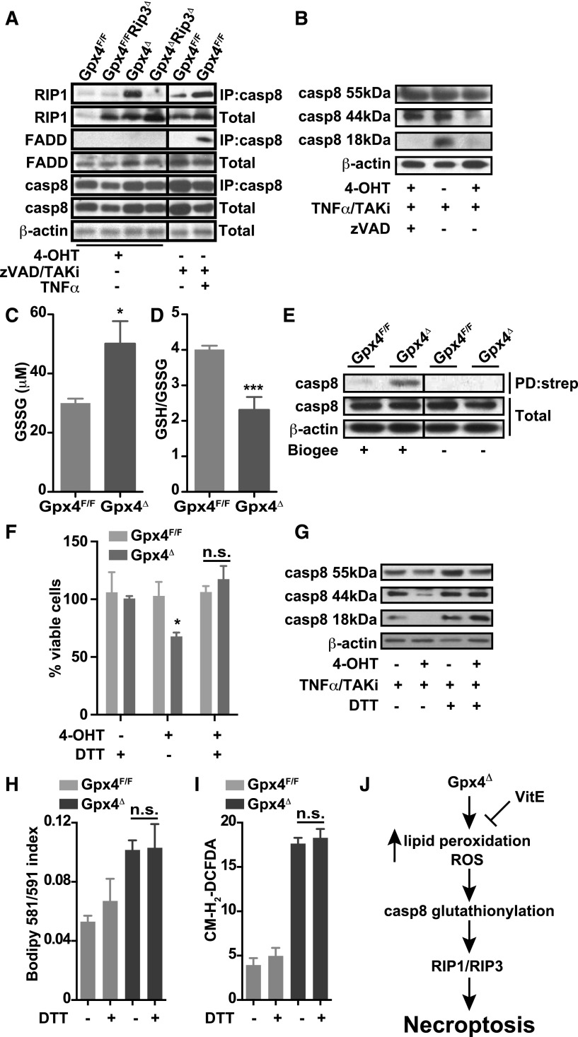
Maintaining cellular redox balance is vital for cell survival and tissue homoeostasis because imbalanced production of reactive oxygen species (ROS) may lead to oxidative stress and cell death. The antioxidant enzyme glutathione peroxidase 4 (Gpx4) is a key regulator of oxidative stress-induced cell death. We show that mice with deletion of Gpx4 in hematopoietic cells develop anemia and that Gpx4 is essential for preventing receptor-interacting protein 3 (RIP3)-dependent necroptosis in erythroid precursor cells. Absence of Gpx4 leads to functional inactivation of caspase 8 by glutathionylation, resulting in necroptosis, which occurs independently of tumor necrosis factor α activation. Although genetic ablation of Rip3 normalizes reticulocyte maturation and prevents anemia, ROS accumulation and lipid peroxidation in Gpx4-deficient cells remain high. Our results demonstrate that ROS and lipid hydroperoxides function as not-yet-recognized unconventional upstream signaling activators of RIP3-dependent necroptosis.


Figures

Figure 1
Figure 1
Loss of Gpx4 in hematopoietic cells induces anemia that is compensated by increased erythropoiesis. (A) Immunoblot analysis of Gpx4 in peripheral erythrocytes (TER119+) and bone marrow erythroid progenitors (CD71+/TER119+). Red blood cell counts (B), hemoglobin levels (C), and hematocrit (D) are decreased in Gpx4Δ mice, whereas the number of reticulocytes is increased (E). Data are mean ± SE; n ≥ 20. Elevated serum EPO levels (F) and enlarged spleens (G) in Gpx4Δ mice. Data are mean ± SE; n ≥ 10. (H-I) Immunohistochemistry staining of BrdU incorporation in the spleen and nuclear counterstain hematoxylin. Image acquisition was performed using a Zeiss Axio Imager M2 with a 20×/0.5 EC Plan Neofluar objective (magnification ×200). (J) Quantification of splenic BrdU+/TER119+ cells determined by flow cytometry. Data are mean ± SE; n ≥ 3. **P < .01, ***P < .001 by Student t test. BM, bone marrow; SE, standard error.
Figure 2
Figure 2
Increased lipid peroxidation and oxidative stress in Gpx4Δ erythroid cells does not impair their life span in the periphery. (A) Measurement of lipid peroxidation in unchallenged peripheral TER119+ erythrocytes using the lipophilic redox-sensitive dye BODIPY 581/591, which upon oxidation, shifts its fluorescence from red to green. Data are mean ± SE; n ≥ 7. (B) ROS in unchallenged peripheral TER119+ erythrocytes using the redox-sensitive dye 5-(and-6)-chloromethyl-2′,7′-dichlorodihydrofluorescein diacetate, acetyl ester (CM-H2-DCFDA) (n ≥ 7). (C) Survival of biotin-labeled peripheral TER119+ erythrocytes. Data are mean ± SE; n ≥ 6. (D) Increased number of peripheral CD71+/TER119+ reticulocytes in Gpx4Δ mice within the first 4 weeks after poly(I:C) administration. Data are mean ± SE; n ≥ 5. (E-F) Representative images of TUNEL assay in spleen sections and 4,6 diamidino-2-phenylindole (DAPI) as a counterstain showing increased cell death in Gpx4Δ mice. Image acquisition was performed using a Zeiss Axio Imager M2 with 40×/0.95 korr Apochromat objective (magnification ×400). Flow cytometry analysis using PI to determine the number of nonviable CD71+ cells in bone marrow (G) and in spleen (H). Data are mean ± SE; n ≥ 3. (I) Formation of o-dianisidine–positive erythroid colonies from bone marrow in methylcellulose semisolid media. Red colony formation of Gpx4Δ bone marrow cells could be rescued by addition of α-tocopherol to the medium. Data are mean ± SE; n ≥ 3. *P < .05, **P < .01, ***P < .001 by Student t test. α-Toc, α-tocopherol; n.s., not significant; PI, propidium iodide.
Figure 3
Figure 3
Increased erythropoiesis in Gpx4Δ mice depends on vitamin E. (A) Schematic overview of treatment: bone marrow from Gpx4F/F or Mx1-Cre/Gpx4F/F mice was transplanted (BMT) into wild-type recipients; mice were kept on a vitamin E–depleted diet (VitEΔ) after the recovery; and deletion was induced by poly(I:C). Red blood cell counts (B), hemoglobin levels (C), hematocrit (D), and reticulocyte counts (E). Data are mean ± SE; n ≥ 4. Enlarged spleens (F) and elevated serum EPO levels (G) in transplanted mice. Data are mean ± SE; n ≥ 6. ***P < .001, **P < .01, *P < .05 by Student t test.
Figure 4
Figure 4
RIP3-dependent necroptosis, but not ferroptosis, causes cell death in Gpx4-deficient erythroid progenitor cells. Erythroid cells were differentiated in vitro from Gpx4F/F and Rosa26-CreERT2/Gpx4F/F bone marrow, and deletion was induced by 4-OHT after 24 hours of culture in the presence of EPO. Microscopic images of in vitro–differentiated erythroid cells from Gpx4F/F (A) and Rosa26-CreERT2/Gpx4F/F (Gpx4Δ) (B) bone marrow 48 hours after the induction of deletion with tamoxifen. (C) Schematic illustration of programmed cell death pathways. Ferroptosis (left) is triggered by an iron-dependent accumulation of lethal ROS and lipid peroxides in cells, which can be inhibited via iron (Fe) chelators such as DFO. Ferroptosis can be induced by erastin, which inhibits cellular cysteine (Cys) uptake and thus limits the production of intracellular GSH, or by Ras synthetic lethality molecule 3 (RSL3) via inhibition of Gpx4, leading to increased lipid peroxidation and ROS accumulation. Fer-1 and Lip-1 inhibit ferroptosis via inhibiting the lipid peroxidation. Apoptosis and necroptosis (right) are mainly regulated via TNFR1 signaling. Upon TNF binding, TNFR1 undergoes a conformational change, activating 2 possible cell death execution mechanisms: caspase-dependent or caspase-independent. Normally, caspase 8 triggers apoptosis by activating the classical caspase cascade. It also cleaves, and hence inactivates, RIP1 and RIP3. If caspase 8 is inhibited (eg, via zVAD), phosphorylated RIP1 and RIP3 engage the effector mechanisms of necroptosis. (D) Percentage of viable in vitro–differentiated erythroid cells counted via trypan blue exclusion 48 hours after the induction of deletion in the presence of α-tocopherol (the most prominent member of the vitamin E family), the ferroptosis inhibitors Fer-1 and Lip-1, the iron chelator DFO, the pan-caspase inhibitor zVAD, the RIP1 kinase inhibitor nec-1, or recombinant soluble TNFR2 (etanercept). Data are mean ± SE; n ≥ 6; *P < .05 by Student t test. (E) Absence of Gpx4 and RIP3 is verified by immunoblot analysis. (F) Percentage of viable in vitro–differentiated erythroid cells from Gpx4F/F, Rip3−/− (Gpx4F/FRip3Δ), Rosa26-CreERT2/Gpx4F/F (Gpx4Δ), and Rip3−/−/Rosa26-CreERT2/Gpx4F/F (Gpx4ΔRip3Δ) 48 hours after the induction of deletion. Data are mean ± SE; n ≥ 8; ***P < .001 by ANOVA/Bonferroni. (G) Flow cytometry analysis of in vitro–cultured erythroid cells 36 hours after the 4-OHT treatment to analyze necrotic cells (AnnexinVPI+). Data are mean ± SE; n ≥ 4; *P < .05 by ANOVA/Bonferroni. (H) Deletion of Rip3 significantly improved the formation of erythroid o-dianisidine–positive Gpx4Δ colonies, similar to α-Toc supplementation. Data are mean ± SE; n ≥ 3; *P < .05, **P < .01, ***P < .001 by ANOVA/Bonferroni. ANOVA, analysis of variance.
Figure 5
Figure 5
Genetic deletion of Rip3 normalizes red cell parameters and rescues anemia. Normalization of red blood cell counts (A), hemoglobin levels (B), and hematocrit (C), and reduction of reticulocyte numbers (D). Enlarged spleens (E) and elevated serum EPO levels (F) in Gpx4Δ mice are normalized upon deletion of Rip3. Data are mean ± SE; n ≥ 9. (G) Quantification of TUNEL+ cells in spleen sections. (H) Flow cytometry analysis of AnnexinVPI+ erythroid progenitor cells in the spleen. Data are mean ± SE; n ≥ 4. *P < .05, **P < .01, ***P < .001 by ANOVA/Bonferroni.
Figure 6
Figure 6
Necroptosis in Gpx4Δ mice is triggered independently of TNFR or CD95 engagement and PARP activation. Lipid peroxidation (A) and ROS accumulation (B) in peripheral blood erythroid cells (TER119+) are not significantly affected upon Rip3 deletion. Data are mean ± SE; n ≥ 4; *P < .05, **P < .01, ***P < .001 by ANOVA/Bonferroni. The number of peripheral CD71+/TER119+ cells remains unaffected (C), and there is no change in red blood cell counts (D), hemoglobin levels (E), hematocrit (F), and reticulocyte counts (G) in Gpx4Δ mice when treated with etanercept (5 mg/kg) or anti-CD95L neutralizing antibody (50 μg) for 2 weeks. Data are mean ± SE; n ≥ 5. Immunohistochemistry of phospho-H2Ax (pH2Ax) and nuclear counterstain hematoxylin in the spleen (H-I) and the quantification of phospho-H2Ax+ foci (J). Image acquisition was performed using a Zeiss Axio Imager M2 with a 20×/0.5 EC Plan Neofluar objective (magnification ×200). Data are mean ± SE; n ≥ 3; ***P < .001 by Student t test. The number of peripheral CD71+/TER119+ cells remains unaffected (K), and there is no change in red blood cell counts (L), hemoglobin levels (M), hematocrit (N), and reticulocyte counts (O) in Gpx4Δ mice when treated with the PARP inhibitor olaparib (5 mg/kg) for 2 weeks. Data are mean ± SE; n ≥ 5. DMSO, dimethylsulfoxide; PBS, phosphate-buffered saline.
Figure 7
Figure 7
Caspase 8 is inactivated in Gpx4-deficient cells. (A) Immunoprecipitation of caspase 8 (IP:casp8) and immunoblot analysis of RIP1 and FADD in in vitro erythroid cultures treated with 4-OHT for 36 hours. Classical activation of necroptosis using zVAD/TAKi/TNF-α treatment in wild-type in vitro–cultured erythroid cells shows a strong interaction of caspase 8 with RIP1 and FADD upon 2 hours of stimulation. (B) Cleavage of caspase 8 (casp8) is blocked in in vitro–cultured Gpx4Δ erythroid cells 36 hours after 4-OHT treatment, determined by immunoblot of caspase 8 when stimulated 2 hours with TNF-α in the presence of the TAK1 inhibitor 5Z-7- oxozeaenol (TAKi), whereas zVAD treatment completely inhibits caspase 8 cleavage. Concentration of GSSG (C) and the GSH/GSSG ratio (D) in peripheral blood cells of Gpx4Δ mice and control littermates. (E) Detection of glutathionylated caspase 8 in peripheral CD71+/TER119+ cells from Gpx4Δ mice. Cells were loaded with BioGEE, and immunoblot analysis was performed after immunoprecipitation with streptavidin (PD:strep). Data shown is representative of 4 independently analyzed mice of each genotype. DTT supplementation rescues cell death in erythroid cells (F) and restores caspase 8 cleavage upon TNF-α stimulation (G). DTT treatment does not inhibit lipid peroxidation (H) or ROS accumulation (I) in cultured erythroid cells of either genotype. (J) Our model proposes that in the absence of Gpx4, lipid peroxides and ROS can act as signaling molecules upstream of the necrosome independently of TNFR/FAS signaling. Loss of Gpx4 in the erythroid lineage leads to inactivation of caspase 8 via glutathionylation. ROS and lipid peroxides activate the RIP1/RIP3–containing necrosome and trigger necroptotic cell death. The presence of vitamin E (VitE) can compensate for Gpx4 deficiency.

Comment in

References

    1. D’Autréaux B, Toledano MB. ROS as signalling molecules: mechanisms that generate specificity in ROS homeostasis. Nat Rev Mol Cell Biol. 2007;8(10):813–824. - PubMed
    1. Vanden Berghe T, Vanlangenakker N, Parthoens E, et al. Necroptosis, necrosis and secondary necrosis converge on similar cellular disintegration features. Cell Death Differ. 2010;17(6):922–930. - PubMed
    1. Fiers W, Beyaert R, Declercq W, Vandenabeele P. More than one way to die: apoptosis, necrosis and reactive oxygen damage. Oncogene. 1999;18(54):7719–7730. - PubMed
    1. Beutler E, Luzzatto L. Hemolytic anemia. Semin Hematol. 1999;36(4 Suppl 7):38–47. - PubMed
    1. Johnson RM, Ho Y-S, Yu D-Y, Kuypers FA, Ravindranath Y, Goyette GW. The effects of disruption of genes for peroxiredoxin-2, glutathione peroxidase-1, and catalase on erythrocyte oxidative metabolism. Free Radic Biol Med. 2010;48(4):519–525. - PMC - PubMed

Publication types

Substances

LinkOut - more resources