. 2015 Dec 4;427(24):3921-34.
doi: 10.1016/j.jmb.2015.10.003. Epub 2015 Oct 20.

Structure of Full-Length Human PDGFRβ Bound to Its Activating Ligand PDGF-B as Determined by Negative-Stain Electron Microscopy

Affiliations

Structure of Full-Length Human PDGFRβ Bound to Its Activating Ligand PDGF-B as Determined by Negative-Stain Electron Microscopy

Po-Han Chen et al. J Mol Biol. .

Abstract

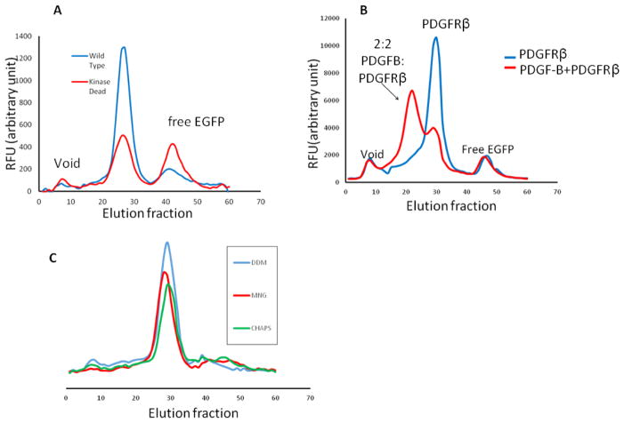
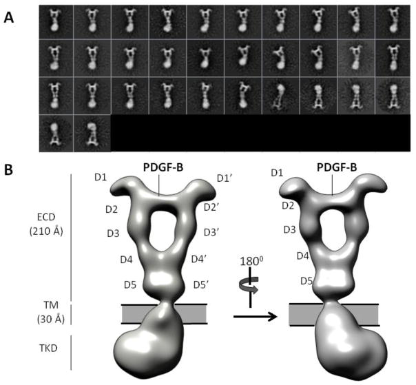
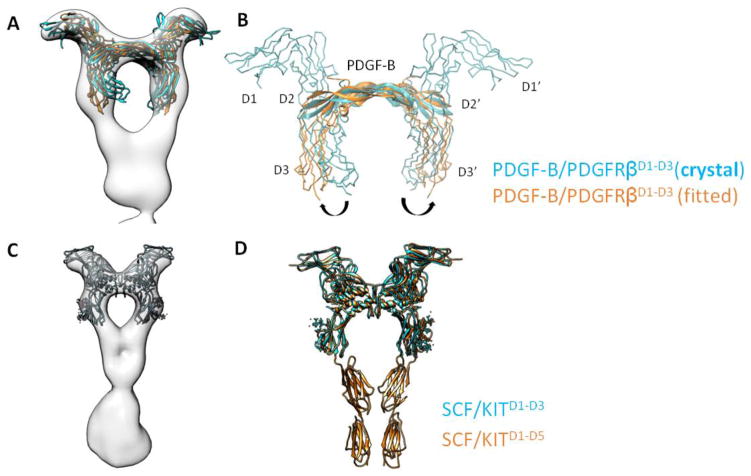
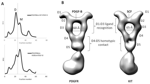
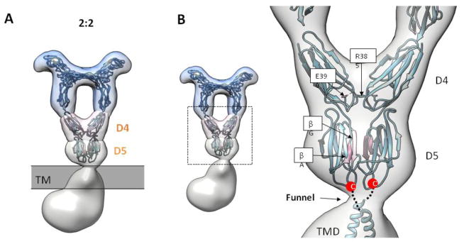
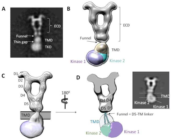
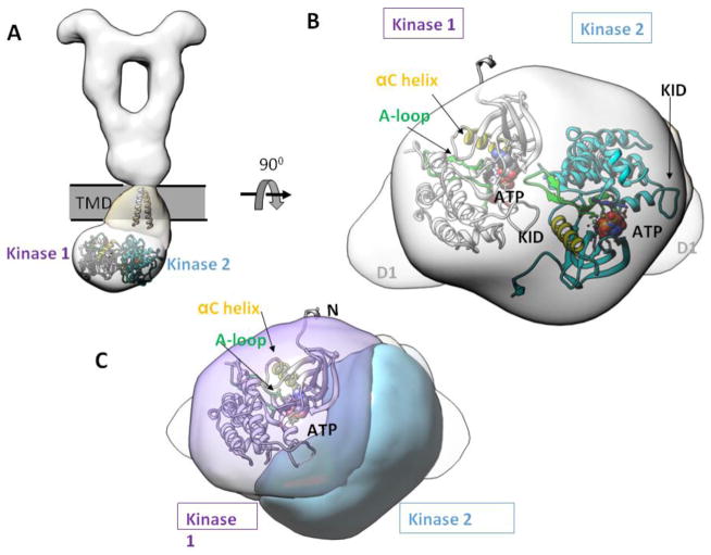
Members of the receptor tyrosine kinases (RTKs) regulate important cellular functions such as cell growth and migration, which are key steps in angiogenesis, in organ morphogenesis and in the unregulated states, cancer formation. One long-standing puzzle regarding RTKs centers on how the extracellular domain (ECD), which detects and binds to growth factors, is coupled with the intracellular domain kinase activation. While extensive structural works on the soluble portions of RTKs have provided critical insights into RTK structures and functions, lack of a full-length receptor structure has hindered a comprehensive overview of RTK activation. In this study, we successfully purified and determined a 27-Å-resolution structure of PDGFRβ [a full-length human platelet-derived growth factor receptor], in complex with its ligand PDGF-B. In the ligand-stimulated complex, two PDGFRβs assemble into a dimer via an extensive interface essentially running along the full-length of the receptor, suggesting that the membrane-proximal region, the transmembrane helix and the kinase domain of PDGFRβ are involved in dimerization. Major structural differences are seen between the full-length and soluble ECD structures, rationalizing previous experimental data on how membrane-proximal domains modulate receptor ligand-binding affinity and dimerization efficiency. Also, in contrast to the 2-fold symmetry of the ECD, the intracellular kinase domains adopt an asymmetric dimer arrangement, in agreement with prior observations for the closely related KIT receptor. In essence, the structure provides a first glimpse into how platelet-derived growth factor receptor ECD, upon ligand stimulation, is coupled to its intracellular domain kinase activation.

Keywords: cancer; electron microscopy; membrane protein structure; receptor tyrosine kinase; signal transduction.


Figures

Figure 1
Figure 1. FSEC screening of PDGFRβ
(A) WT versus kinase-dead mutant of PDGFRβ. (B) PDGF-B induced dimerization probed by FSEC. (C) Detergent screen of PDGFRβ.
Figure 2
Figure 2. Overall architecture of PDGF-B/PDGFRβFL
(A) Representative 2D class averages. (B) 3D Reconstruction.
Figure 3
Figure 3. Conformation of D2-D3 in full-length receptor
(A and B) Comparison between PDGFRβD1-D3 in crystal structure (cyan) versus PDGFRβD1-D3 after MDFF fitting in EM map (orange); the latter is representative of D2-D3 conformation in a full-length receptor. (C and D) Similar comparison for KIT receptor.
Figure 4
Figure 4. Influence of D4 on receptor dimerization, and common geometric requirement for Class III RTK dimerization
(A) Influence of R385A that disrupts D4-D4 homotypic contact on receptor dimerization. D = ligand-induced dimer. M = receptor monomer. (B) PDGFR (left) versus KIT (right) full-length structure. Whereas D1-D3 are different for the two receptors to accommodate different ligands, the D4-D5 homotypic contact is conserved.
Figure 5
Figure 5. PDGFR D5 homotypic contact
(A) Full-length PDGF-B/PDGFRβ structure stratified into different layers. The D4 and D5 layers highlight the homotypic contact established by MDFF simulation. (B) Closed-up view for the MDFF simulation, with the interface strands highlighted in pink.
Figure 6
Figure 6. Conformation of TMD-TKD layer
(A) 2D class average showing features of PDGF-B/PDGFRβ TMD-TKD. (B) EM reconstruction agrees with 2D class average. (C) Fitting of ECD and TMD atomic models. (D) Slice-through view of 3D map. The “funnel” likely represents the linker between ECD and the beginning of TM domain, and the TM helices likely cross each other near their N-terminal portions (near the funnel) and separate at the C-termini in order to connect to the two kinases located on each side of the TKD density. Inset: 2D view.
Figure 7
Figure 7. Asymmetric dimer of TKD
(A) The atomic models of TM and TKD are fitted to show spatial relationships. (B) Bottom view of TKD asymmetric dimer. (C) Same view as in (B), but highlighting the catalytic site position of kinase 1, where the ATP binding pocket, the A-loop, and the αC helix are in close proximity to the neighboring kinase 2. KID = kinase insert domain. αC = control α-helix. A-loop = activation loop.

References

    1. Kohler N, Lipton A. Platelets as a source of fibroblast growth-promoting activity. Exp Cell Res. 1974;87:297–301. - PubMed
    1. Ross R, Glomset J, Kariya B, Harker L. A platelet-dependent serum factor that stimulates the proliferation of arterial smooth muscle cells in vitro. Proc Natl Acad Sci U S A. 1974;71:1207–10. - PMC - PubMed
    1. Andrae J, Gallini R, Betsholtz C. Role of platelet-derived growth factors in physiology and medicine. Genes Dev. 2008;22:1276–312. - PMC - PubMed
    1. Ostman A. PDGF receptors-mediators of autocrine tumor growth and regulators of tumor vasculature and stroma. Cytokine Growth Factor Rev. 2004;15:275–86. - PubMed
    1. Lemmon MA, Schlessinger J. Cell signaling by receptor tyrosine kinases. Cell. 2010;141:1117–34. - PMC - PubMed

Publication types

MeSH terms

Substances

LinkOut - more resources