

Review
doi: 10.1021/acs.chemrev.5b00135.
Epub 2015 Aug 4.
Polymeric Nanostructures for Imaging and Therapy
Affiliations
- PMID: 26463640
- PMCID: PMC4610256
- DOI: 10.1021/acs.chemrev.5b00135
Item in Clipboard
Review
Polymeric Nanostructures for Imaging and Therapy
Chem Rev.
.
No abstract available
Figures

Examples of therapeutics of various sizes, solubilities and structures, and diagnostics (fluorophores, radioactive probes, quantum dots, etc.), that are commonly utilized for therapy and imaging. Labeling of various therapeutics with imaging probes is also exploited for simultaneous therapy and diagnosis (theranostics). It is observed in the body inset that random biodistribution usually occurs after administration of these agents.

The main challenges towards the use of various therapeutics, diagnostics and theranostics, during formulation, administration and the resulting biodistribution, with no control over their characteristics and navigation within the biological system.

Intracellular trafficking pathways: (1) Hydrophilic molecules and macromolecules usually have low cellular uptake and, thus, overall cationic charge or decoration with targeting ligands is required for efficient cellular uptake; (2) Endocytosis starts with the internalization of the nanoparticles into vesicles, that later are pinched off to form membrane-bound vesicles of different sizes, compositions and internal environments, called endosomes, phagosomes or macropinosomes, depending on the energy dependent or independent, and receptor dependent or independent internalization pathways;- (3) Degradation of nanoparticles or their cargoes usually occurs with catalysis by the acidic pH and/or enzymes found in the lysosomes; (4-6) Escape of nanoparticles or their payloads from these vesicles into the cytoplasm is essential for them to reach the targeted subcellular organelles (steps 4 and 5 represent the escape of nanoparticles or some of their components from a leaky endosomes); (7) Nanoparticles can also be cleared from the cells via exocytosis. Adapted with permission from Reference . Copyright 2012 Royal Society of Chemistry.

Nanomedicine-based therapeutic targets in atherosclerosis. (A) Summary of mechanisms underlying atherogenesis, atheroprogression, and plaque destabilization. The leakiness of endothelial cell junctions in areas of low shear stress permits low-density lipoprotein (LDL) to enter the intima, where it is oxidized (oxLDL) (1). LDL mediates the upregulation of adhesion molecules, such as P-selectin, intercellular adhesion molecule 1 (ICAM1), and vascular cell adhesion molecule 1 (VCAM1), to recruit leukocytes, such as monocytes and neutrophils (2). Recruited macrophages engulf oxLDL via scavenger receptors and give rise to foam cells (3). Atheroprogression is characterized by further accumulation of leukocytes by local proliferation, ongoing recruitment, and hampered egress (4). The fate of atherosclerotic plaques is determined by the failed clearance of apoptotic cells, which leads to secondary necrosis and plaque destabilization (5). The plaque is shielded from the bloodstream by a matrix-containing fibrous cap that is covered by endothelial cells. At late stages, the fibrous cap is weakened by matrix-degrading proteases from macrophages, leading to plaque rupture and the exposure of thrombogenic material to the bloodstream, causing platelet activation and blood clotting, which is clinically observed as myocardial infarction or stroke (6). (B) To intervene in leukocyte recruitment, circulating monocytes can be targeted to deliver nanoparticles to the lesion as ‘Trojan horses’ or to knock down surface receptors, such as CC-chemokine receptor 2 (CCR2), which is crucial for the adhesion to endothelial cells, by siRNA. (C) Natural ligands (as well as mimetics or antibodies) of adhesion molecules can be used as targeting entities to direct nanoparticles to atherosclerotic tissue. (D) Particles such as high-density lipoprotein (HDL) and LDL naturally home to atherosclerotic lesions, and synthetic equivalents or mimetics can be used as nanocarriers for drug delivery or as cholesterol acceptors to stimulate cholesterol efflux. The increased permeability of endothelial cells or neovessels not only allows lipoproteins to enter the lesion but also permits the entry of (untargeted, long-circulating) nanocarriers within a certain size range. (E) The fate of the stability of atherosclerotic lesion is determined by defects in the clearance of apoptotic cells. Inducing this clearance by anti-inflammatory and pro-resolving drugs [such as dexamethasone (DXM)] encapsulated into liposomes could therefore stabilize the lesion. Reprinted with permission from Reference . Copyright 2014 Elsevier Ltd.

Compositional versatility of multifunctional nanoparticles for biomedical delivery applications, illustrated generically for a solid core-shell polymer nanoparticle scaffold. (a) Targeting: Clusters of targeting moieties are important for multivalent binding to receptors for enhanced cellular uptake; the use of various ligands (antibody, antibody fragment, peptide, etc.) depends on the therapeutic application and disease type. (b) Shell: The length, spacing and crosslinking of the shell polymers are critical parameters that dictate the blood circulation time and stability of nanoparticles with ~1 nm spacing found to be efficient in preventing protein adsorption. (c) Core: The nature of the core dictates the type of the drug to be encapsulated. Crosslinking and conjugation of drugs to the core-forming polymer are common strategies for enhancing the stability of nanoparticles and drug-encapsulation efficiency, respectively. (d) Cargo: A wide range of imaging agents and/or therapeutics can be packaged, ranging from small molecules to macromolecular cargoes. Adapted with permission from References ,. Copyright 2012 and 2013 Royal Society of Chemistry.

Passive and active targeting features of multifunctional nanomaterials. In passive targeting, nanoparticles accumulate into pathological sites with leaky vasculature (e.g. tumor) due to the enhanced permeability and retention effect. In active targeting, the targeting ligands on the surface of nanoparticles enhance cellular uptake by binding to specific receptors overexpressed on the diseased cells. Targeting may also be achieved via facilitating escape from endosomes/lysosomes and/or enhancing nuclear translocation. Multifunctional nanoparticles have additional functionalities to deliver more than one cargo (e.g. more than one type of therapeutic and/or diagnostic agent), or combine more than one targeting mechanism (i.e. passive and active targeting). Adapted with permission from Reference . Copyright 2012 Royal Society of Chemistry.
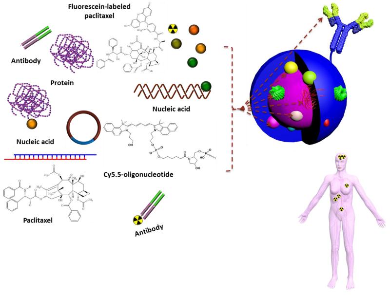
Possibilities of incorporating various therapeutic and/or diagnostic agents into one set of nanomaterial. It is observed in the body inset that encapsulation of these agents into nanomaterials may re-direct their biodistribution to specific organ (brain, for example) for targeted delivery to the sites of the diseases, as compared to the free drug (vide supra, Figure 1).

Main types of polymeric nanoparticles that have been utilized for therapy and/or imaging. Adapted with permission from Reference . Copyright 2013 Royal Society of Chemistry.

Building blocks of various types of polymeric nanoparticles with examples of some commonly used polymers and linkages. The main building blocks of polymeric nanoparticles are usually comprised of core-forming polymer; hydrophobic or charged (a), shell-forming polymer; neutral, hydrophilic and flexible properties are important for stealth nanoparticles (b), targeting ligand for selective cellular uptake and accumulation at target sites (c), and linkages between the shell and core and/or targeting moieties (d). Stimuli-responsiveness (pH, temperature, enzymatic, reductive or oxidative, etc.) can be imparted into the core, shell and/or the linkages. Shell or core-crosslinking can be also utilized to enhance the stability of nanoparticles (e). Adapted with permission from Reference . Copyright 2012 Royal Society of Chemistry.
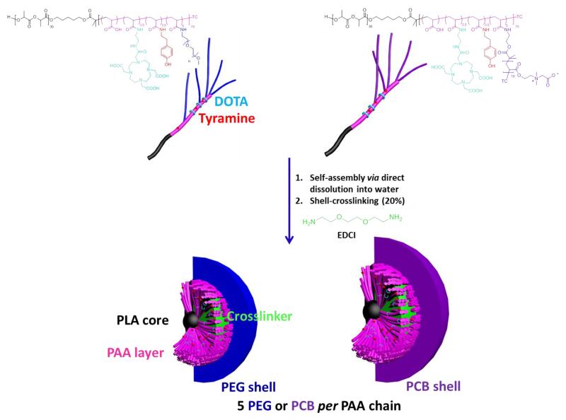
Chemical structures of DOTA- and tyramine-functionalized PEG- and PCB-g-PAA-b-PLA copolymers, their self-assembly in water and crosslinking to form SCKs, with PLA degradable cores, PAA crosslinked shells, DOTA and tyramine available functionalities, and a hydrophilic shell of either PEG or PCB. TC = SC(=S)SC12H25, trithiocarbonate chain end from the RAFT polymerization chemistry. Reproduced with permission from Reference . Copyright 2013 Elsevier Ltd.
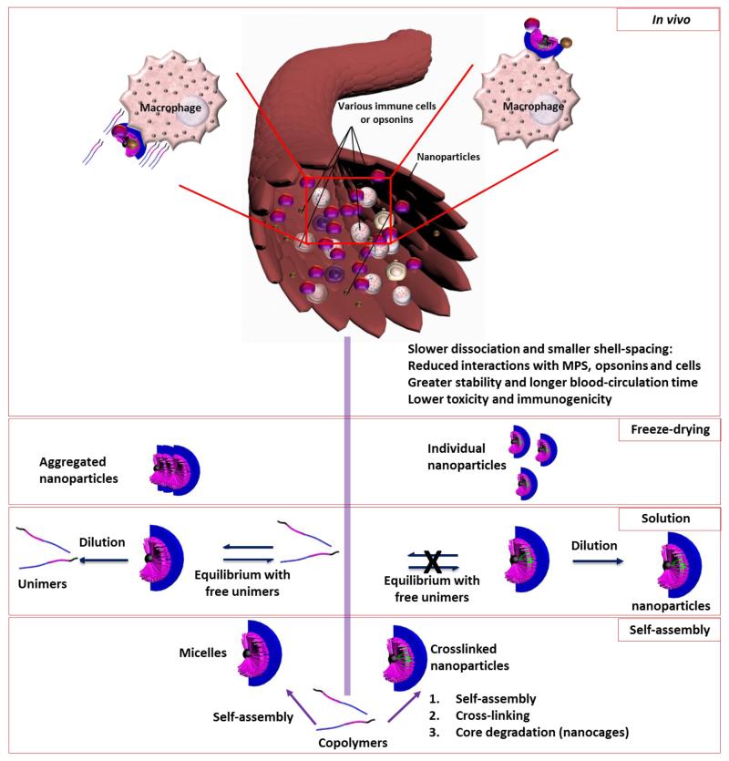
Crosslinking imparts superior characteristics and features to the nanoparticles compared to the non-crosslinked ones, such as, higher stability in solution and in contact with biological interfaces, which reduces the premature cargo release and cyto/immunotoxicities, higher kinetic- and blood-stability, and resistance to aggregation during lyophilization and resuspension processes.

The potential of scope PET atherosclerosis imaging. Inflammation and related pathogenic processes occurring within high-risk plaques can be imaged in vivo using specifically targeted radiolabelled PET tracers. 18F-FDG is the most-widely studied and validated tracer, which is taken up by active macrophages where it is metabolically trapped and accumulates in proportion to intracellular demands. However, the 18F-FDG arterial signal is also influenced by local hypoxia and uptake by other resident cell types. Alternative PET tracers, including 18F-FMCH, 68Ga-DOTATATE, and 11C-PK11195 could be more-specific for macrophage activity (and, therefore, for inflammation) than 18F-FDG. These tracers also seem to have lower background myocardial cell uptake than 18F-FDG, which makes them preferable for coronary artery imaging. Within an inflamed plaque, hypoxia, neoangiogenesis, and microcalcification also contribute to plaque vulnerability; these processes can potentially be imaged with PET using novel tracers, such as 18F-FMISO, 68Ga-NOTA-RGD, and 18F-NaF, respectively. Abbreviations: DOTATATE, [1,4,7,10-tetraazacyclododecane-N,N′,N″,N′″-tetraacetic acid]-d -Phe1,Tyr3-octreotate; FDG, fluorodeoxyglucose; FMCH, fluoromethylcholine; FMISO, fluoromisonidazole; GLUT, solute carrier family 2, facilitated glucose transporter member; NaF, sodium fluoride; NOTA-RGD, 1,4,7-triazacyclononane-1,4,7-triacetic acid-Arg–Gly–Asp; SSTR2, somatostatin receptor type 2; TSPO, translocator protein. Reprinted with permission from Reference . Copyright 2014 Macmillan Publishers Ltd.

(a) Schematic representation of hyperbranched polymer (HBP) used in folate-targeting experiments and MRI and optical images of the mouse subcutaneous B16 melanoma model with HBP. (b) MRI images of bladder, kidney, liver, or tumor (circled in image) in the tumor-bearing mice 1 h following intravenous injection of 100 μL of folate-conjugated or unconjugated (control) HBP (20 mg/mL in PBS). The high-resolution 1H MR image is overlaid with the 19F image. Experiments were performed under isofluorane. (c) Fluorescence images of mice following injection of the same two compounds at the same concentration. The fluorescence images are co-registered with X-ray images of the mice 1 h following subcutaneous injection. (d) Synthesis of a hyperbranched polymer using RAFT mediated polymerization and subsequent end group modifications to introduce amine functionality, followed by attachment of PET ligand and optical NIR dye to synthesize multimodal imaging agent. PET/optical images of C57 Bl/6J mice with subcutaneous B16 melanoma tumor, injected with HBP 2A (e-f). (e) PET/CT image 24 h post injection, showing significant uptake in the tumor (white circle). (f) 72 h time course of optical imaging, top row: non-tumor bearing flank, bottom row: tumor bearing flank. (g) Optical imaging of excised organs (L: liver, K: kidneys, B: blood, Lu: lungs, H: heart, S: spleen, G: gut, and T: tumor). Adapted with permission from References ,. Copyright 2014, Royal Society of Chemistry and American Chemical Society, respectively.

Redox-sensitive dual-modality imaging mechanism. (a) Schematic for dual-modality molecular imaging in response to nitroxide reduction. (b) Emission behavior of OF1 and a control polymer with Cy5.5 and no nitroxides upon exposure to varied amounts of ascorbate or glutathione (GSH) in PBS buffer. The solution pH before and after addition of 60 equiv. ascorbic acid was 7.0 and 6.31, respectively; this pH change has no effect on Cy5.5 absorbance/emission. Reprinted with permission from Reference . Copyright 2014 Macmillan Publishers Ltd.
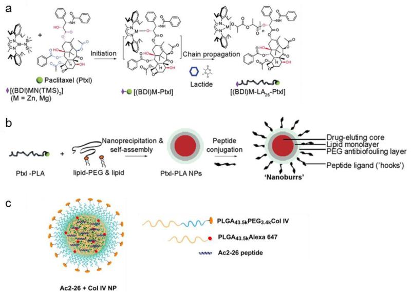
Nanoparticle core-shell design and synthesis. (a) Schematic of paclitaxel-polylactide (Ptxl–PLA) biomaterial synthesis. Ptxl was mixed with equimolar amounts of [(BDI)ZnN(TMS)2]; the (BDI)Zn–Ptxl complex formed in situ initiated and completed the polymerization of lactide. For the nanoburr core, we synthesized Ptxl–PLA25 drug conjugates, which have approximately 25 DL-lactide monomer units. (b) Schematic of nanoburr synthesis by nanoprecipitation and self-assembly. Ptxl–PLA in acetone was added dropwise to a heated lipid solution, vortexed vigorously, and allowed to self-assemble for 2 h to form NPs. The NPs were peptide-functionalized using maleimide-thiol chemistry. Nanoburrs have a drug-eluting polymeric core, a lipid monolayer, a PEG antibiofouling layer, and peptide ligands (hooks) to adhere to the exposed basement membrane during vascular injury. (c) Targeted (Col IV) NPs encapsulating the Ac2-26 peptide was developed using biodegradable polymers via a single-step nanoprecipitation method. The synthesized polymer and Ac2-26 peptide were dissolved in acetonitrile (total polymer 3 mg/mL), and 25 (wt/wt) of the fluorescent PLGA-Alexa 647 was added to the formulation. The NP sample contained 4% (wt/wt) peptide and 5% (wt/wt) of the Col IV peptide-conjugated targeting polymer. The organic mixture containing the polymers and peptide was then added dropwise to nuclease-free water (10 mL). The solution was stirred for 2-4 h, and the particles were filtered, washed, and resuspended in water or PBS. BDI = 2-((2,6-diisopropylphenyl)amino)-4-((2,6-diisopropylphenyl)imino)-2-pentene, TMS = trimethylsilyl. Adapted with permission from References -. Copyright 2010 National Academy of Sciences.

(A) Schematic diagram for mitochondrial delivery of cisplatin prodrug using a targeted NP and the mechanism of action. (B) Synthesis of mitochondria-targeted Pt(IV) prodrug Platin-M. Reprinted with permission from Reference . Copyright 2014 National Academy of Sciences.
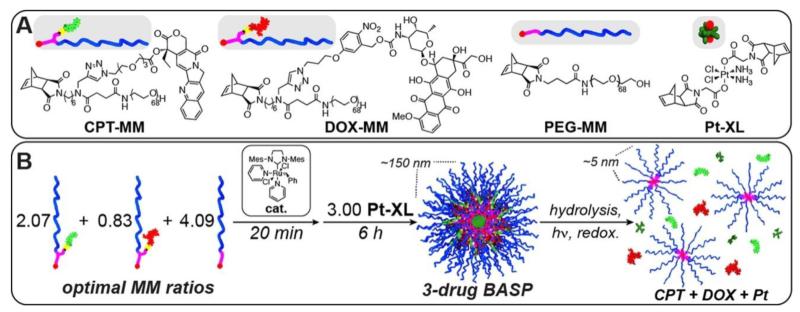
(A) Structures of monomers used in the controlled loading and release of multiple anticancer drugs. (B) Schematic for synthesis of three-drug-loaded BASP. Drug release occurs in response to three distinct triggers. Reprinted with permission from Reference . Copyright 2014 American Chemical Society.

Construction of the DPc-TPM and light-responsive gene transfer. (a) Design of the DPc-TPM. First, a three-layered polyplex micelle is prepared by mixing PEG-PAsp(DET)-PLys triblock copolymer and pDNA; the polyplex micelle is composed of a PEG shell, an intermediate PAsp(DET) layer and a PLys/pDNA core. The DPc-TPM is constructed by adding DPc to the PAsp(DET) intermediate layer. (b) Chemical structure of DPc. (c) Scheme showing the delivery at systemic and intracellular levels. At the systemic level, DPc-TPM circulates in the blood stream; non-specific interaction with biological components is prevented after intravenous injection. In the target tissue (a solid tumor), DPc-TPMs are taken up by cells via endocytosis and entrapped in endo-/lysosomes. In response to the low pH prevalent in the endo-/lysosome, DPc is released from the DPc-TPMs owing to the protonation of the peripheral carboxyl groups and interacts with the endo-/lysosomal membrane through hydrophobic interactions. Upon photoirradiation, DPc generates ROS that destabilize the endo-/lysosomal membrane, facilitating endo-/lysosomal escape. Reprinted with permission from Reference . Copyright 2014 Macmillan Publishers Ltd.

(A) Preparative procedure to obtain PS-carrying BVqMAA micelles in water. A BVT triblock terpolymer is quaternized and hydrolyzed in dioxane to give amphiphilic BVqMAA. After PS addition, self-assembly to micelles takes place through the exchange of solvent from dioxane to PBS buffer. (B) Complexation with PLL-b-PEG diblock copolymers and subsequent crosslinking of PMAA with PLL yields PEGylated micelles (BVqMAA/PLL-b-PEG). Reprinted with permission from Reference . Copyright 2014 American Chemical Society.
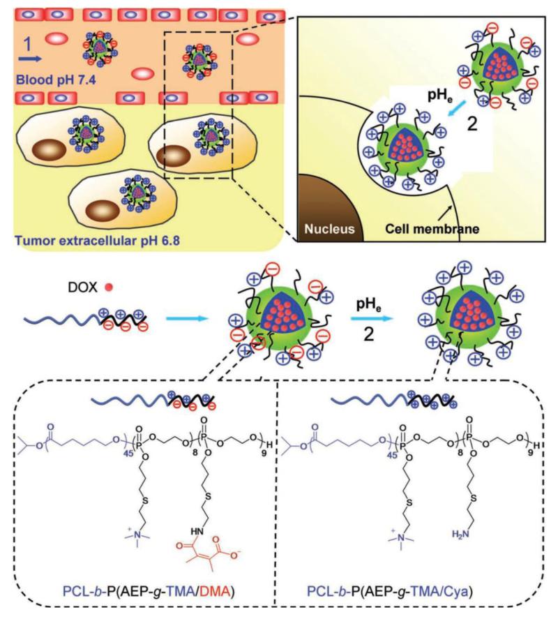
Schematic illustration of doxorubicin (DOX)-loaded zwitterionic polymer-based nanoparticles and the changing of surface charge property in response to the tumor acidity (pHe). 1) Amphiphilic zwitterionic block copolymer PCL-b-P(AEP-g-TMA/DMA) self-assembles into nanoparticles in aqueous solution with DOX encapsulation. During circulation in blood, the nanoparticles show prolonged circulation time and can leak into tumor sites through the EPR effect. 2) Responding to the pHe, the zwitterionic polymer diminishes its anionic part, forming PCL-b-P(AEP-g-TMA/Cya), and the formed nanoparticles are activated to be positively charged and become recognizable by tumor cells. Reprinted with permission from Reference . Copyright 2012 John Wiley & sons, Inc.
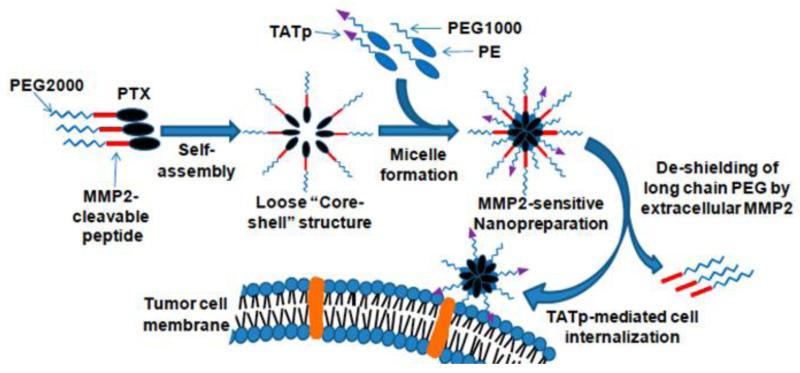
Drug delivery strategy of the MMP2-sensitive nanopreparation. Adapted with permission from Reference . Copyright 2013 National Academy of Sciences.

Synthesis and micelle formation of cationic amphiphilic polycarbonates. a, Cationic amphiphilic polycarbonates were synthesized with a well-defined structure and narrow molecular weight distribution. Based on light scattering, zeta potential, TEM and simulation analyses, these polymers easily formed cationic micelles by direct dissolution in water. b,c, The formation of micelles was simulated through molecular modeling using Materials Studio Software (b) (in the polymer molecule: red, O; white, H; grey, C; blue, N), and was observed in a TEM image of polymer 3 (c) (scale bar, 0.2 μm). Reprinted with permission from Reference . Copyright 2011 Macmillan Publishers Ltd.
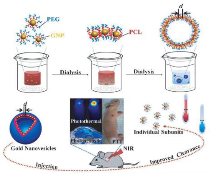
Self-assembly of biodegradable gold vesicles (BGVs) composed of poly(ethylene glycol)-b-poly(ε-caprolactone) (PEG-b-PCL)-tethered GNPs through the dot–line–plane–vesicle mode during the dialysis process. BGVs with an ultrastrong plasmonic coupling effect are superior photoacoustic (PA) imaging and photothermal therapy (PTT) agents with improved clearance after the dissociation of the assemblies. The PA signal and PTT efficiency of BGVs are increased as the distance (d) between adjacent GNPs decreases. Reprinted with permission from Reference . Copyright 2014 John Wiley & Sons, Inc.
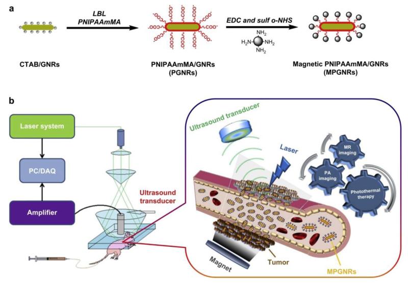
(a) Schematic illustration of the synthesis and structure of PGNRs and MPGNRs. (b) The mechanism of action of MPGNRs for targeted photothermal therapy and dual MR/PA imaging. Abbreviations: PGNRs, PEGylated gold nanorods; MPGNRs, magnetic PGNRs; MR/PA imaging, magnetic resonance/photoacoustic imaging. Reprinted with permission from Reference . Copyright 2013 Elsevier Ltd.
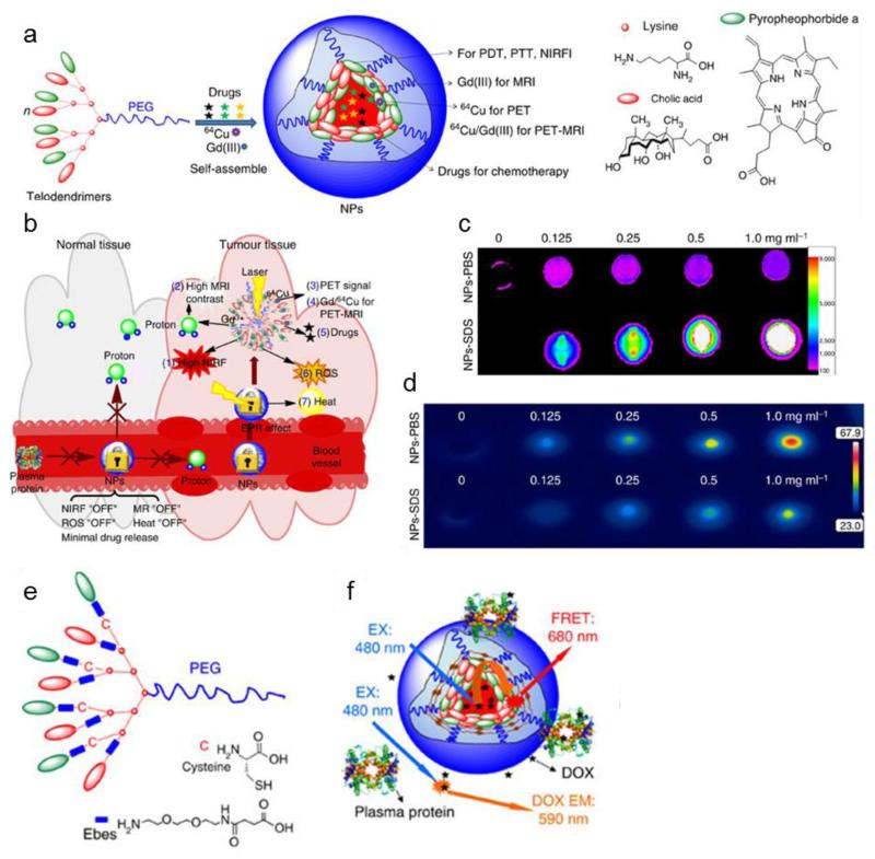
Design, synthesis and characterizations of NPs and disulfide-crosslinked NPs. (a) Schematic illustration of a multifunctional NP self-assembled by a representative porphyrin–telodendrimer, PEG5k-Por4-CA4, composed of four pyropheophorbide-a molecules and four cholic acids attached to the terminal end of a linear PEG chain. (b) Schematic illustration of NPs as a smart ‘all-in-one’ nanomedicine platform against cancers. (c) Near-infrared fluorescence imaging of NP solution (10 μL) in the absence and in the presence of SDS with an excitation bandpass filter at 625/20 nm and an emission filter at 700/35 nm. Concentration-dependent photothermal transduction of NPs: (d) thermal images after irradiation with NIR laser (690 nm) at 1.25 w cm−2 for 20 s. (e) Schematic illustration of a representative crosslinkable porphyrin–telodendrimer (PEG5k-Cys4-Por4-CA4), composed of four cysteines, four pyropheophorbide-a molecules and four cholic acids attached to the terminal end of a linear PEG chain. (f) Schematic illustration of FRET-based approach for study of the real-time release of doxorubicin from nanoporphyrins in human plasma. Adapted with permission from Reference . Copyright 2014 Macmillan Publishers Ltd.
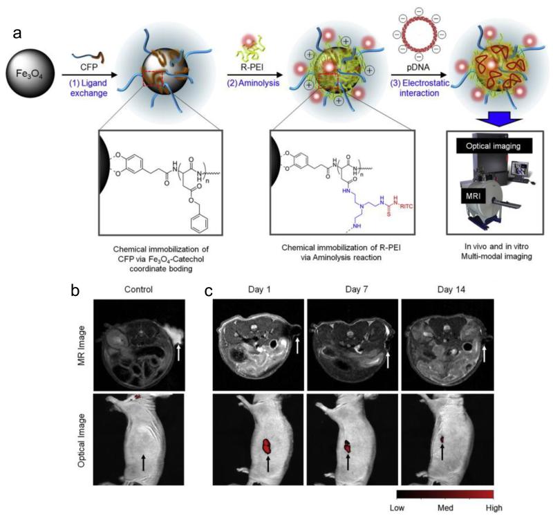
(a) Fabrication process of multi-modal transfection agents (MTA) and in vitro and in vivo multi-modal imaging of MTA in hMSCs. (b) No hyperintense signal (arrow) of MR and optical fluorescence was detected in mice transplanted with MTA-untransfected hMSCs. (c) Hyperintense signals (arrows) of MR and optical fluorescence were detected in mice transplanted with MTA-transfected hMSCs and were still visible 14 days after transplantation. Adapted with permission from Reference . Copyright 2014 Elsevier Ltd.

Design of the multifunctional pH-sensitive polymeric nanoparticle system. Reprinted with permission from Reference . Copyright 2014 Royal Society of Chemistry.

Design and synthesis of target-selective theranostic nanoparticles mimicking virus entry into cells. a) Schematic illustration and SEM image of the filamentous virus-like theranostic nanoparticles composed of poly(γ-glutamic acid)-graft-cetylester (γ-PGA-g-cetylester), pH-switchable fluorophores (pSF), therapeutic drugs (paclitaxel), and targeting antibody (Herceptin). b) Target-selective theranosis: receptor-mediated entry of γ-PGA-g-cetylester [pSF/paclitaxel] nanoparticles followed by pH-dependent signal-on, hydrophobicity-induced membrane-disruption and cytosolic delivery of therapeutic drugs. Reprinted with permission from Reference . Copyright 2014 John Wiley & Sons, Inc.
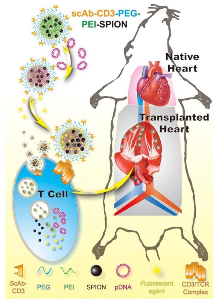
Schematic diagram of therapeutic process of magnetic targeting polyplex scAbCD3-PEG-g-PEI-SPION in vitro and in vivo. Reprinted with permission from Reference . Copyright 2012 American Chemical Society.
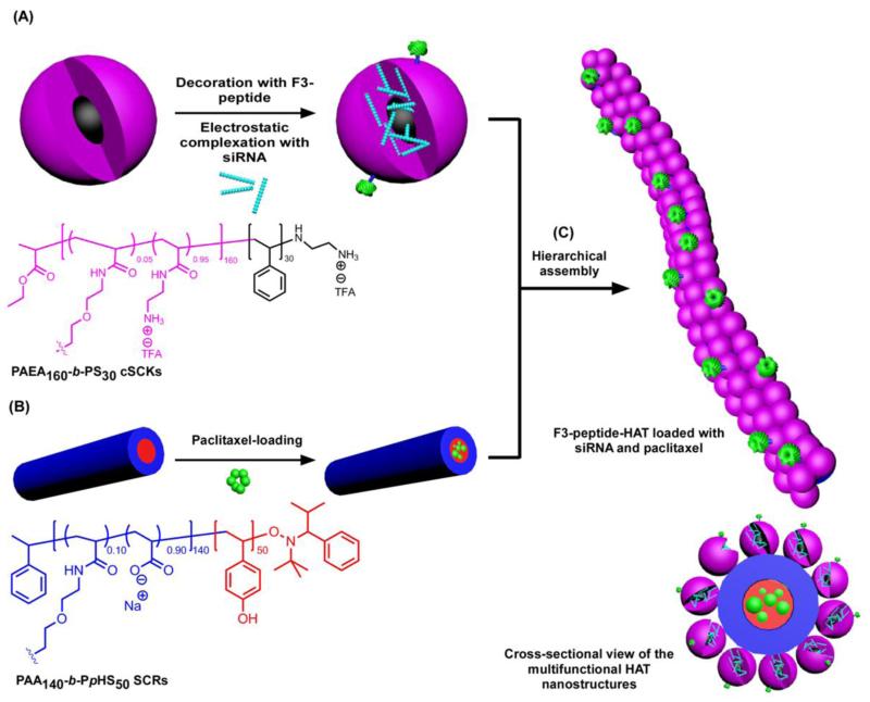
Construction of HAT nanostructures as a template for the co-delivery of siRNA and paclitaxel: (A) Electrostatic complexation of PAEA160-b-PS30 cSCKs and nucleic acids (e.g. siRNA) and decoration of the surface of nanoparticles with targeting ligands (e.g. F3 peptides); (B) Loading of hydrophobic drugs (e.g. paclitaxel) into SCRs composed of PAA140-b-PpHS50. (C) Hierarchical-assembly of cSCKs and SCRs to form the HAT nanoassemblies and the cross-sectional view of the multifunctional HAT nanostructures. Reprinted with permission from Reference . Copyright 2013 American Chemical Society.
References
-
- Toumey C. Plenty of room, plenty of history. Nat. Nanotechnol. 2009;4:783–784. - PubMed
-
- Taniguchi N. On the basic concept of nano-technology; Proc. Intl. Conf. Prod. Eng. Tokyo, Part II, Japan Society of Precision Engineering; 1974.
-
- Devadasu VR, Bhardwaj V, Kumar MN. Can controversial nanotechnology promise drug delivery? Chem. Rev. 2013;113:1686–1735. - PubMed
-
- Brambilla D, Luciani P, Leroux JC. Breakthrough discoveries in drug delivery technologies: the next 30 years. J. Control. Release. 2014;190:9–14. - PubMed
-
- Pahlm O, Wagner G. Multimodal Cardiovascular Imaging: Principles and Clinical Applications. McGraw-Hill; New York: 2011.
Publication types
MeSH terms
Substances
Grants and funding
LinkOut - more resources
Full Text Sources
Other Literature Sources
Medical
