Comparative Study
. 2015 Dec 2;43(21):10143-56.
doi: 10.1093/nar/gkv1028. Epub 2015 Oct 12.

Explaining the striking difference in twist-stretch coupling between DNA and RNA: A comparative molecular dynamics analysis

Affiliations
Comparative Study

Explaining the striking difference in twist-stretch coupling between DNA and RNA: A comparative molecular dynamics analysis

Korbinian Liebl et al. Nucleic Acids Res. .

Abstract

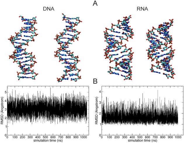
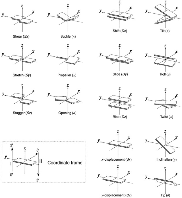
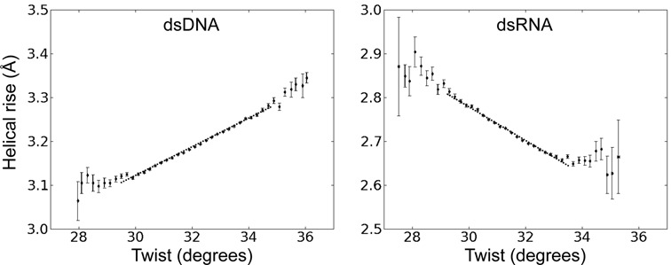
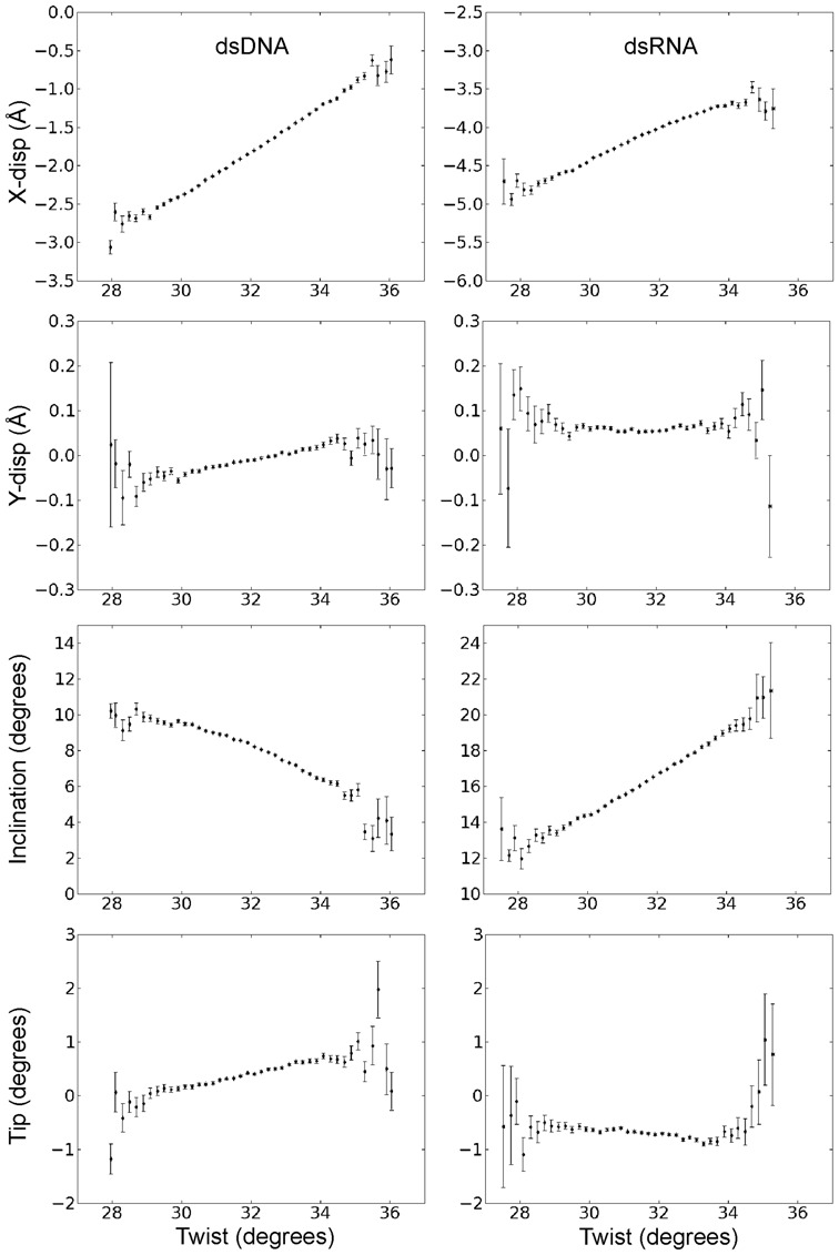
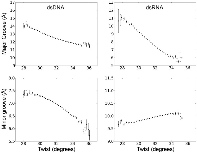
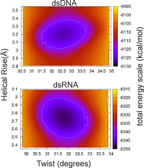
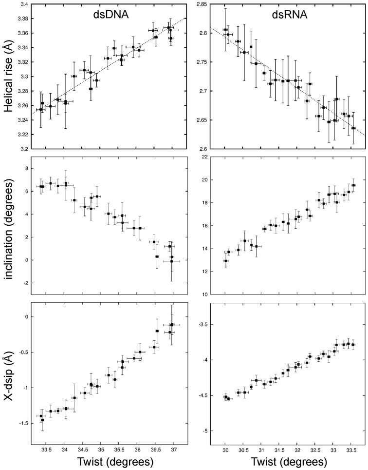
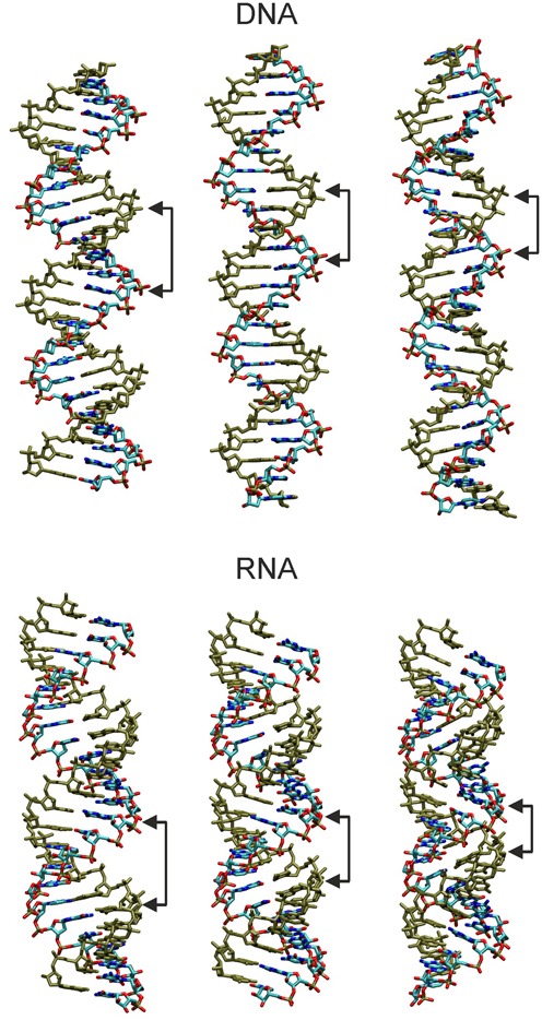
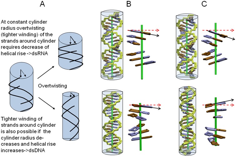
Double stranded helical DNA and RNA are flexible molecules that can undergo global conformational fluctuations. Their bending, twisting and stretching deformabilities are of similar magnitude. However, recent single-molecule experiments revealed a striking qualitative difference indicating an opposite sign for the twist-stretch couplings of dsDNA and dsRNA [Lipfert et al. 2014. Proc. Natl. Acad. Sci. U.S.A. 111, 15408] that is not explained by existing models. Employing unconstrained Molecular Dynamics (MD) simulations we are able to reproduce the qualitatively different twist-stretch coupling for dsDNA and dsRNA in semi-quantitative agreement with experiment. Similar results are also found in simulations that include an external torque to induce over- or unwinding of DNA and RNA. Detailed analysis of the helical deformations coupled to twist indicate that the interplay of helical rise, base pair inclination and displacement from the helix axis upon twist changes are responsible for the different twist-stretch correlations. Overwinding of RNA results in more compact conformations with a narrower major groove and consequently reduced helical extension. Overwinding of DNA decreases the size of the minor groove and the resulting positive base pair inclination leads to a slender and more extended helical structure.


Figures

Figure 1.
Figure 1.
(A) Snapshots of undertwisted (left) and overtwisted (right) dsDNA and dsRNA extracted from 1 μs Molecular Dynamics simulations. In case of DNA the overtwisted snapshot indicates a greater extension in the helical (z) axis direction and a smaller diameter compared to the unwound snapshot. For RNA the overtwisted snapshot shows a reduced extension in the z-direction, a more closed major groove and also a reduced diameter compared to an unwound snapshot. (B) Root mean square deviation (RMSD) of all heavy atoms of the central 10 base pairs with respect to standard B-form DNA (left plot) and standard A-form RNA (right plot) sampled during the entire data gathering simulations.
Figure 2.
Figure 2.
Definition of local base pair parameters (left panels), local base pair step parameters (right panels) and helical base pair axis parameters (lower right panels, in this description helical twist and helical rise correspond to a rotation and translation along a helical axis, respectively) and coordinate frame. Note, that both the set of local base pair step parameters as well as the helical base pair axis parameters offer a complete description of the base pair placements in space (60).
Figure 3.
Figure 3.
Coupling of helical rise and helical twist variation in unrestraint simulations of dsDNA and dsRNA. The plots were generated from a total of ∼100 000 regularly spaced snapshots (every 10 ps) taken during ∼1 μs unrestraint MD simulations. Recorded helical twist and rise of the 10 central base pair steps were analyzed as the mean helical rise and helical twist within intervals of 0.2°. Error bars for twist and rise were obtained as standard errors of the mean in each interval. The slope of the correlation was extracted from a linear fit to the data for a range of ±1.5° with respect to the average twist (over the entire range of twist values, indicated as dotted line). The slope was 0.032 Å·deg−1 in case of DNA and −0.037 Å·deg−1 in case of RNA.
Figure 4.
Figure 4.
Correlation of helical base pair axis parameters and helical twist during unrestrained MD simulations. The plots were generated in the same way as described in the legend of Figure 3.
Figure 5.
Figure 5.
Correlation of major and minor groove width with helical twist during unrestraint MD simulations.
Figure 6.
Figure 6.
Histogram of the calculated total energy (using an implicit solvent model, see Methods) of sampled dsDNA and dsRNA conformations (in unrestraint simulations) versus mean helical twist and mean helical rise. The color-coded total energy is shown for the range of ∼35 kcal·mol-1 with respect to the lowest energy (same color for larger energies).
Figure 7.
Figure 7.
Variation of helical rise, inclination and x-disp during simulations with a torque restraint on the total twist of the DNA (left panels) and RNA (right panels). The mean twist per base pair step was changed in step of ∼0.15° employing a soft harmonic potential on the total twist (see Methods for details) and the resulting mean rise, inclination and x-disp were recorded. Errors were estimated by splitting the data (10 ns simulation time per twist step) into five intervals and separate calculation of mean helical parameters and the standard deviation over these intervals.
Figure 8.
Figure 8.
Illustration of twist-stretch coupling in DNA and RNA induced by locking base pair inclination and x-disp. Energy minimized structures were generated in helical and nucleic acid backbone coordinates using Jumna (61) keeping x-disp and inclination locked (see Table 1). Structures in the middle correspond to relaxed/minimized canonical B-form (DNA) and A-form (RNA) structures (keeping x-disp and inclination locked to canonical values). (left panels) DNA locked to negative x-disp and positive inclination (producing underwound and shortened duplex, see Table 1); RNA locked to more negative x-disp and reduced (positive) inclination (resulting in unwinding and a more extended helix with increased major groove).(right panels) DNA locked to positive x-disp and negative inclination (yielding reduced minor groove width, increased helical rise and increased twist; opposite effect seen for RNA (less negative x-disp and increased inclination produces increased twist and shrinking of the helical extension).
Figure 9.
Figure 9.
(A) Effect of tighter wrapping (overtwisting) of two simplified nucleic acid strands (black lines) around a cylinder (in light blue) at constant radius and with decreasing cylinder radius. The simplified RNA and DNA representations have been generated based on the helical parameters sampled at low twist values (∼28°, B) and high twist values (∼34°, C). Backbone is shown in yellow and bases of one strand in blue and brown, respectively. Enlarged blocks of three base pairs separated by approximately half a helical turn are shown without the backbone for clarity. In case of RNA (upper panels in B, C) the diameter of the double helix decreases only slightly and the length of the helix decreases with increasing twist. This is achieved with a minimum of sterical strain by a more acute Inclination (compare black and red arrows in B, C) and a slide motion (without disturbing the base pair geometry) resulting in a more narrow minor groove and compaction along the helical axis (black arrows in C). For DNA the increase of twist results in a significant narrowing of the helix (reflected by the large change in x-disp). With a minimum of sterical strain this is achieved by a change of inclination in the opposite direction compared to RNA (compare black and red reference arrows in B, C) which extends the helix (black arrows in C).

References

    1. Rohs R., Jin X., West S.M., Joshi R., Honig B., Mann R.S. Origins of specificity in protein-DNA recognition. Annu. Rev. Biochem. 2010;79:233–269. - PMC - PubMed
    1. Alexander R.W., Eargle J., Luthey-Schulten Z. Experimental and computational determination of tRNA dynamics. FEBS Lett. 2010;584:376–386. - PubMed
    1. Lee G., Bratkowski M.A., Ding F., Ke A., Ha T. Elastic coupling between RNA degradation and unwinding by an exoribonuclease. Science. 2012;336:1726–1729. - PMC - PubMed
    1. Pinheiro A.V., Han D., Shih W.M., Yan H. Challenges and opportunities for structural DNA nanotechnology. Nat. Nanotechnol. 2011;6:763–772. - PMC - PubMed
    1. Marko J.F., Siggia E.D. Bending and twisting elasticity of DNA. Macromolecules. 1994;27:881–998.

Publication types