. 2015 Oct 27;112(43):E5805-14.
doi: 10.1073/pnas.1517952112. Epub 2015 Oct 13.

Structural insights into species-specific features of the ribosome from the pathogen Staphylococcus aureus

Affiliations

Structural insights into species-specific features of the ribosome from the pathogen Staphylococcus aureus

Zohar Eyal et al. Proc Natl Acad Sci U S A. .

Abstract

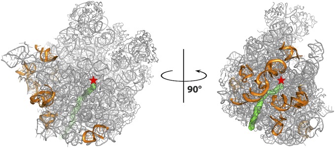
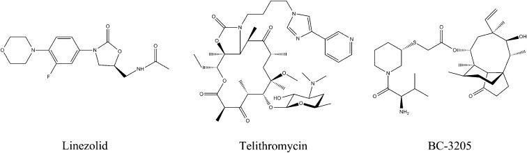
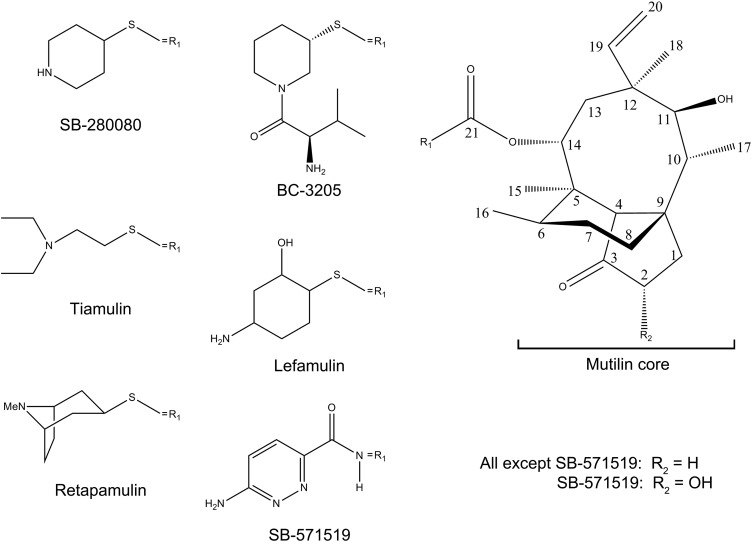
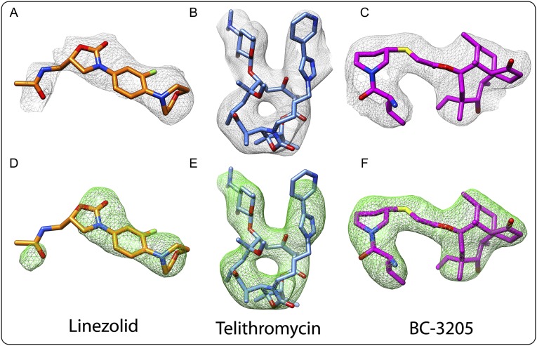
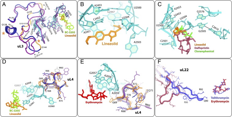
The emergence of bacterial multidrug resistance to antibiotics threatens to cause regression to the preantibiotic era. Here we present the crystal structure of the large ribosomal subunit from Staphylococcus aureus, a versatile Gram-positive aggressive pathogen, and its complexes with the known antibiotics linezolid and telithromycin, as well as with a new, highly potent pleuromutilin derivative, BC-3205. These crystal structures shed light on specific structural motifs of the S. aureus ribosome and the binding modes of the aforementioned antibiotics. Moreover, by analyzing the ribosome structure and comparing it with those of nonpathogenic bacterial models, we identified some unique internal and peripheral structural motifs that may be potential candidates for improving known antibiotics and for use in the design of selective antibiotic drugs against S. aureus.

Keywords: antibiotic resistance; potential advanced pleuoromutilin; protein biosynthesis; species specificity.


Conflict of interest statement

The authors declare no conflict of interest.

Figures

Fig. 1.
Fig. 1.
The structure of the large ribosomal subunit of SA50S is shown in gray, the polypeptide exit tunnel is shown in green, and the PTC location is marked by a red star. The rRNA regions with fold variability compared with all other known structures (see below) on the SA50S are shown in orange and are rotated 90° to one another.
Fig. 2.
Fig. 2.
Chemical structures of the oxazolidinone linezolid, the ketolide telithromycin, and the pleuromutilin BC-3205.
Fig. S1.
Fig. S1.
Chemical formulas of selected pleuromutilin derivatives. Typically, the tricyclic mutilin core (Right) is conserved among pleuromutilin derivatives. Note the variability of the C14 extension (R1). Chemical formulas for all mentioned pleuromutilins are shown.
Fig. S2.
Fig. S2.
Electron density of linezolid, telithromycin, and BC-3205 within their complexes with SA. (A–C) Weighted 2Fo-Fc electron density maps contoured at 1.0 σ. (DF) Weighted Fo-Fc electron density maps contoured at 3.0 σ.
Fig. 3.
Fig. 3.
Details of structural variability in the rRNA backbone among the four known eubacteria structures. SA50S is in teal, E. coli (E70S) is in purple, D. radiodurans (D50S) is in gray, and T. thermophilus (T70S) is in orange. (A) H25. (B) H63. (C) H79. (D) H10 and H15-16. (E) H68. (F) H28.
Fig. S3.
Fig. S3.
(A–D) Electron density of 23S rRNA. Weighted 2Fo-Fc electron density maps contoured at 1.2 σ. (E–H) Electron density of rProteins. Weighted 2Fo-Fc electron density maps contoured at 1.0 σ. (A) H9. (B) H25. (C) H28. (D) H63. (E) uL3. (F) uL16. (G) bL17. (H) bL32 N terminus.
Fig. 4.
Fig. 4.
Structural differences in rProteins. SA50S is shown in navy, E. coli (E70S) is in purple, D. radiodurans (D50S) is in gray, and T. thermophilus (T70S) is in orange. (A) The N-terminal end of protein bL32 that resides in the second shell around the erythromycin-binding pocket is shorter in S. aureus (SA50S, navy) and E. coli (E70S, purple) compared with in D. radiodurans (D50S, gray) and T. thermophilus (T70S orange), and thus may provide a space for a specific extension of erythromycin (Fig. S5). (B) SA50S rProtein bL3 has a unique extended A58-A69 loop compared with D50S, T70S, and E70S. (C) SA50S uL16 points opposite to E70S and thus may provide a potential specific binding site (Fig. S5) for a compound that may interfere with binding of the A-site tRNA acceptor stem (blue). P-site tRNA (green) interacts with the loop K77-V90, which has some structural variability. (D) SA50S bL17 has an extended (T65-A81) loop that is unique to SA50S. Because it is exposed on the surface of the SA50S subunit, it may provide a potential specific binding site (Fig. S5) for a compound that may interfere with the two subunits association. Its C terminus is ∼10 aa longer in E70S relative to the other three organisms. (E) In D50S, bL27 reaches the position of the acceptor stem of the P-site tRNA (green), whereas in T70S it reaches the PTC, in the proximity of the CCA-end of the P-site tRNA. (F) bL28 is located near the 50S surface, close to the position of the CCA 5′ of E-site tRNA (yellow). in D50S and T70S, it has a 15-residue extended loop (S19-K27) This loop is shorter in SA50S and E70S and does not exist in D50S and T70S, and thus may provide a potential site for a compound that may interfere with the binding of the E-site tRNA acceptor stem (yellow) (Fig. S5).
Fig. S4.
Fig. S4.
Additional structural differences identified in rProteins. A possible path of the backbone of a modeled nascent protein chain is indicated in yellow. The SA50S structure is shown in teal, the E. coli (E70S) structure is in purple, the D. radiodurans (D50S) structure is in gray, and the T. thermophilus (T70S) structure is in orange. (A) The bL32 N terminus in D50S and T70S is elongated relative to that in SA50S and E70S, reaching the rims of the erythromycin-binding site. (B) View into the exit tunnel opening, showing structural variability of the rProteins uL23, uL24, and uL29. The polypeptide chain was modeled in the tunnel for clarity (lime). (C) uL24 in all four structures. (D) The uL18 N-terminal domain is elongated in S. aureus relative to D50S, T70S, and E70S reaching the other side of the central protuberance. (E) bL25 domains, as well as structural variability, in the fold of G11-L26 and F79- I86 loops. The uL16 C terminus is longer in T70S than in SA50S, D50S, and E70S. (F) Zoom-in view of the hinge between the bL25 domains. The uL16 C terminus is longer in T70S than in SA50S, D50S, and E70S. It penetrates into the proximity of bL25, thereby changing the angle between bL25 domains. (G) bL27 R79-K85 loop and C-terminal fold variability among the four structures.
Fig. S5.
Fig. S5.
Structural differences in rProteins. SA50S is shown in navy, E. coli (E70S) is in purple, D. radiodurans (D50S) is in gray, and T. thermophilus (T70S) is in orange. (A) The N-terminal end of protein bL32 that resides in the second shell around the erythromycin-binding pocket is shorter in S. aureus (SA50S, navy) and E. coli (E70S, purple) compared with that in D. radiodurans (D50S, gray) and T. thermophilus (T70S, orange), thus may provide a space for a specific extension of erythromycin (encircled in pink). (B) SA50S rProtein bL3 has a unique extended A58-A69 loop compared with that in D50S, T70S, and E70S. (C) SA50S uL16 is pointing opposite compared with E70S thus may provide a potential specific binding site (encircled in pink) for a compound that may interfere with binding of the A-site tRNA acceptor stem (blue). P-site tRNA (green) interacts with the loop K77-V90 that has some structural variability. (D) SA50S bL17 has an extended (T65-A81) loop that is unique to SA50S. Because it is exposed on the surface of the SA50S subunit, it may provide a potential specific binding site (encircled in pink) for a compound that may interfere with the two subunits association. Its C-terminal is ∼10 aa longer in E70S relative to the other three organisms. (E) bL27 in D50S reaches the position of the acceptor stem of the P-site tRNA (green), whereas in T70S it reaches the PTC, in the proximity of the CCA-end of the P-site tRNA. (F) bL28 is located near the 50S surface; close to the position of the CCA 5′ of E-site tRNA (yellow). In D50S and T70S, it has a 15-residue extended loop (S19–K27). This loop is shorter in SA50S and E70S and does not exist in D50S and T70S, and thus may provide a potential site for a compound that may interfere with the binding of the E-site tRNA acceptor stem (yellow) (encircled in pink).
Fig. S6.
Fig. S6.
(A) Flexible nucleotides at the PTC and at the exit tunnel in U2506, U2585, A2062, A2602, and U2491, where the P-site tRNA (green surface) and A-site tRNA (blue surface) should bind. S. aureus 23S RNA backbone and nucleotides are shown in teal. D. radiodurans, T. thermophilus, and E. coli nucleotides are shown in gray, orange, and purple, respectively. (B) Flexible nucleotides toward the tunnel opening. A90, A91, and A508 are located in the ribosomal exit tunnel, and were detected with different conformations in all four structures. A possible path of the backbone of a modeled nascent polyalanine chain is shown in yellow.
Fig. S7.
Fig. S7.
Structural differences in rProteins at the subunit surface. (A) Length variations detected in the V6-I17 surface loop of protein uL4 and the N terminus of uL15 located in its vicinity. Most extend in SA50S and less in E70S and D50S, and have a different orientation in T70S. Shown are the uL15 N termini in E70S, D50S (traced from the fourth amino acid), T70S (traced from the fifth amino acid), and SA50S (the shortest, traced from the first amino acid). (B and C) The C and N terminals of uL4 have different backbone folds in the four eubacterial structures. (D) SA50S L15 loops I69-T89 and T89-V97 have different structures compared with the corresponding loops in D50S, T70S, and E70S. (E) Structural variability in the M38-A53 and G126-D144 loops of uL5 that participate in the B1b intersubunit bridge with the 30S subunit (16S in gray) with rProtein uS13 (dark green).
Fig. S8.
Fig. S8.
Surface of the SA50S structure indicating the locations of the globular regions of the rProteins. rRNA is shown in gray, and rProteins are shown in different colors. (Upper, Left) View from the SA50S intersubunit surface. (Upper, Right) View from the SA50S outer surface. (Lower) +900 and −900 vertical rotation of the intersubunit surface view.
Fig. 5.
Fig. 5.
The linezolid-binding site as identified in the SA50Slin complex structure (A and B), with hydrogen bonds between the bound drugs and 23S rRNA shown in black dashes, the telithromycin-binding site as identified in the SA50Stel complex structure (C and D), and the BC3205-binding site as identified in the SA50S-BC3205 complex structure (E and F). (A) Comparison of native SA50S PTC (teal) and SA50Slin complex (pale purple) structures. (B) Overlay of the structures of various ribosome-linezolid complexes, including SA50Slin (pale purple; this study), H50Slin (green) + CCA-Phe substrate analog (teal) (PDB ID code 3CPW) (47), and D50Slin (gray) (PDB ID code 3DLL) (46), and of the model of E70Slin (pink) (48). The color coding of the rRNA components of the various linezolid-binding pockets is the same as that of the corresponding linezolid molecules. (C) Comparison of the structures of native SA50S PTC (teal) and SA50Stel complex (red). (D) Structural overlay of telithromycin conformations observed in various ribosome-telithromycin complex structures: SA50Stel (slate; this study), D50Stel (orange) (PDB ID ID code 1P9X) (27), H50Stel (gray) (PDB ID code 1YIJ) (28), E70S-tel (pink) (PDB ID code 3OAT) (29), and T70Stel (green) (PDB ID code 3OI3) (30). The color coding of the rRNA components of the various telithromycin-binding pockets is in a brighter tone than the corresponding telithromycin molecules. (E) Comparison of the PTC structure in native SA50S (teal) and in SA50S-BC3205 complex (purple). The arrows indicate the movements of nucleotides U2585 and U2506 in the bound vs. native structure. (F) Structural overlay of various pleuromutilins in their binding pockets: SA50S-BC3205 (violet; this study), D50S-SB571519 (green) (PDB ID code 2OGM), D50S-retapamulin (cyan) (PDB ID code 2OGO), D50S-tiamulin (slate) (PDB ID code 1XBP), and D50S-SB280080 (lemon) (PDB ID code 2OGN). Only one hydrogen bond between BC-3205 (violet) and 23S rRNA is shown here (as black dashes) for increased clarity.
Fig. 6.
Fig. 6.
Resistance and cross-resistance mechanisms in S. aureus. SA50S rRNA and rProteins are colored in navy. D50S (gray), E70S (purple), and T70S (orange) structures are superimposed on the SA50S structure for comparative analysis. rRNA nucleotides of S. aureus are shown only in regions in which they can be well aligned with the corresponding nucleotides in all other structures used for the comparisons. (A) SA50S rRNA nucleotides of the linezolid-binding sites (orange) and BC-3205–binding sites (green) are superimposed on the corresponding E70S rRNA (purple). G2576 is located in the second shell around the linezolid- and BC-3205–binding sites, in proximity to the first shell nucleotides G2505 and U2506; thus, its mutations may cause alterations in them. The locations of the uL3 mutations acquiring resistance to S. aureus are marked on the protein chain (yellow). For comparison, the structure of E. coli uL3 (purple) is superposed on SA50S uL3 (navy). Key structural differences are marked by arrows. The orange stars indicate deletions. (B and C) Linezolid (orange), chloramphenicol (green), and dalfopristin (streptogramins A, in raspberry) are shown in the SA50S rRNA-binding pocket. (D) uL4 rProtein is in the vicinity of S. aureus linezolid-binding (orange) and S. aureus BC-3205–binding (green) pockets. The structural variability of its loop (W65-Q75) in four species is shown. (E) SA50S rRNA A2058 and A2059 are main binding determinants of MLSBK family of antibiotics, represented here by erythromycin (PDB ID code 3OFR) (red). uL4 and bL32 rProteins form a second shell around the erythromycin-binding pocket next to A2058 and A2059. Structural variability of uL4 among D50S, E70S, T70S, and SA50S is shown. (F) S. aureus telithromycin (slate) conformation within its S. aureus complex, where its alkyl-aryl arm is folded such that it overlaps the desosamine sugar of erythromycin (red) in its complex with D50S. SA50tel uL22 (blue) is superimposed on the same rProtein in H. marismortui large ribosomal subunit with a deletion mutation Δ82–84 in uL22 (red) that allows protein progression even though it binds erythromycin (PDB ID code 1YJ9).

References

    1. Lowy FD. Staphylococcus aureus infections. N Engl J Med. 1998;339(8):520–532. - PubMed
    1. Kurosu M, Siricilla S, Mitachi K. Advances in MRSA drug discovery: Where are we and where do we need to be? Expert Opin Drug Discov. 2013;8(9):1095–1116. - PMC - PubMed
    1. World Health Organization . Antimicrobial Resistance: Global Report on Surveillance. World Health Organization; Geneva: 2014.
    1. Chambers HF. The changing epidemiology of Staphylococcus aureus? Emerg Infect Dis. 2001;7(2):178–182. - PMC - PubMed
    1. Schlünzen F, et al. Structural basis for the interaction of antibiotics with the peptidyl transferase center in eubacteria. Nature. 2001;413(6858):814–821. - PubMed

Publication types

MeSH terms