. 2015 Oct 27;112(43):13372-7.
doi: 10.1073/pnas.1516473112. Epub 2015 Oct 13.

Auxin signaling modules regulate maize inflorescence architecture

Affiliations

Auxin signaling modules regulate maize inflorescence architecture

Mary Galli et al. Proc Natl Acad Sci U S A. .

Abstract

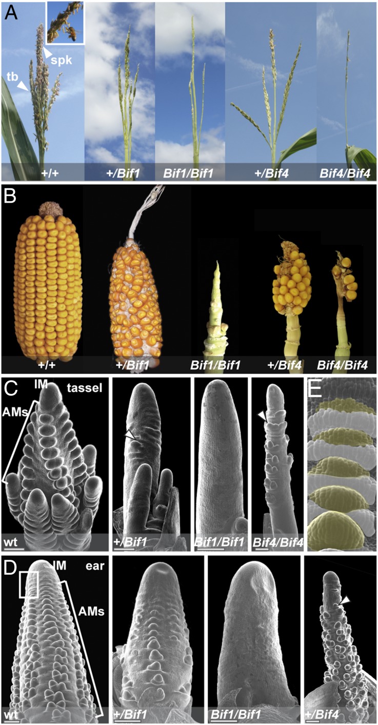
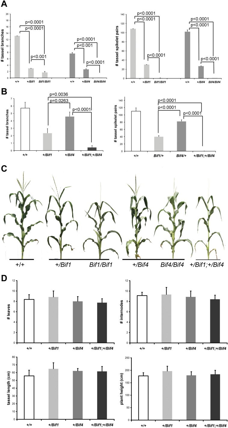
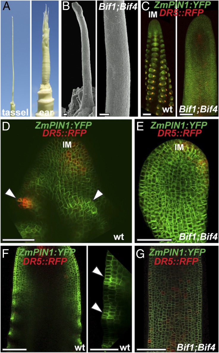
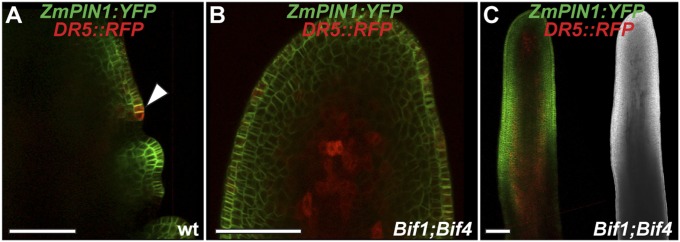
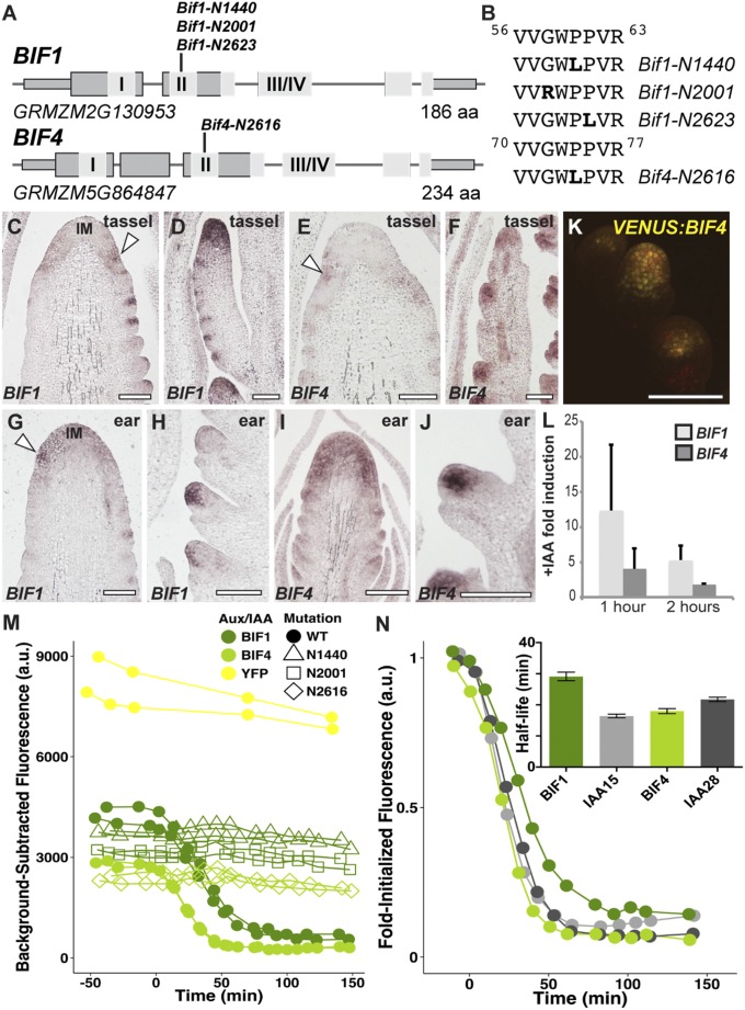
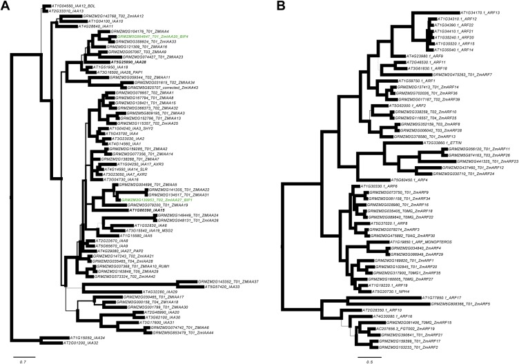
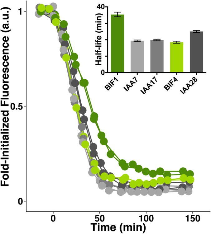
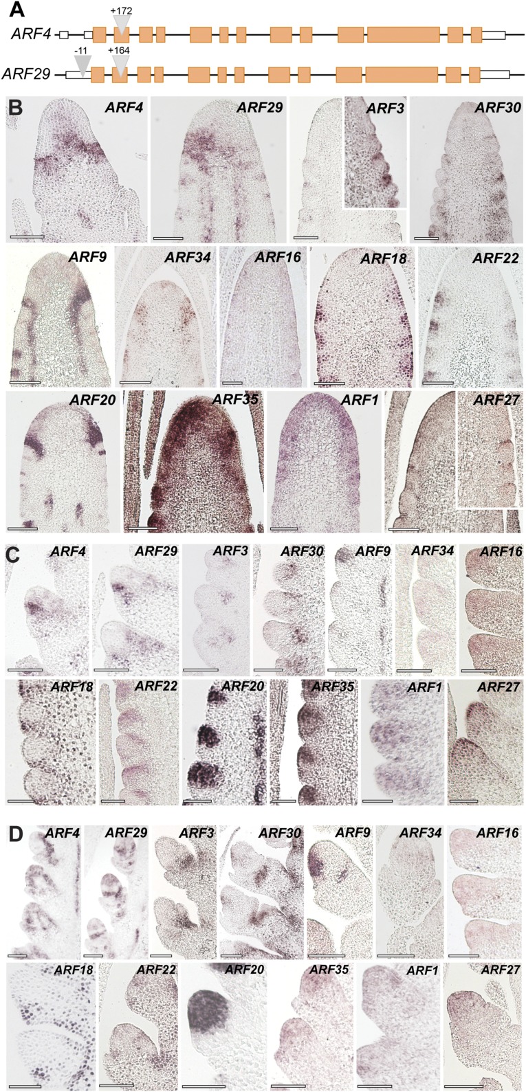
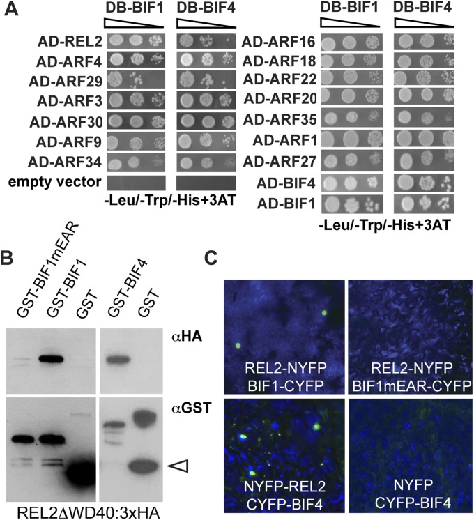
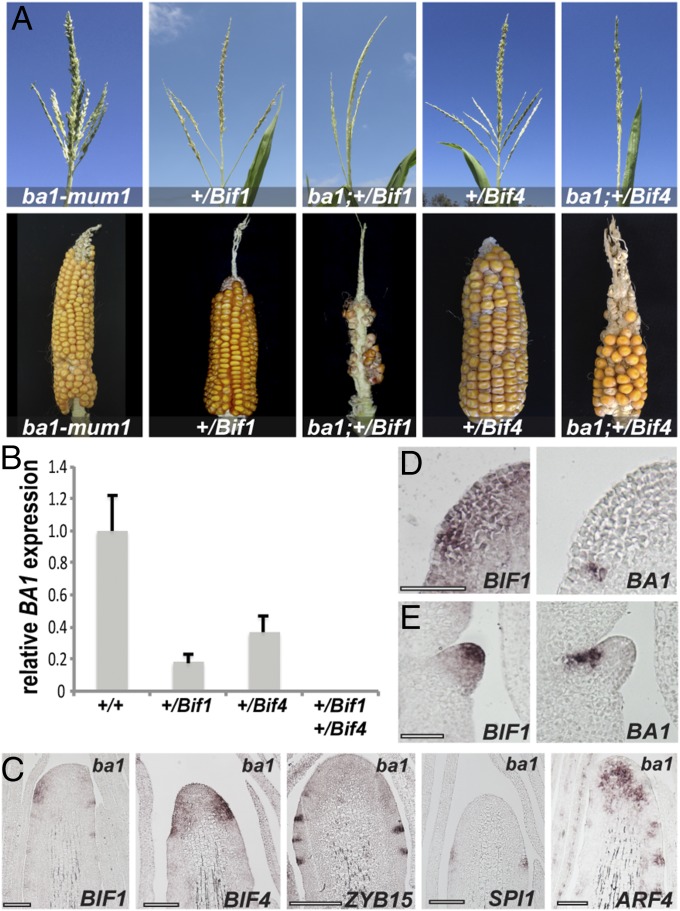
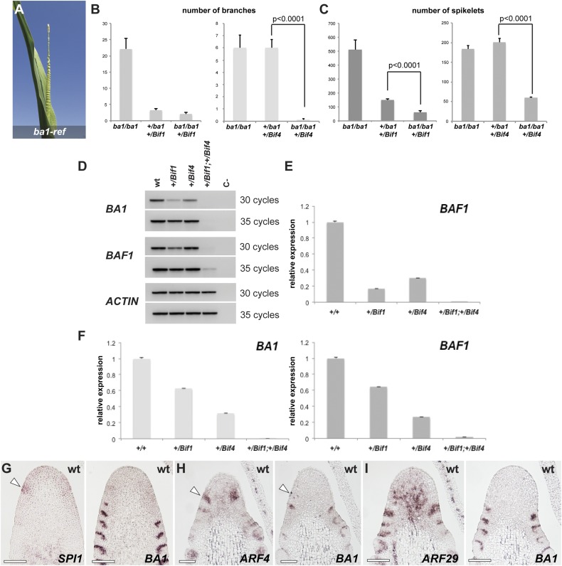
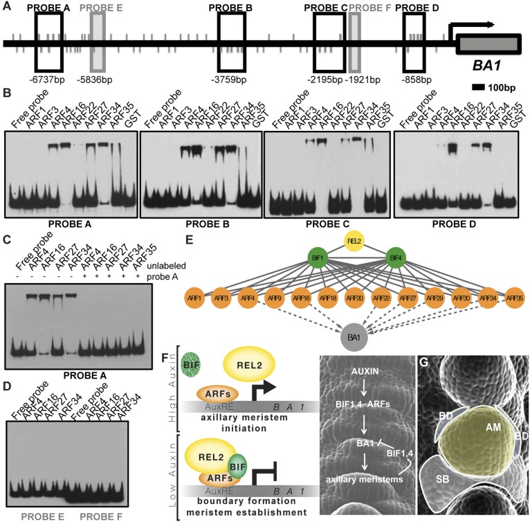
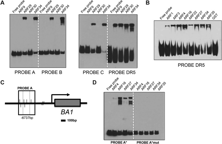
In plants, small groups of pluripotent stem cells called axillary meristems are required for the formation of the branches and flowers that eventually establish shoot architecture and drive reproductive success. To ensure the proper formation of new axillary meristems, the specification of boundary regions is required for coordinating their development. We have identified two maize genes, BARREN INFLORESCENCE1 and BARREN INFLORESCENCE4 (BIF1 and BIF4), that regulate the early steps required for inflorescence formation. BIF1 and BIF4 encode AUXIN/INDOLE-3-ACETIC ACID (Aux/IAA) proteins, which are key components of the auxin hormone signaling pathway that is essential for organogenesis. Here we show that BIF1 and BIF4 are integral to auxin signaling modules that dynamically regulate the expression of BARREN STALK1 (BA1), a basic helix-loop-helix (bHLH) transcriptional regulator necessary for axillary meristem formation that shows a striking boundary expression pattern. These findings suggest that auxin signaling directly controls boundary domains during axillary meristem formation and define a fundamental mechanism that regulates inflorescence architecture in one of the most widely grown crop species.

Keywords: auxin signaling; axillary meristems; boundary domains; inflorescence development; maize.


Conflict of interest statement

The authors declare no conflict of interest.

Figures

Fig. 1.
Fig. 1.
Bif1 and Bif4 mutant phenotype. (A) Mature tassel phenotype. Normal tassels produce spikelets and flowers that are reduced in both mutants. (Inset) Spikelets with protruding anthers. tb, tassel branch. spk, spikelet. (B) Mature ear phenotype. (C–E) Scanning electron microscope image of early tassel and ear development in normal and mutant plants. Arrowheads point to a few axillary meristems forming in mutant plants. Primordia are absent in homozygous Bif1 mutants. Boxed region in D marks the peripheral zone of the IM. (E) Close-up of the peripheral zone of the IM. White and yellow colors mark suppressed bract primordia and AMs, respectively. Note the acropetal development of primordia (from top, younger, to bottom, older).
Fig. S1.
Fig. S1.
Quantification of the phenotypic defects of Bif1 and Bif4 mutants. (A) Reproductive defects in tassels of single mutants in B73 background (n ≥ 7). (B) Analysis of double mutants in A619 background (n ≥ 7). (C) Whole-plant images. (D) Analysis of vegetative and reproductive phenotypes with no detectable difference (n ≥ 6). Error bars show SD.
Fig. 2.
Fig. 2.
BIF1 and BIF4 are required for patterning of primordium initiation. (A) Mature inflorescence phenotype of the double heterozygous Bif1 and Bif4 mutant. (B) Scanning electron microscope image of a young Bif1;Bif4 tassel showing lack of primordium initiation. (Scale bars, 100 μm.) (C) Confocal images of normal and Bif1;Bif4 tassels expressing ZmPIN1a-YFP fusion proteins and DR5::RFP. (D and E) Maximum projections of confocal images of wild-type and Bif1;Bif4 mutant IMs. (F and G) Confocal images of the peripheral zone of immature tassels showing up-regulation of ZmPIN1a-YFP signals in normal tassels (F, arrowheads in close-up right panel) that is missing in double Bif1;Bif4 mutants (G).
Fig. S2.
Fig. S2.
Confocal analysis of ZmPIN1a:YFP and DR5rev::RFP transgenes. (A) Wild-type tassel, showing the peripheral zone of the IM with an emerging primordium (arrowhead). (B and C) +/Bif1;+/Bif4 tassels. In B is a close-up view of the IM. (C, Right) Brightfield image of the confocal sample. (Scale bars, 100 μm.)
Fig. 3.
Fig. 3.
BIF1 and BIF4 encode Aux/IAA proteins. (A) Schematic representation of BIF1 and BIF4 genes. Exons are depicted as gray rectangles. I and II represent the EAR repressor motif and the degron domain; III/IV corresponds to the dimerization domain. (B) The amino acid sequence of the degron domains of BIF1 and BIF4 and the mutations identified. (C–J) mRNA in situ hybridizations of immature inflorescences with BIF1 and BIF4 antisense probes. Arrowheads, localized signals at the peripheral zone of the IM. (D and F) Branch meristems are shown; (H and J) spikelet meristems. (Scale bars, 100 μm.) (K) Confocal image of VENUS-BIF4 in spikelet meristems. (L) Auxin inducibility of BIF1 and BIF4. Error bars show SD. (M and N) Auxin-induced degradation profiles of normal and mutant BIF1 and BIF4 proteins.
Fig. S3.
Fig. S3.
Phylogenetic analysis. (A) Bayesian consensus phylogram of 67 Arabidopsis (At) and maize (GRMZM) Aux/IAA protein sequences. The maize BIF1 and BIF4 sequences and their closest Arabidopsis relatives, ATIAA15, and ATIAA28, respectively, are bolded. Phylogram is rooted using Arabidopsis IAA32 and IAA34 based on Remington (61). Branch widths are proportional to support with bold branches equivalent to greater than or equal to 0.95 clade credibility. (B) Bayesian consensus phylogram of 55 Arabidopsis (At) and maize (GRMZM) ARF protein sequences. Phylogram is rooted using the Arabidopsis ARF10, ARF16, and ARF17 clade, based on Remington (61). Branch widths are proportional to support with bold branches equivalent to greater than or equal to 0.95 clade credibility. (Scale bars, substitution per site.)
Fig. S4.
Fig. S4.
Auxin-induced degradation of wild-type BIF1 and BIF4 proteins in a yeast synthetic system compared with similar Arabidopsis proteins.
Fig. S5.
Fig. S5.
The maize ARF gene family. (A) Gene structure of ZmARF4 and ZmARF29, the maize co-orthologs of Arabidopsis MONOPTEROS. Triangles represent transposon insertions. Orange boxes, exons; white boxes, UTR regions. (B–D) Inflorescence-specific expression of maize-activating ARF genes by mRNA in situ hybridizations in immature tassels. Expression pattern in inflorescence meristems (B), spikelet-pair (C), and spikelet meristems (D). (Scale bars, 100 μm.)
Fig. S6.
Fig. S6.
Protein–protein interaction assays. (A) Y2H analysis of BIF1 and BIF4 with the 13 activating ARFs and REL2. (B) The interaction of BIF1 and BIF4 with REL2 in in vitro pull-down assays. Arrowhead points to GST. (C) BiFC assay by transient expression in tobacco leaves.
Fig. 4.
Fig. 4.
Genetic and expression analysis of ba1 mutants. (A) Double-mutant analysis of Bif1 and Bif4 with ba1-mum1 in A619 background. (B) qRT-PCR of BA1 in double Bif1;Bif4 mutants. Error bars, SD. (C) In situ hybridization of immature ba1-ref tassels with specific markers. (Scale bars, 100 μm.) (D and E) mRNA in situ hybridizations on consecutive sections of immature inflorescences with BIF1 and BA1 antisense probes. (Scale bars, 50 μm.)
Fig. S7.
Fig. S7.
Genetic and expression analysis. (A) ba1-ref tassel. Note the enlarged suppressed bracts. (B and C) Quantification of tassel phenotypes (branch and spikelet-pair number) in Bif1;ba1-mum1 and Bif4;ba1-mum1 double mutants (n ≥ 6). Error bars, SEM. (D) Semiquantitative RT-PCRs of BA1, and BAF1 and ACTIN controls in single- and double-mutant tassels. BAF1 is a boundary-expressed gene that functions upstream of BA1 (50). (E) Quantitative RT-PCR of BAF1 (same samples as in Fig. 4; background A619). (F) Quantitative RT-PCR of BA1 and BAF1 in a B73 background. (G–I) Consecutive sections of young IMs hybridized with different in situ probes. (G) Note that SPI1 expression (arrowhead) appears before BA1. (H and I) ARF expression precedes BA1 expression (arrowheads), but later overlaps with it in slightly broader domains.
Fig. 5.
Fig. 5.
BA1 is a target of BIF/ARF transcriptional regulatory modules. (A) Schematic showing BA1 genomic locus including 7 kb of putative promoter. Promoter fragments used as probes in EMSAs are shown as boxed regions. Values below boxes indicate position relative to BA1 start codon (+1). Gray lines indicate TGTC core AuxRE elements. (B) EMSAs show that various activating ARFs bind to BA1 promoter fragments; GST alone does not. (C) EMSA showing specificity of ARF binding to probe A. Addition of unlabeled probe A outcompetes binding to labeled probe A. (D) EMSA showing ARFs do not bind non-TGTC-enriched promoter fragments E and F. (E) Summary of protein–protein (solid lines) and protein–DNA (dashed lines) interactions identified in this study. (F) Molecular model of organogenesis in the peripheral zone of maize IMs. (G) Diagram of the resulting functional domains (false-colored). SB, suppressed bract; BD, boundary domain; AM, axillary meristem.
Fig. S8.
Fig. S8.
Multiple-activating ARFs directly bind to the BA1 promoter. (A) EMSAs showing that ARF29 and ARF34 bind to probes A, B, C, and DR5. (B) EMSA showing that all ARFs tested bind to a 9xAuxRE DR5 probe. (C) Schematic showing simplified BA1 genomic locus. Promoter fragment used for probe A in EMSAs is shown as boxed region; dashed box in probe A corresponds to 100-bp probe A*. Values below boxes indicate position relative to BA1 start codon (+1). Gray lines indicate TGTC core AuxRE elements. (D) EMSA showing that mutation of core TGTCs in probe A* eliminates binding of ARF4, ARF16, ARF27, and ARF34.

Similar articles

Cited by

References

    1. Doebley JF, Gaut BS, Smith BD. The molecular genetics of crop domestication. Cell. 2006;127(7):1309–1321. - PubMed
    1. Bommert P, Nagasawa NS, Jackson D. Quantitative variation in maize kernel row number is controlled by the FASCIATED EAR2 locus. Nat Genet. 2013;45(3):334–337. - PubMed
    1. Park SJ, et al. Optimization of crop productivity in tomato using induced mutations in the florigen pathway. Nat Genet. 2014;46(12):1337–1342. - PubMed
    1. Gälweiler L, et al. Regulation of polar auxin transport by AtPIN1 in Arabidopsis vascular tissue. Science. 1998;282(5397):2226–2230. - PubMed
    1. Przemeck GK, Mattsson J, Hardtke CS, Sung ZR, Berleth T. Studies on the role of the Arabidopsis gene MONOPTEROS in vascular development and plant cell axialization. Planta. 1996;200(2):229–237. - PubMed

Publication types

MeSH terms

LinkOut - more resources