Review
. 2015 Oct 12:12:28.
doi: 10.1186/s12983-015-0112-2. eCollection 2015.

Comparative morphology of the nervous system in three phylactolaemate bryozoans

Affiliations
Review

Comparative morphology of the nervous system in three phylactolaemate bryozoans

Ksenia V Shunkina et al. Front Zool. .

Abstract

Background: Though some elements of the bryozoan nervous system were discovered 180 years ago, few studies of their neuromorphology have been undertaken since that time. As a result the general picture of the bryozoan nervous system structure is incomplete in respect of details and fragmentary in respect of taxonomic coverage.

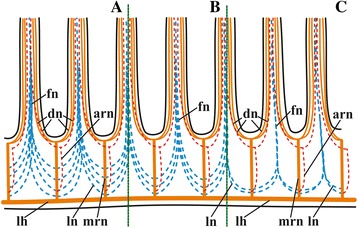
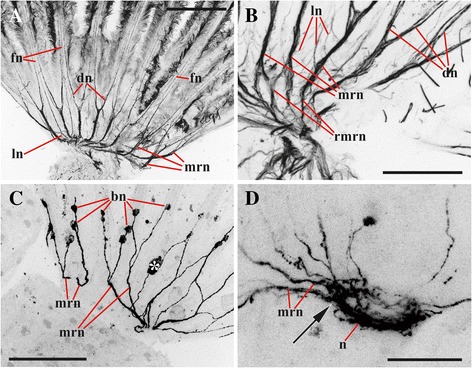
Results: The nervous system of three common European freshwater bryozoans - Cristatella mucedo, Plumatella repens (both with a horseshoe-shaped lophophore) and Fredericella sultana (with a circular lophophore) had numerous differences in the details of the structure but the general neuroarchitecture is similar. The nervous system of the zooid consists of the cerebral ganglion, a circumpharyngeal ring and lophophoral nerve tracts (horns), both sending numerous nerves to the tentacles, and the nerve plexuses of the body wall and of the gut. A number of the important details (distal branching of the additional radial nerve, pattern of distribution of nerve cells and neurites in the ganglion, etc.) were described for the first time. The number and position of the tentacle nerves in Cristatella mucedo was ascertained and suggestions about their function were made. The revealed distribution of various neuromediators in the nervous system allowed us to suggest functional affinities of some major nerves.

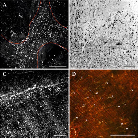
Conclusions: Despite the basic similarity, both the ganglion and the lophophore nervous system in Phylactolaemata have a more complex structure than in marine bryozoans (classes Gymnolaemata and Stenolaemata). First of all, their neuronal network has a denser and more complex branching pattern: most phylactolaemates have two large nerve tracts associated with lophophore arms, they have more nerves in the tentacles, additional and basal branches emitting from the main radial nerves, etc. This, in part, can be explained by the horseshoe shape of the lophophore and a larger size of the polypide in freshwater species. The structure of the nervous system in Fredericella sultana suggests that it underwent a secondary simplification following the reduction of the lophophore arms. Colony locomotion in Cristatella mucedo is based on co-ordinated activity of two perpendicular muscle layers of the sole and the plexus of motor neurons sandwiched between them. The trigger of this activity and the co-ordination mechanism remain enigmatic.

Keywords: Bryozoa; Immunohistochemistry; Nervous system; Neuromediators; Phylactolaemata.


Figures

Fig. 1
Fig. 1
Simplified scheme of zooidal innervation in Cristatella mucedo based on staining with antibodies against acetylated tubulin (only some tentacle bases shown, partially by dashed line). Cavity of the cerebral ganglion shown by asterisk. Abbreviations: a, anus; bwn, nerves of the body wall originating from the basal nerves of cerebral ganglion (partially shown, paired branches on the opposite side are not shown); cnr, circumpharyngeal nerve ring; dn, distal branches of main radial nerve; fn, tentacle frontal nerve; g, cerebral ganglion; lh, lophophore nerve tracts (horns); ln, lateral branches of main radial nerve; m, mouth; mrn, main radial nerve; p, pharynx; pn, nerves of pharynx (paired branch on the opposite side is not shown); r, rectum
Fig. 2
Fig. 2
Lophophore innervation in Cristatella mucedo visualized after staining with antibodies against acetylated tubulin (a, b arm view from above, double distal dichotomy of the main radial nerves not seen in this plane; c, d lateral view, double dichotomy of the main radial nerves well seen in d). a Proximal part of the arm with basal parts of the tentacles (mouth region seen on the left); b distal part of the arm; c distal part of the arm with basal parts of the tentacles (lophophore horn is behind the main radial nerves); d fronto-lateral part of the lophophore showing basal parts of tentacles. Abbreviations: arn, additional radial nerve; brn, basal radial nerve; dn, distal branches of main radial nerve; fn, tentacle frontal nerve; lh, lophophore nerve tract (horn); ln, lateral branches of main radial nerve; m, mouth; mrn, main radial nerve. Scale bars: 100 μm
Fig. 3
Fig. 3
Simplified scheme of lophophore base innervation in Cristatella mucedo (a) Plumatella repens (b) and Fredericella sultana (c) based on staining with antibodies against acetylated tubulin. Abbreviations: arn, additional radial nerve; dn, distal branches of main radial nerves; fn, frontal nerve; lh, lophophore nerve tracts (horns); ln, lateral branches of main radial nerves; mrn, main radial nerves
Fig. 4
Fig. 4
Schematic 3D-reconstruction of lophophore and tentacle innervation in Cristatella mucedo based on staining with antibodies against acetylated tubulin and TEM. Abbreviations: afn, tentacle abfrontal nerve; arn, additional radial nerve; brn, basal radial nerve; dn, distal branches of main radial nerve; fln, tentacle frontolateral nerve; fn, tentacle frontal nerve; itm, intertentacular membrane; lh, lophophore nerve tract (horn); ln, lateral branches of main radial nerves; mrn, main radial nerve (abfrontolateral nerves not shown)
Fig. 5
Fig. 5
Details of lophophore innervation and cerebral ganglion in Cristatella mucedo. a, b cerebral ganglion and lophophoral nerves (view from above, bipolar neurons situated near oral and lophophore arms tentacle bases, in some of them the main neurites merge before joining main radial nerves in a) (staining with antibodies against serotonin); c tentacle tips with sensory bipolar neurons which central neurite descends with tentacle frontal nerve (staining with antibodies against FMRFamide); d innervation of lophophore arm (view from above, bipolar neurons situated near tentacle bases, arrows point to the places where additional radial nerves join main radial nerves) (staining with antibodies against serotonin shown in green and against α-tubulin in violet). Abbreviations: arn, additional radial nerve; bn, bipolar neurons; bsn, bipolar sensory neurons situated near the tentacle tip; cnr, serotonin-like immunoreactive neurites of circumpharyngeal nerve ring; co, commissure; dn, distal branches of main radial nerve; lh, lophophore nerve tract (horn); mrn, main radial nerve; n, neuropil of cerebral ganglion. Scale bars: 100 μm
Fig. 6
Fig. 6
Details of lophophore innervation and cerebral ganglion in Cristatella mucedo. a, b cerebral ganglion (a view from above; b lateral view) (staining with antibodies against FMRFamide); c lophophore innervation (lateral view; epistome is outlined by blue colour) (catecholamine staining). Abbreviations: afn, tentacle abfrontal nerve; an, accumulations of neurons near bases of lateral tentacles; bn, bipolar neurons of tentacles; cnr, circumpharyngeal nerve ring; dn, distal branches of main radial nerve; fln, tentacle frontolateral nerve; fn, tentacle frontal nerve; g, cerebral ganglion; lh, lophophore nerve tract (horn); ln, lateral branches of main radial nerves; mrn, main radial nerve; npp, nerve plexus of pharynx; npr, nerve plexus of rectum; nr, nerve strands emitting from cerebral ganglion to rectum. Scale bars: 100 μm
Fig. 7
Fig. 7
Lophophore innervation in Plumatella repens visualized after staining with antibodies against acetylated tubulin. a, lophophore view from above with the mouth area seen below; b lateral view of arm; c lophophore view from frontal side showing ‘closure’ of circumpharyngeal nerve ring (main radial nerves are emitted from circumpharyngeal nerve ring). Abbreviations: arn, additional radial nerve; cnr, circumpharyngeal nerve ring; dn, distal branches of main radial nerve; en, nerves of epistome; fln, tentacle frontolateral nerve; lh, lophophore nerve tract (horn); ln, lateral branches of main radial nerve; m, area of mouth; mrn, main radial nerve. Scale bars: 100 μm
Fig. 8
Fig. 8
Details of lophophore innervation and cerebral ganglion in Plumatella repens and Fredericella sultana. a, b, cerebral ganglion and lophophoral nerves in P. repens (view from above) (staining with antibodies against serotonin); c, cerebral ganglion and accumulations of neurons at the tentacle bases in P. repens (lateral view) (staining with antibodies against FMRFamide); d, cerebral ganglion and accumulations of neurons at the tentacle bases in F. sultana (lateral view) (staining with antibodies against FMRFamide). Abbreviations: an, accumulations of neurons at tentacle bases; bn, bipolar neurons situated near oral and lophophore arms tentacle bases; co, commissure; g, cerebral ganglion; inp, nerve plexus of introvert; lh, lophophore nerve tract (horn); n, neuropil of cerebral ganglion. Scale bars: 100 μm
Fig. 9
Fig. 9
Details of lophophore innervation and cerebral ganglion in Fredericella sultana. a, b innervation of lophophore base and proximal parts of tentacles (lateral view) (in b roots of main radial nerves are clearly seen) (staining with antibodies against acetylated tubulin); c lophophore innervation (view from oral side showing that circumpharyngeal nerve ring is incomplete and that main neurites of the adjacent bipolar nerves merge together before joining the radial nerves) (staining with antibodies against serotonin); d cerebral ganglion (view from anal side, arrow shows the local area corresponding to lophophore horn from which main radial nerves are emitted) (staining with antibodies against serotonin). Abbreviations: bn, bipolar neurons situated inside tentacle bases; dn, distal branches of main radial nerve; fn, tentacle frontal nerve; ln, lateral branches of main radial nerve; mrn, main radial nerve; n, neuropil of cerebral ganglion; rmrn, roots of main radial nerves. *indicates a visual artifact. Scale bars: (a, c) 100 μm, (b, d) 50 μm
Fig. 10
Fig. 10
Innervation of lophophore and introvert in Cristatella mucedo (viewed from frontal side; pharynx with its ciliature outlined by dashed red line) (staining with antibodies against acetylated tubulin). Abbreviations: d, duplicature; i, introvert; inp, nerve plexus of introvert; la, lophophore arm; lh, lophophore nerve tract (horn); m, mouth; mrn, main radial nerve; zc, zooidal coelom. Scale bar: 200 μm
Fig. 11
Fig. 11
Innervation of introvert and cystid wall in Plumatella repens visualized after staining with antibodies against FMRFamide (a, b, d), acetylated tubulin (c, e) and FMRFamide and acetylated tubulin together (f). a, nerve plexus of the body wall (partially protruded introvert (above) separated from the cystid by dashed red line); b, nerve plexus of cystid wall; c, d, multipolar neurons in the introvert wall (marked by red asterisks); e, f, innervation of introvert wall (multipolar neurons shown by arrows). Note that FMRFamide elements often correspond to those revealed by tubulin staining. Scale bars: a, b, d, 100 μm, c, e, f, 50 μm
Fig. 12
Fig. 12
Innervation of introvert and lophophore in Fredericella sultana and introvert wall in Plumatella repens. a, Nervous network of introvert wall in F. sultana (lateral view) (staining with antibodies against acetylated tubulin); b, cerebral ganglion and nervous network of introvert wall in F. sultana (abfrontal view) (staining with antibodies against FMRFamide); c, innervation of introvert wall in P. repens (staining with antibodies against FMRFamide) (ring muscle layer of the body wall stained by phalloidin is seen); d, cerebral ganglion and innervation of introvert wall and lophophore in F. sultana (lateral view) (staining with antibodies against FMRFamide). Abbreviations: ag, accumulations of FMRFamide-like immunoreactive neurons in cerebral ganglion near bases of lophophore horns; an, accumulations of neurons at tentacle bases; g, cerebral ganglion; inp, nerve plexus of introvert; lm, longitudinal musculature of introvert wall; mrn, main radial nerve; rm, ring musculature of introvert wall; tn, tentacle nerve. Scale bars: 100 μm
Fig. 13
Fig. 13
Innervation of cystid wall and sole in Cristatella mucedo. a, nerve plexus of frontal cystid wall (colony view from above, duplicatures of neighbouring zooids are outlined by dashed red line) (staining with antibodies against FMRFamide); b, nerve plexus of sole (bipolar neurons are mostly seen) (staining with antibodies against FMRFamide); c, part of sole surrounded by a peripheral rim visible as a thick diagonal line (nerve plexus and two perpendicular muscle layers are clearly seen) (staining with antibodies against acetylated tubulin); d, multipolar neurons (marked by asterisks) sandwiched between two perpendicular muscular layers (some of them innervate fibers of both layers) (staining with antibodies against acetylated tubulin and phalloidin). Scale bars: 100 μm
Fig. 14
Fig. 14
Gut innervation. a, nerve plexus of pharynx in Plumatella repens (staining with antibodies against acetylated tubulin); b, nerve plexus of pharynx (top) and stomach of Cristatella mucedo (staining with antibodies against FMRFamide); c, two nerve branches emitting from cerebral ganglion and innervating rectum in C. mucedo (staining with antibodies against serotonin). Abbreviations: g, cerebral ganglion; p, pharynx; rnb, nerve branches of rectum; s, stomach. Scale bars: 100 μm
Fig. 15
Fig. 15
Simplified schemes of lophophore innervation in Fredericella sultana (a) and Cristatella-Plumatella (b) based on staining with antibodies against acetylated tubulin. Dashed line corresponds to the fronto-abfrontal (oral-anal) axis. In A only basal parts of tentacles are shown. In B only tentacle bases are shown on the internal sides of lophophore arms. Abbreviations: cnr, circumpharyngeal nerve ring; dn, distal branches of main radial nerve; g, cerebral ganglion; lh, lophophore nerve tract (horn); m, mouth; mrn, main radial nerve
Fig. 16
Fig. 16
a-f, Simplified schemes of lophophore innervation in Cristatella mucedo (a, b), Plumatella repens (c, d) and Fredericella sultana (e, f) based on staining with antibodies against serotonin (a, c, e) and FMRFamide (b, d, f) (in (ad) only tentacle bases are shown on the internal sides of lophophore arms, intertentacular membrane shown by dashed line; in (e, f), only tentacle bases are shown). Only perikaryon and central neurite of bipolar neurons at/below tentacle bases are shown. Abbreviations: bsn, bipolar sensory neurons; cnr, circumpharyngeal nerve ring; g, cerebral ganglion; lh, lophophore nerve tract (horn); m, mouth; mrn, main radial nerve
Fig. 17
Fig. 17
Transverse section of tentacle of Cristatella mucedo made across its upper half (TEM). Sections of tentacle nerves are encircled by red. Abbreviations: afln, abfrontolateral nerve; afn, abfrontal nerve; bm, basal membrane; fln, frontolateral nerve, fn, frontal nerve; tc, tentacle coelom. Scale bar: 5 μm

References

    1. Ryland JS. Bryozoa: an introductory overview. In: Wöss, E, editor. Moostiere (Bryozoa). Linz: Oberösterreichisches Landesmuseum; Denisia; 2005, NS 28, 16:9–20.
    1. Reed CG. Bryozoa. In: Giese AC, Pearse JS, Pearse VB, editors. Reproduction of Marine Invertebrates – Echinoderms and Lophophorates. Pacific Grove: The Boxwood Press; 1991. pp. 85–245.
    1. Mukai H, Terakado K, Reed CG. Bryozoa. In: Harrison FW, editor. Microscopic Anatomy of Invertebrates. New York: Wiley-Liss; 1997. pp. 45–206.
    1. Allman G. On the homology of the organs of the Tunicata and of the Polyzoa. Trans R Irish Acad. 1852;22:275–289.
    1. Hyman LH. The invertebrates: Smaller coelomate groups. New York: McGraw-Hill; 1959.

LinkOut - more resources