. 2015 Oct 13:10:29.
doi: 10.1186/s13020-015-0061-x. eCollection 2015.

Qing-dai powder promotes recovery of colitis by inhibiting inflammatory responses of colonic macrophages in dextran sulfate sodium-treated mice

Affiliations

Qing-dai powder promotes recovery of colitis by inhibiting inflammatory responses of colonic macrophages in dextran sulfate sodium-treated mice

Hai-Tao Xiao et al. Chin Med. .

Abstract

Background: Qing-dai powder (QDP), comprising Indigo naturalis (Qing-dai) and dried alum (Ku-fan), was used in Chinese medicine to treat the conditions associated with mucosal hemorrhage, such as ulcerative colitis (UC). This study aims to investigate the effects and potential mechanism of QDP on dextran sulfate sodium (DSS)-induced acute colitis in mice and to examine the regulatory effects of QDP on macrophages.

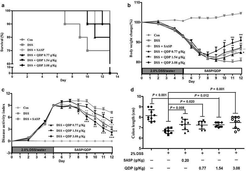
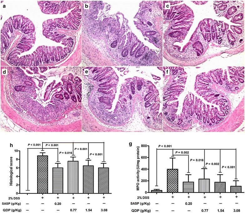
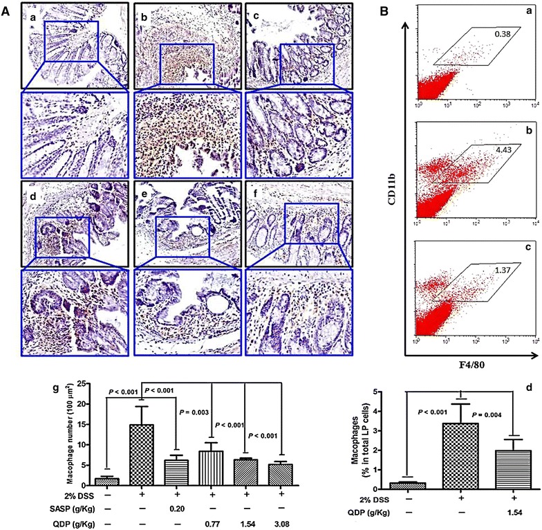
Methods: Seven- to eight-week-old male C57BL/6 mice were challenged with 2.0 % DSS in drinking water for 5 days and then the colitic mice were arbitrarily allocated into five groups (n = 10 for each group). QDP (0.77, 1.54 and 3.08 g/kg) and sulfasalazine (SASP) (0.20 g/kg) were orally administered for 7 days. The disease activity index was determined by scores of body weight loss, diarrhea and rectal bleeding; histological signs of damage was analyzed by H&E staining; myeloperoxidase activity was measured by colorimetric method, levels of proinflammatory cytokines were determined by ELISA; changes in macrophages in the colon were analyzed by immunohistochemistry (IHC) and flow cytometry. Lipopolysaccharide (LPS)-induced RAW264.7 cells were treated with or without QDP, then the production of TNF-α and IL-6 were measured by ELISA; and protein molecules such as COX-2, iNOS, IкB-α were determined by Western blot.

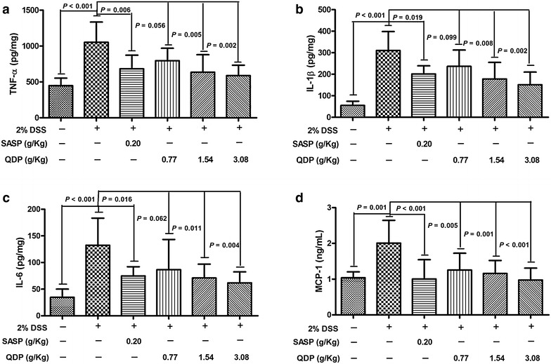
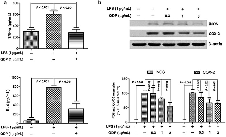
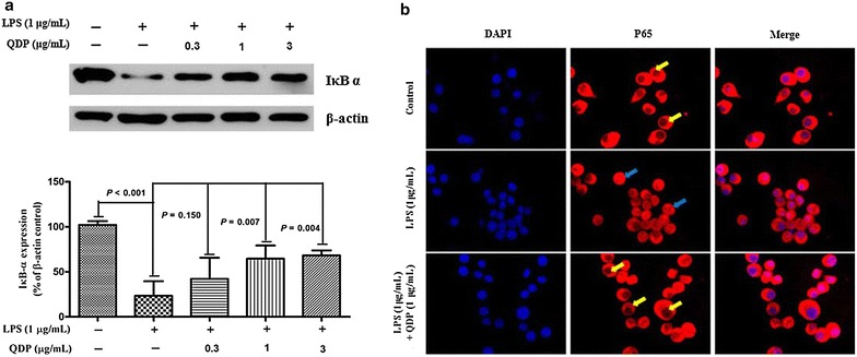
Results: Oral administration of QDP at dosages of 1.54 and 3.08 g/kg significantly reduced disease activity index on day 12 (P < 0.001 for 1.54 g/kg and P < 0.0008 for 3.08 g/kg), colon shortening (P = 0.012 for 1.54 g/kg, P = 0.001 for 3.08 g/kg), histological damage (P < 0.001 for 1.54 g/kg, P < 0.001 for 3.08 g/kg) and colonic myeloperoxidase activity (P = 0.002 for 1.54 g/kg, P < 0.001 for 3.08 g/kg) of DSS-treated mice. Moreover, QDP treatment (1.54 and 3.08 g/kg) significantly decreased DSS-induced infiltration of macrophages, and production of TNF-α (P = 0.005 for 1.54 g/kg, P = 0.002 for 3.08 g/kg), IL-1β (P = 0.008 for 1.54 g/kg, P = 0.002 for 3.08 g/kg) and IL-6 (P = 0.011 for 1.54 g/kg, P = 0.004 for 3.08 g/kg) in colonic tissues, and also reduced serum MCP-1 levels (P = 0.001 for 1.54 g/kg, P < 0.001 for 3.08 g/kg). In RAW264.7 cells, QDP significantly suppressed LPS-induced production of TNF-α and IL-6 (Both P < 0.001 for 1.0 μg/mL QDP treatment) and expression levels of COX-2 (P = 0.002 and P = 0.001 for 1 and 3 μg/mL QDP treatment, respectively) and iNOS (P < 0.001 for 3 μg/mL QDP treatment) by inhibiting IкB-α degradation (P = 0.007 and P = 0.004 for 1 and 3 μg/mL QDP treatment, respectively) and NF-кB p65 nuclear translocation.

Conclusion: QDP suppressed the inflammatory responses of colonic macrophages in DSS-induced UC in mice and LPS-induced RAW264.7 cells.


Figures

Fig. 1
Fig. 1
Elementary particle flow graph (BPI) chromatogram monitored in positive ion mode for QDP
Fig. 2
Fig. 2
Effects of QDP on mortality a body weight change; b disease activity index; c and colon length; and d of mice with DSS-induced colitis. Colitis was induced in all groups except control group. QDP and SASP were administered to mice from day 6 to 12. The change in body weight was taken as the difference between the body weight before induction of colitis and that immediately before sacrifice on day 13. Disease activity index was determined by combining scores of (1) body weight loss; (2) stool consistency; and (3) stool blood. On day 13, mice were sacrificed, and colon length was measured. Survival data of challenged mice were collected from 9 to 10 mice respectively, and other data were expressed as mean ± SD (n = 7–9). ### P < 0.001, compared with control group; *P < 0.05, **P < 0.01 and ***P < 0.001, compared with DSS model group
Fig. 3
Fig. 3
Effects of QDP on histopathological changes and MPO activity in colon of mice with DSS-induced colitis. a control; b DSS model; c SASP; d 0.77 g/kg QDP; e 1.54 g/kg QDP; f 3.08 g/kg QDP (magnification, ×100); g histological score; and h MPO activity. Colitis was induced in all groups except control group. QDP and SASP were administered to mice from day 6 to 12. On day 13, mice were sacrificed, and colonic tissue damage was evaluated by histopathological analysis (H&E staining). MPO activity was determined in colonic homogenates. Data were expressed as mean ± SD (n = 7–9)
Fig. 4
Fig. 4
Effects of QDP on macrophage infiltration in colons of DSS-treated mice. A Immunohistochemical (IHC) analysis of colonic macrophages by F4/80 marker [af representative images (a control; b DSS model; c SASP; d 0.77 g/kg QDP; e 1.54 g/kg QDP; and f 3.08 g/kg QDP), g number of macrophages]; B flow cytometric analysis of macrophages in the colon lamina propria (LP) (a control; b DSS model; c 1.54 g/kg QDP; and d percentage of macrophages in total LP cells). Colitis was induced in all groups except control group. QDP and SASP were administered to mice from day 6 to 12. On day 13, mice were sacrificed. The colon section was evaluated by IHC analysis with F4/80 marker (n = 7–9). For flow cytometric analysis, the population of macrophage in lamina propria mononuclear cells from whole colonic tissue was determined by CD11b and F4/80 markers (n = 5 for normal control group, and n = 6 for DSS-treated groups). Data were expressed as mean ± SD
Fig. 5
Fig. 5
Effects of QDP on colonic production of pro-inflammatory cytokines and serum MCP-1 level in DSS-treated mice (a colonic TNF-α; b colonic IL-6; c colonic IL-1β; and d serum MCP-1). QDP and SASP were administered to mice from day 6 to 12. On day 13, mice were sacrificed, amounts of various cytokines in colonic homogenates and MCP-1 levels in serum were determined by ELISA. Data were expressed as mean ± SD (n = 7–9)
Fig. 6
Fig. 6
Effects of QDP on LPS-induced production of TNF-α and IL-6 and expression of iNOS and COX-2 in RAW264.7 cells. Cells were treated with QDP for 1 h, followed by continuous incubation with LPS (1 μg/mL) for 24 h. a Concentrations of TNF-α and IL-6 in culture medium were monitored by ELISA. b Cells were lysed and expression of iNOS and COX-2 were determined by Western blotting. Data represent the mean ± SD (n = 3). The images shown are representatives of three independent experiments
Fig. 7
Fig. 7
Effects of QDP on LPS-induced IκB α degradation and p65 nuclear translocation. a Cells were pretreated with the indicated concentrations of QDP for 1 h and incubated with LPS (1 μg/mL) for another 30 min. Cells were lysed and IκB α expression was determined by Western blot. b Cells were pretreated with 1 μg/mL QDP for 1 h prior to stimulation with LPS (1 μg/mL) for 1 h. The nuclear localization of p65 was determined using fluorescence microscopy after staining with DAPI, anti-p65, and FITC-labeled anti-rabbit IgG antibody (p65 sequestered in the cytoplasm were indicated by yellow arrows and p65 translocated into nuclear were indicated by blue arrows). Data represent the mean ± SD (n = 3). The images shown are the representatives of three independent experiments

Similar articles

Cited by

References

    1. Kaser A, Zeissig S, Blumberg RS. Inflammatory bowel disease. Annu Rev Immunol. 2010;28:573–621. doi: 10.1146/annurev-immunol-030409-101225. - DOI - PMC - PubMed
    1. Chow DK, Leong RW, Tsoi KK, Ng SS, Leung WK, Wu JC, Wong VW, Chan FK, Sung JJ. Long-term follow-up of ulcerative colitis in the Chinese population. Am J Gastroenterol. 2009;104:647–654. doi: 10.1038/ajg.2008.74. - DOI - PubMed
    1. Wang YF, Ouyang Q, Hu RW. Progression of inflammatory bowel disease in China. J Dig Dise. 2010;11:76–82. doi: 10.1111/j.1751-2980.2010.00421.x. - DOI - PubMed
    1. Macdermott RP, Green JA. Refractory ulcerative colitis treatment. Gastroenterol Hepatol (N Y) 2007;3:64–69. - PMC - PubMed
    1. Nanda K, Moss AC. Update on the management of ulcerative colitis: treatment and maintenance approaches focused on MMX((R)) mesalamine. Clin Pharmacol. 2012;4:41–50. - PMC - PubMed