

Correlation Between Clinical Findings of Temporomandibular Disorders and MRI Characteristics of Disc Displacement
- PMID: 26464595
- PMCID: PMC4598384
- DOI: 10.2174/1874210601509010273
Correlation Between Clinical Findings of Temporomandibular Disorders and MRI Characteristics of Disc Displacement
Abstract
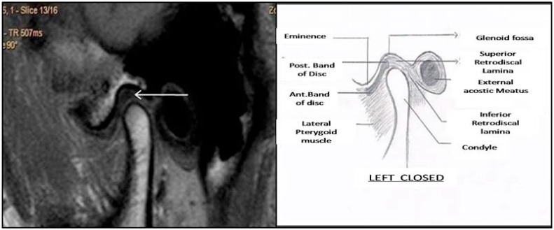
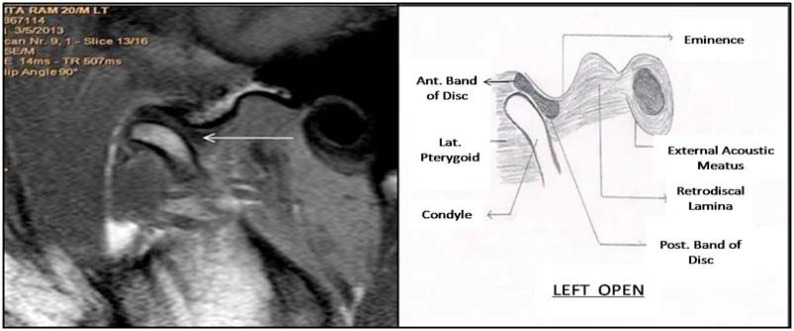
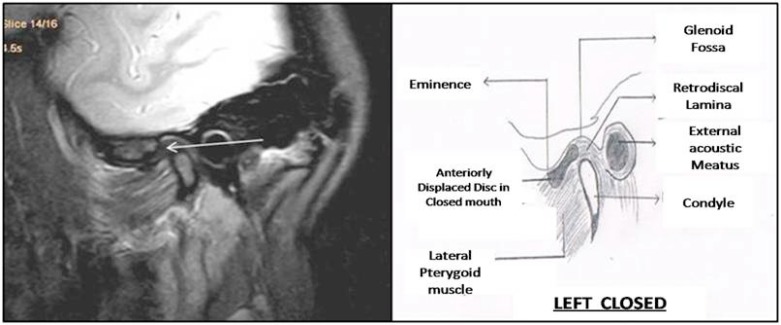
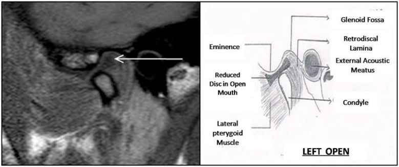
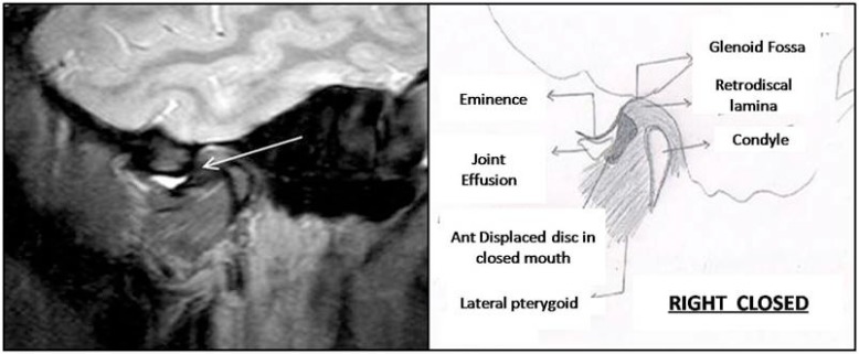
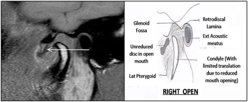
Temporomandibular joint (TMJ) dysfunction is a common condition that is best evaluated with magnetic resonance (MR) imaging. The first step in MR imaging of the TMJ is to evaluate the articular disk, or meniscus, in terms of its morphologic features and its location relative to the condyle in both closed- and open-mouth positions. Disk location is of prime importance because the presence of a displaced disk is a critical sign of TMJ dysfunction. However, disk displacement is also frequently seen in asymptomatic volunteers. It is important for the maxillofacial radiologist to detect early MR imaging signs of dysfunction, thereby avoiding the evolution of this condition to its advanced and irreversible phase which is characterized by osteoarthritic changes such as condylar flattening or osteophytes. Further the MR imaging techniques will allow a better understanding of the sources of TMJ pain and of any discrepancy between imaging findings and patient symptoms. Henceforth, the aim of the study was to evaluate whether MRI findings of various degrees of disk displacement could be correlated with the presence or absence of clinical signs and symptoms of temporomandibular disorders in symptomatic and asymptomatic subjects. Materials and Methods : In this clinical study, 44 patients (88 TMJs) were examined clinically and divided into two groups. Group 1 consisted of 22 patients with clinical signs and symptoms of TMDs either unilaterally or bilaterally and considered as study group. Group 2 consisted of 22 patients with no signs and symptoms of TMDs and considered as control group. MRI was done for both the TMJs of each patient. Displacement of the posterior band of articular disc in relation to the condyle was quantified as anterior disc displacement with reduction (ADDR), anterior disc displacement without reduction (ADDWR), posterior disc displacement (PDD). Results : Disk displacement was found in 18 (81.8%) patients of 22 symptomatic subjects in Group 1 on MRI and 4 (18.1%) were diagnosed normal with no disc displacement. In Group 2, 2 (9.1%) of 22 asymptomatic patients were diagnosed with disc displacement while 20 (90.1%) were normal. Sensitivity and Specificity tests were applied in both the groups to correlate clinical findings of TMD and MRI characterstics of disc displacement and results showed Sensitivity of 90% and Specificity of 83.3%. Conclusion : Disk displacement on MRI correlated well with presence or absence of clinical signs and symptoms of temporomandibular disorders with high Sensitivity and Specificity of 90% and 83.3% respectively.
Keywords: Disc displacement; magnetic resonance imaging; temporomandibular joint disorders.
Figures
Similar articles
-
MR imaging of temporomandibular joint dysfunction: a pictorial review.Radiographics. 2006 May-Jun;26(3):765-81. doi: 10.1148/rg.263055091. Radiographics. 2006. PMID: 16702453 Review.
-
Displacement of the temporomandibular joint disk: correlation between clinical findings and MRI characteristics.J Can Dent Assoc. 2010;76:a3. J Can Dent Assoc. 2010. PMID: 20633336
-
Dynamic MR imaging of temporomandibular joint: an initial assessment with fast imaging employing steady-state acquisition sequence.Magn Reson Imaging. 2015 Apr;33(3):270-5. doi: 10.1016/j.mri.2014.10.013. Epub 2014 Nov 24. Magn Reson Imaging. 2015. PMID: 25461305
-
Role of Dynamic 3 Tesla MRI in the Evaluation of Temporomandibular Joint Dysfunction.Cureus. 2023 Mar 25;15(3):e36681. doi: 10.7759/cureus.36681. eCollection 2023 Mar. Cureus. 2023. PMID: 37113366 Free PMC article.
-
Role of magnetic resonance imaging in the clinical diagnosis of the temporomandibular joint.Cells Tissues Organs. 2005;180(1):6-21. doi: 10.1159/000086194. Cells Tissues Organs. 2005. PMID: 16088129 Review.
Cited by
-
Nonlinear Relationship between Temporomandibular Joint Disc Displacement Distance and Disc Length: A Magnetic Resonance Imaging Analysis.J Clin Med. 2022 Dec 1;11(23):7160. doi: 10.3390/jcm11237160. J Clin Med. 2022. PMID: 36498733 Free PMC article.
-
Counselling treatment versus counselling associated with jaw exercises in patients with disc displacement with reduction-a single-blinded, randomized, controlled clinical trial.BMC Oral Health. 2023 Jun 14;23(1):389. doi: 10.1186/s12903-023-03096-7. BMC Oral Health. 2023. PMID: 37316791 Free PMC article. Clinical Trial.
-
Temporomandibular joint pain and associated magnetic resonance findings: a retrospective study with a control group.Acta Radiol Open. 2020 Sep 30;9(9):2058460120938738. doi: 10.1177/2058460120938738. eCollection 2020 Sep. Acta Radiol Open. 2020. PMID: 33088591 Free PMC article.
-
Correlation of Condylar Translation During Maximal Mouth Opening with Presence of Signs of Temporomandibular Joint Disorders in an Asymptomatic Population of 18-25 Years Age Group of Northern India.Open Dent J. 2018 Sep 28;12:770-781. doi: 10.2174/1745017901814010770. eCollection 2018. Open Dent J. 2018. PMID: 30369987 Free PMC article.
-
Internal derangement as a predictor of provoked pain on mouth opening: A magnetic resonance imaging study.Imaging Sci Dent. 2017 Dec;47(4):219-226. doi: 10.5624/isd.2017.47.4.219. Epub 2017 Dec 12. Imaging Sci Dent. 2017. PMID: 29279820 Free PMC article.
References
-
- Okeson J.P. Functional anatomy and biomechanics of masticatory system. 2013.
-
- Kannan A., Sathasivasubramanian S. Comparative study of clinical and Magnetic resonance imaging diagnosis in patients with internal derangement of temporomandibular joint. J Indian Acad Oral Med Radiol. 2011;23:569–575. doi: 10.5005/jp-journals-10011-1224. - DOI
-
- Maizlin Z.V., Nutiu N., Dent P.B., Vos P.M., Fenton D.M., Kirby J.M., Vora P., Gillies J.H., Clement J.J. Displacement of the temporomandibular joint disk: correlation between clinical findings and MRI characteristics. J. Can. Dent. Assoc. 2010;76:a3. - PubMed
LinkOut - more resources
Full Text Sources
Other Literature Sources
Medical