

Squamosamide derivative FLZ inhibits TNF-α-induced ICAM-1 expression via down-regulation of the NF-κB signaling pathway in ARPE-19 cells
- PMID: 26464656
- PMCID: PMC4583888
Squamosamide derivative FLZ inhibits TNF-α-induced ICAM-1 expression via down-regulation of the NF-κB signaling pathway in ARPE-19 cells
Abstract
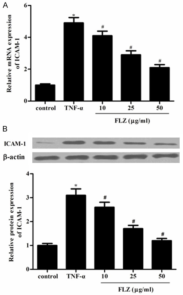
Dysfunction of the retinal pigment epithelium (RPE) resulting from chronic inflammation is implicated in the pathogenesis of age-related macular degeneration (AMD). It has been reported that tumor necrosis factor-α (TNF-α) could induce intercellular adhesion molecule-1 (ICAM-1) expression in RPE cells. FLZ, a novel synthetic squamosamide derivative from a Chinese herb, Annona glabra, has displayed significant anti-inflammatory activity. However, the effects of FLZ on TNF-α-induced ICAM-1 expression in RPE cells remain unknown. Therefore, in the present study, we evaluated the effects of FLZ on TNF-α-induced ICAM-1 expression in RPE cells. We found that FLZ prevented TNF-α-induced ICAM-1 expression and the ability of monocytes to adhere to ARPE-19 cells induced by TNF-α. Furthermore, FLZ inhibited TNF-α-induced NF-κB p65 expression, as well as phosphorylation of IκBα in ARPE-19 cells. Taken together, these results suggest that FLZ inhibited TNF-α-induced ICAM-1 expression through blocking NF-κB signaling pathway in ARPE-19 cells. Thus, FLZ could be used for designing novel therapeutic agents against AMD.
Keywords: Squamosamide derivative FLZ; age-related macular degeneration (AMD); intercellular adhesion molecule-1 (ICAM-1); retinal pigment epithelium (RPE); tumor necrosis factor-α (TNF-α).
Figures


References
-
- Congdon N, O’Colmain B, Klaver CC, Klein R, Muñoz B, Friedman DS, Kempen J, Taylor HR, Mitchell P Eye Diseases Prevalence Research Group. Causes and prevalence of visual impairment among adults in the United States. Arch Ophthalmol. 2004;122:477–485. - PubMed
-
- van Lookeren Campagne M, LeCouter J, Yaspan BL, Ye W. Mechanisms of age-related macular degeneration and therapeutic opportunities. J Pathol. 2014;232:151–164. - PubMed
-
- Zech JC, Pouvreau I, Cotinet A, Goureau O, Le Varlet B, De Kozak Y. Effect of cytokines and nitric oxide on tight junctions in cultured rat retinal pigment epithelium. Invest Ophthalmol Vis Sci. 1998;39:1600–1608. - PubMed
-
- Abe T, Sugano E, Saigo Y, Tamai M. Interleukin-1β and barrier function of retinal pigment epithelial cells (ARPE-19): aberrant expression of junctional complex molecules. Invest Ophthalmol Vis Sci. 2003;44:4097–4104. - PubMed
Publication types
MeSH terms
Substances
LinkOut - more resources
Full Text Sources
Miscellaneous