. 2015 Oct 14;10(10):e0140055.
doi: 10.1371/journal.pone.0140055. eCollection 2015.

Growth Hormone Ameliorates the Radiotherapy-Induced Ovarian Follicular Loss in Rats: Impact on Oxidative Stress, Apoptosis and IGF-1/IGF-1R Axis

Affiliations

Growth Hormone Ameliorates the Radiotherapy-Induced Ovarian Follicular Loss in Rats: Impact on Oxidative Stress, Apoptosis and IGF-1/IGF-1R Axis

Yasmen F Mahran et al. PLoS One. .

Abstract

Radiotherapy is one of the standard cytotoxic therapies for cancer. However, it has a profound impact on ovarian function leading to premature ovarian failure and infertility. Since none of the currently available methods for fertility preservation guarantees future fertility, the need for an effective radioprotective agent is highly intensified. The present study investigated the mechanisms of the potential radioprotective effect of growth hormone (GH) on γ irradiation-induced ovarian failure and the impact of the insulin like growth factor 1 (IGF-1) in the underlying protection. Immature female Sprague-Dawley rats were either exposed to single whole body irradiation (3.2 Gy) and/or treated with GH (1 mg/kg s.c). Experimental γ-irradiation produced an array of ovarian dysfunction that was evident by assessment of hormonal changes, follicular development, proliferation marker (PCNA), oxidative stress as well as apoptotic markers. In addition, IGF-1/IGF-1R axis expression was assessed using real-time PCR and immunolocalization techniques. Furthermore, after full maturity, fertility assessment was performed. GH significantly enhanced follicular development and restored anti-Mullerian hormone serum level as compared with the irradiated group. In addition, GH significantly ameliorated the deleterious effects of irradiation on oxidative status, PCNA and apoptosis. Interestingly, GH was shown to enhance the ovarian IGF-1 at transcription and translation levels, a property that contributes significantly to its radioprotective effect. Finally, GH regained the fertility that was lost following irradiation. In conclusion, GH showed a radioprotective effect and rescued the ovarian reserve through increasing local IGF-1 level and counteracting the oxidative stress-mediated apoptosis.


Conflict of interest statement

Competing Interests: The authors have declared that no competing interests exist.

Figures

Fig 1
Fig 1. Schematic diagram of the experimental design.
Fig 2
Fig 2. Follicular proliferation.
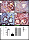
Immunohistochemical localization of PCNA in ovarian follicles was studied 4 days after irradiation. (A): Expression of PCNA in the control ovaries shows a large antral follicle (AnF) with positively stained (dark brown) granulosa cells (GC) and oocyte (O) and a cluster of primordial follicles with positively stained oocytes and stain-free pregranulosa cells around (oval). (B): Expression of PCNA in ovaries of rats subjected to γ-irradiation (3.2 Gy) shows minimal expression. (C) & (D): Expression of PCNA in ovaries of rats treated with GH 1 mg/kg for one week [either exposed to γ-radiation (C) or not (D)] shows a high PCNA expression of oocyte (O) and Granulosa cells (GC) (brown color). Scale bar, 20 μm. (E): Quantitative Immunohistochemical staining of PCNA expressed as a percentage of immunopositive cells against the total number of granulosa cells across six high power fields (40×) from each rat section. Bars represent the Mean ± SEM of at least three independent experiments. a or b: Statistically significant from control or radiation group, respectively at P<0.05 using one-way ANOVA followed by Tukey–Kramer as a post-hoc test.
Fig 3
Fig 3. Photomicrographs of ovarian sections stained by hematoxylin and eosin.
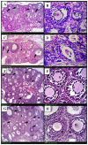
(A)& (B): histological sections from control ovaries shows normal histopathological structure with multiple follicles of different stages (black arrows), intact oocytes (O) and granulosa cells (g). (C)& (D): γ- irradiated ovarian sections shows few, if any, healthy follicles (arrows) with hemorrhage in the cortex (circle). Many small primary follicles are atretic (a) with degenerating oocytes and granulosa cells in irradiated ovaries. (E)& (F): GH-irradiated ovarian sections shows similar organization to the control group. (G) & (H): ovaries from animals treated with GH alone shows multiple growing follicles (arrows) with intact oocytes (O) and granulosa cells (g). Scale bar, 20 μm. gf: Graffian follicle, S: Stroma.
Fig 4
Fig 4. Photomicrographs of uterine sections stained by hematoxylin and eosin.
(A): uterus from control rats shows normal histopathological structure of the mucosal lining epithelium (m) and the underlying lamina propria (p) with normal glandular structure (G). (B): uterine sections from rats subjected to γ- irradiation shows marked degeneration and stratification of the mucosal lining epithelium with multiple vacuoles (v) and thickening in the lamina propria (C) & (D): uterine sections taken from rats following GH treatment [either γ- irradiated (C) or not (D)] shows normal histological structure. Scale bar, 20 μm.
Fig 5
Fig 5. Immunohistochemical localization of cytochrome c.
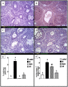
(A): section of ovary obtained from the control rats shows minimal degree of Cyto c expression (brown color). (B): section of ovary obtained from rats exposed to γ- irradiation (3.2 Gy) shows extensive Cyto c expression (brown color) in granulosa cells of all follicles. (C): section of ovary obtained from rats treated with GH (1mg/kg) and exposed to γ- irradiation (3.2 Gy) shows limited Cyto c expression (brow color). (D): section of ovary obtained from rats treated with GH (1mg/kg) alone for one week shows minimal Cyto c expression of granulosa cells (brown color). Scale bar, 20 μm. (E): Quantification of ovarian Cyto c staining represents the percentage of immunopositive cells to the total area of the microscopic high power field (20×); was averaged across 6 fields for each rat section of at least three independent experiments. (F): ovarian caspase-3 activity expressed as nmol/ mg protein. Each bar represents the Mean ± SEM for a group of 6 rats. a or b: Statistically significant from control or radiation group, respectively at P<0.05 using one way ANOVA followed by Tukey–Kramer as a post-hoc test.
Fig 6
Fig 6. Immunohistochemical localization and Real-time quantitative Rt-PCR of insulin like growth factor-1 receptor (IGF-1R).
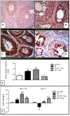
(A): section of ovary obtained from the control rats shows modest degree of IGF-1R expression (brown color). (B): section of ovary obtained from rats subjected to γ- irradiation (3.2 Gy) shows extensive IGF-1R expression (brown color) in granulosa cells of almost all follicles. (C): section of an ovary obtained from rats treated with GH and exposed to γ –irradiation (3.2 Gy) shows high IGF-1R expression (brown color). (D): section of ovary obtained from rats treated with GH alone (1mg/kg; once daily for one week) shows modest IGF-1R expression similar to control ovaries (brown color).Scale bar, 20 μm. (E): Semi-quantitative expression of ovarian IGF-1R staining represents the mean optical density of immunopositive cells of 6 high power fields (20×) of at least three independent experiments. (F): Real-time quantitative RT-PCR of ovarian IGF-1 and IGF-1R genes expressed as fold changes relative to control group. Each bar represents the Mean ± SEM for a group of 3 rats. a or b: Statistically significant from control or radiation group, respectively at P<0.05 using one-way ANOVA followed by Tukey–Kramer as a post-hoc test.

Similar articles

Cited by

References

    1. Steel GG. Basic clinical radiobiology (3rdedn.), Edward Arnold Publishers; London; 2002.
    1. Nair CK, Parida DK, Nomura T. Radioprotectors in radiotherapy. J Radiat Res. 2001; 42(1): 21–37. - PubMed
    1. Meirow D. Reproduction post-chemotherapy in young cancer patients. Mol Cell Endocrinol. 2000; 169, 123–131. - PubMed
    1. Pacey AA. Fertility issues in survivors from adolescent cancers. Cancer Treat Rev. 2007; 33:646–655. - PubMed
    1. Stroud JS, Mutch D, Rader J, Powell M, Thaker PH, Grigsby PW. Effects of cancer treatment on ovarian function. Fertil Steril. 2009; 92:417–427. 10.1016/j.fertnstert.2008.07.1714 - DOI - PubMed

MeSH terms