Review
. 2015 Nov;16(11):651-64.
doi: 10.1038/nrm4069. Epub 2015 Oct 14.

Ribosome profiling reveals the what, when, where and how of protein synthesis

Affiliations
Review

Ribosome profiling reveals the what, when, where and how of protein synthesis

Gloria A Brar et al. Nat Rev Mol Cell Biol. 2015 Nov.

Abstract

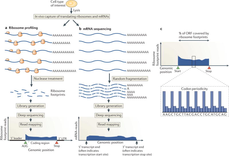
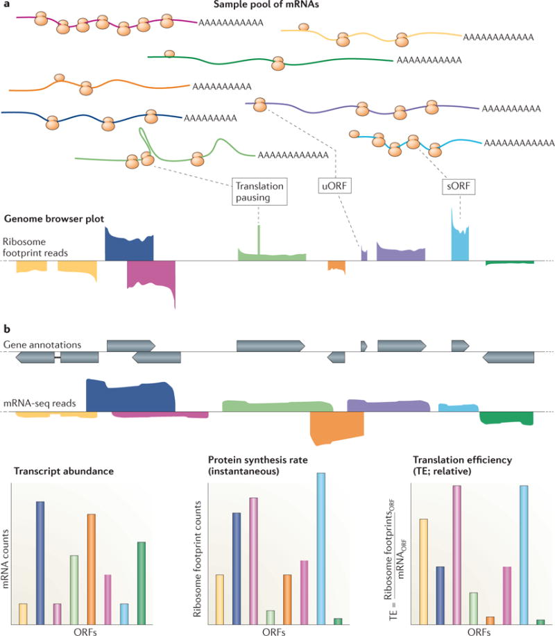
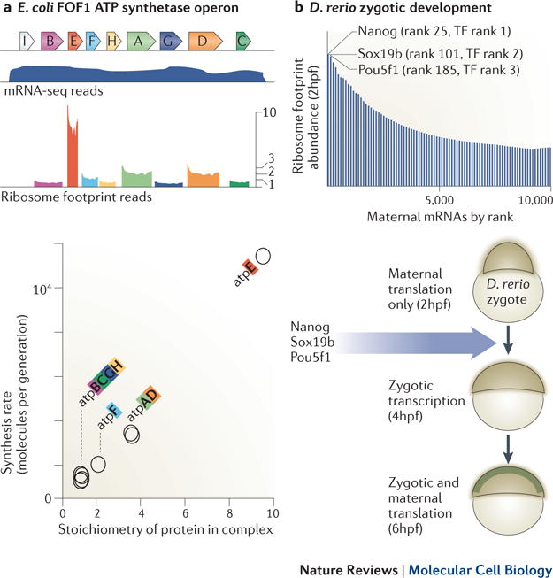
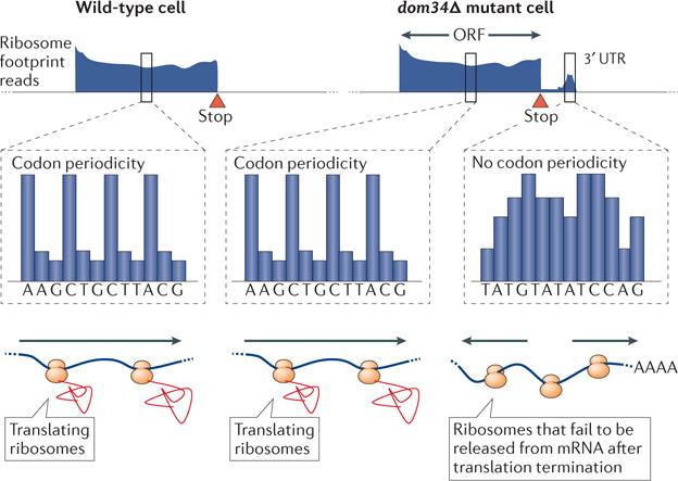
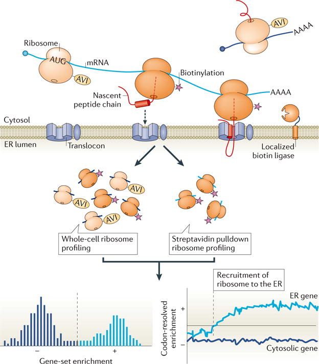
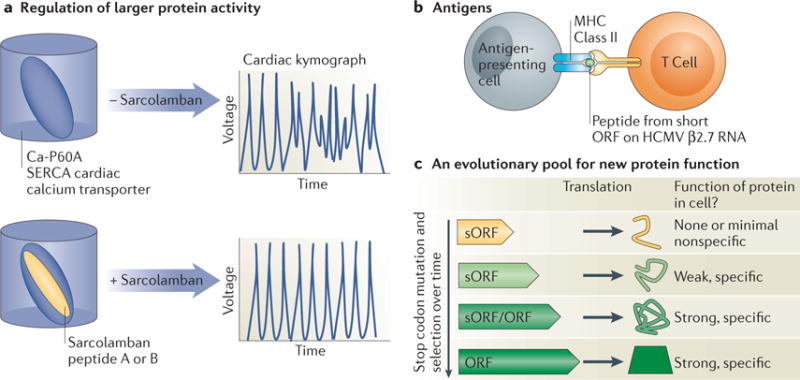
Ribosome profiling, which involves the deep sequencing of ribosome-protected mRNA fragments, is a powerful tool for globally monitoring translation in vivo. The method has facilitated discovery of the regulation of gene expression underlying diverse and complex biological processes, of important aspects of the mechanism of protein synthesis, and even of new proteins, by providing a systematic approach for experimental annotation of coding regions. Here, we introduce the methodology of ribosome profiling and discuss examples in which this approach has been a key factor in guiding biological discovery, including its prominent role in identifying thousands of novel translated short open reading frames and alternative translation products.


Conflict of interest statement

Competing Interests Statement: JSW is an inventor on patent application for ribosome profiling.

Figures

Figure 1
Figure 1
An overview of ribosome profiling. a) Ribosome-bound mRNAs are isolated by size and treated with nonspecific nuclease (typically RNAse I or micrococcal nuclease), resulting in protected mRNA fragments or ‘footprints’. These ribosome footprints are isolated and converted to a library for deep sequencing. The ribosome footprints typically show precise positioning between the start and stop codon of a gene, which enables global and experimental genomic coding region identification. b) By comparison, mRNA sequencing captures random fragments covering the entire mRNA transcript. The positional information determined by standard mRNA sequencing enables approximate determination of transcript boundaries, but is less precise than ribosome profiling due to the loss of 5′ and 3′ ends during the fragment generation method typically used. c) Translated open reading frames (ORFs) house a stereotyped organization of ribosome footprints. Ribosome density over ORFs begins sharply at the start codon, ends sharply at the stop codon, and shows evidence of codon periodicity. True translated regions tend to show ribosome footprint coverage over the majority of the ORF and not typically in the regions before the putative start codon and after the putative stop codon.
Figure 2
Figure 2
Qualitative and quantitative data provided by ribosome profiling. a) A diverse sample pool of mRNAs, distinguished by color, are shown together with a corresponding representative genome browser plot of ribosome profiling data derived from this pool. Note that ribosome profiling enables experimental determination of translated regions, including short Open Reading Frames (sORFs), which may be an important new source of cellular peptides, and upstream ORFs (uORFs), which are thought to be largely regulatory. Pausing during translation elongation may result in peaks in ribosome footprints within ORFs. b) Overlaid gene annotations and mRNA-seq data for the examples shown in (a). c) Examples of quantitative data derived from b). Note that transcript abundances may not correlate closely with the instantaneous protein synthesis rates. The collection of quantitative data for both transcript abundances and their protein synthesis rates enables inference of the relative translation efficiencies. These can vary over several orders of magnitude within a given organism in a given state. The translation efficiency can also change over time for a given mRNA, reflecting dynamic regulation at the level of translation.
Figure 3
Figure 3
Ribosome profiling enables quantitative proteomic discovery in diverse systems. a) Bacterial cells translate components of multi-member protein complexes in ratios that are proportional to their stoichiometry in these complexes. A notable example is the FoF1 ATP synthetase, which is composed of 8 different proteins (A to H) translated from a single operon. mRNA abundance for each gene is thus similar, but ribosome profiling reveals intricate translational control. Modified with permission from refb) Zebrafish zygotic development requires the initiation of zygotic transcription 2 hours post fertilization (hpf), although the specific transcription factors responsible for this transcription have been unclear. Ribosome profiling of embryos at 2 hpf showed the three most highly translated transcription factors (TFs) from maternal messages were Nanog, Sox19b, and Pou5f1 and subsequent experiments confirmed that these three proteins drive zygotic activation. Modified with permission from ref
Figure 4
Figure 4
Dom34 facilitates the release of 80S ribosomes from a subset of 3′UTRs. Ribosome footprints indicative of assembled 80S ribosomes are seen in a subset of 3′UTR regions in dom34Δ mutant cells. Unlike 80S ribosome footprints from ORFs, however, these do not show periodicity and represent ribosomes that have failed to properly release following translation termination.
Figure 5
Figure 5
Proximity specific ribosome profiling at the ER. A ribosome subunit is fused to a biotin-acceptor (AVI) tag and BirA biotin ligase is fused to a localization element that spatially restricts its activity, for example, to the ER. Only ribosomes that orient AVI towards the ER surface, as seen during their close association with the ER membrane during translocation, are biotinylated when a controlled pulse of biotin is applied to cells. Cells are then frozen and ribosomes are collected. Ribosome profiling is carried out on all ribosomes and also only on ribosomes pulled down with streptavidin. The pulldown-enriched message population (light blue) represents genes that are greatly enriched for translation at the ER. The positional data from these analyses also reveals the point in the message when a translating ribosome is recruited to the ER. Modified with permission from ref.
Figure 6
Figure 6
Proposed cellular roles for the peptide products of translated short ORFs identified by ribosome profiling. a) The two Sarcolamban peptides are 28 and 29 amino acids in length, conserved from fruit flies to human, and regulate normal heart function in flies through direct binding to the Ca-P60A SERCA Calcium transporter in cardiac tissue. Modified with permission from ref. b) Sera from HCMV positive blood donors identified a specific and robust antigenic response against multiple short peptides translated from the β 2.7 RNA, previously thought to act as a noncoding RNA. c) Spurious translation of short regions may produce a pool of peptides with weak or no cellular function. New protein domains may evolve through selection for maintenance of peptides with weak cellular function, followed by stop codon mutation and further selection for increasingly specific and important cellular function over time.

References

    1. McCann KL, Baserga SJ. Genetics. Mysterious ribosomopathies. Science. 2013;341:849–850. - PMC - PubMed
    1. Cleary JD, Ranum LPW. Repeat-associated non-ATG (RAN) translation in neurological disease. Hum Mol Genet. 2013;22:R45–51. - PMC - PubMed
    1. Ellis SR. Nucleolar stress in Diamond Blackfan anemia pathophysiology. Biochim Biophys Acta. 2014;1842:765–768. - PubMed
    1. Trainor PA, Merrill AE. Ribosome biogenesis in skeletal development and the pathogenesis of skeletal disorders. Biochim Biophys Acta. 2014;1842:769–778. - PMC - PubMed
    1. Bolze A, et al. Ribosomal protein SA haploinsufficiency in humans with isolated congenital asplenia. Science. 2013;340:976–978. - PMC - PubMed

Publication types