. 2015 Oct 14;10(10):e0139652.
doi: 10.1371/journal.pone.0139652. eCollection 2015.

Cell-Penetrating Peptides Selectively Cross the Blood-Brain Barrier In Vivo

Affiliations

Cell-Penetrating Peptides Selectively Cross the Blood-Brain Barrier In Vivo

Sofie Stalmans et al. PLoS One. .

Abstract

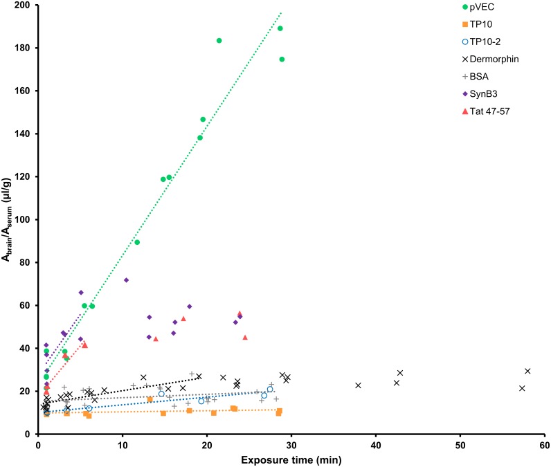
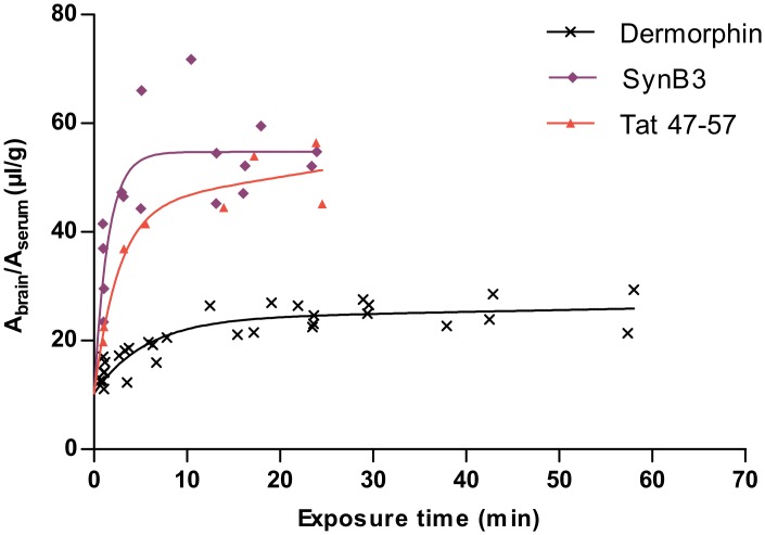
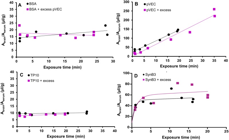
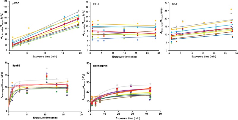
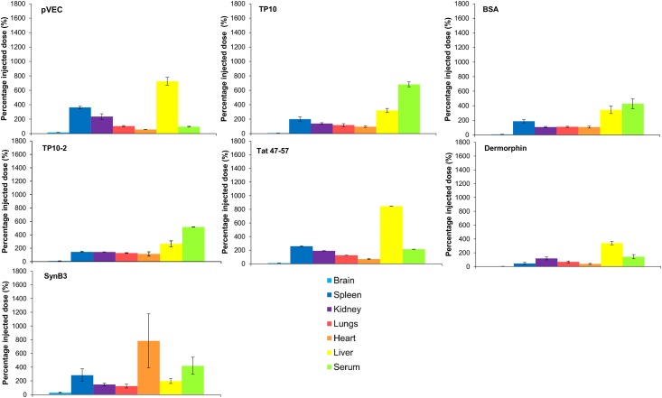
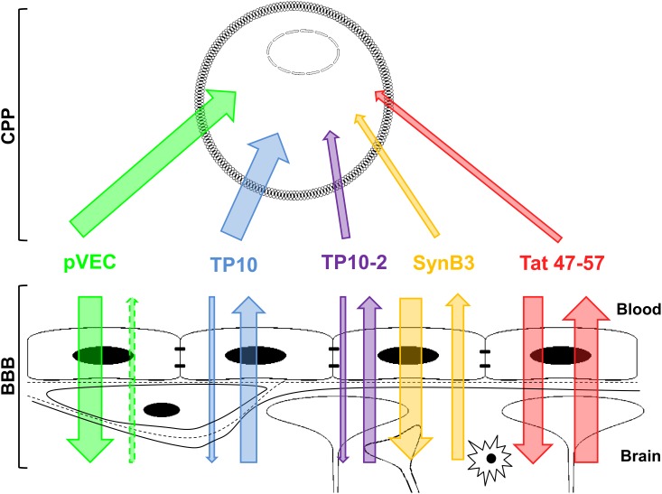
Cell-penetrating peptides (CPPs) are a group of peptides, which have the ability to cross cell membrane bilayers. CPPs themselves can exert biological activity and can be formed endogenously. Fragmentary studies demonstrate their ability to enhance transport of different cargoes across the blood-brain barrier (BBB). However, comparative, quantitative data on the BBB permeability of different CPPs are currently lacking. Therefore, the in vivo BBB transport characteristics of five chemically diverse CPPs, i.e. pVEC, SynB3, Tat 47-57, transportan 10 (TP10) and TP10-2, were determined. The results of the multiple time regression (MTR) analysis revealed that CPPs show divergent BBB influx properties: Tat 47-57, SynB3, and especially pVEC showed very high unidirectional influx rates of 4.73 μl/(g × min), 5.63 μl/(g × min) and 6.02 μl/(g × min), respectively, while the transportan analogs showed a negligible to low brain influx. Using capillary depletion, it was found that 80% of the influxed peptides effectively reached the brain parenchyma. Except for pVEC, all peptides showed a significant efflux out of the brain. Co-injection of pVEC with radioiodinated bovine serum albumin (BSA) did not enhance the brain influx of radiodionated BSA, indicating that pVEC does not itself significantly alter the BBB properties. A saturable mechanism could not be demonstrated by co-injecting an excess dose of non-radiolabeled CPP. No significant regional differences in brain influx were observed, with the exception for pVEC, for which the regional variations were only marginal. The observed BBB influx transport properties cannot be correlated with their cell-penetrating ability, and therefore, good CPP properties do not imply efficient brain influx.


Conflict of interest statement

Competing Interests: The authors have declared that no competing interests exist.

Figures

Fig 1
Fig 1. Results of the multiple time regression analysis experiment of the five CPP using the linear model.
Fig 2
Fig 2. Result of the multiple time regression analysis experiment of SynB3, Tat 47–57 and dermorphin using the biphasic model.
The ratio of the brain-to-serum activity is plotted versus the exposure time and fitted using the biphasic model.
Fig 3
Fig 3. Evaluation of the BBB permeability after injection of pVEC and used brain influx mechanism of pVEC, TP10 and SynB3.
(A) Evaluation of the BBB permeability after IV injection of pVEC: ratio of brain-to-serum radioactivity versus exposure time of radioiodinated BSA with (purple squares) and without (black dots) an excess dose of pVEC (20 μg). (B-D) Evaluation of the saturability of the BBB influx mechanism of pVEC, TP10 and SynB3, respectively: ratio of brain-to-serum radioactivity versus exposure time with (purple squares) and without (black dots) an excess dose of the CPP (10 μg). Data are fitted using the linear Gjedde-Patlak model, except for the data of SynB3, which are fitted using the biphasic model.
Fig 4
Fig 4. Regional variations in brain influx of pVEC, TP10, SynB3, dermorphin and radioiodinated BSA.
The data of pVEC, TP10 and radioiodinated BSA are fitted using the linear Gjedde-Patlak model; the data of SynB3 and dermorphin are fitted using the biphasic model. Grey = whole brain, yellow = frontal cortex, purple = occipital + parietal cortex, light blue = cerebellum, dark blue = striatum, brown = thalamus + hypothalamus, orange = pons medulla, green = hippocampus and red = midbrain.
Fig 5
Fig 5. Relative tissue distribution of the radiolabeled CPPs and the controls dermorphin and radioiodinated BSA 15 min post IV injection expressed as the percentage of the injected dose (± SEM, n = 2).
From the left to the right: brain (light blue), spleen (dark blue), kidneys (purple), lungs (red), heart (orange), liver (yellow) and serum (light green).
Fig 6
Fig 6. Schematic overview of the relationship between cell-penetrating and BBB-penetrating properties of the five investigated CPPs.
The thickness of the arrows indicates the extent of influx and/or efflux.

References

    1. Stalmans S, Wynendaele E, Bracke N, Gevaert B, D’Hondt M, Peremans K, et al. Chemical-Functional Diversity in Cell-Penetrating Peptides. PLOS One 2013;8: e71752 10.1371/journal.pone.0071752 - DOI - PMC - PubMed
    1. Madani F, Lindberg S, Langel Ü, Futaki S, Gräslund A. Mechanisms of Cellular Uptake of Cell-Penetrating Peptides. J. Biophys. 2011;2011: 414729 10.1155/2011/414729 - DOI - PMC - PubMed
    1. Copolovici DM, Langel K, Eriste E, Langel Ü. Cell-Penetrating Peptides: Design, Synthesis, and Applications. ACS Nano 2014;8: 1972–1994. 10.1021/nn4057269 - DOI - PubMed
    1. Bechara C, Sagan S. Cell-penetrating peptides: 20 years later, where do we stand? FEBS Lett. 2013;587: 1693–1702. 10.1016/j.febslet.2013.04.031 - DOI - PubMed
    1. Milletti F. Cell-penetrating peptides: classes, origin, and current landscape. Drug Discov. Today 2012;17: 850–860. 10.1016/j.drudis.2012.03.002 - DOI - PubMed

Publication types