. 2015 Oct 14;10(10):e0140363.
doi: 10.1371/journal.pone.0140363. eCollection 2015.

Absence of the Yeast Hsp31 Chaperones of the DJ-1 Superfamily Perturbs Cytoplasmic Protein Quality Control in Late Growth Phase

Affiliations

Absence of the Yeast Hsp31 Chaperones of the DJ-1 Superfamily Perturbs Cytoplasmic Protein Quality Control in Late Growth Phase

Ingo Amm et al. PLoS One. .

Abstract

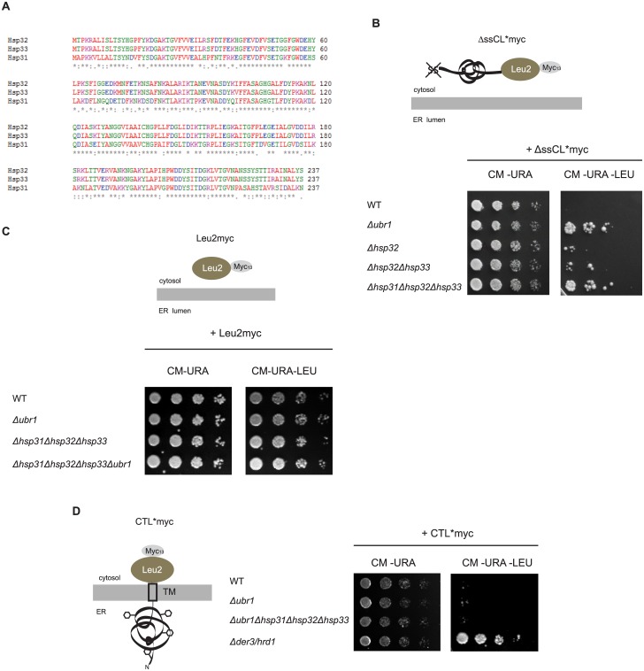
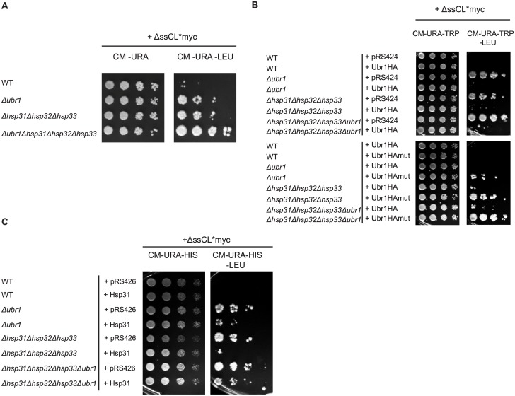
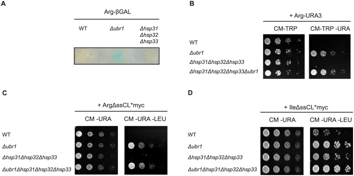
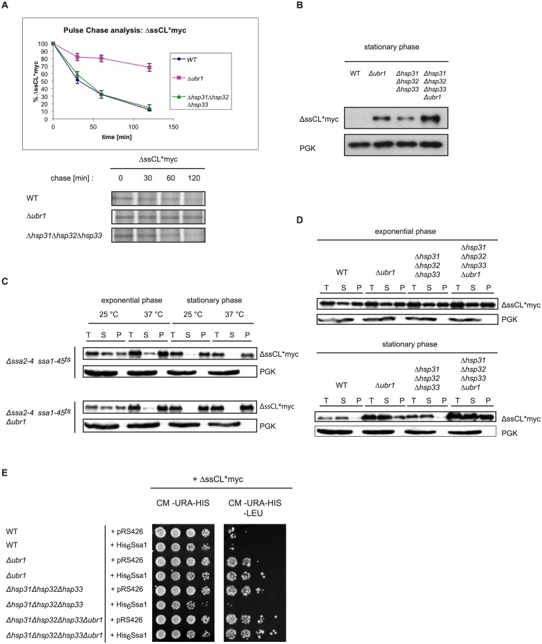
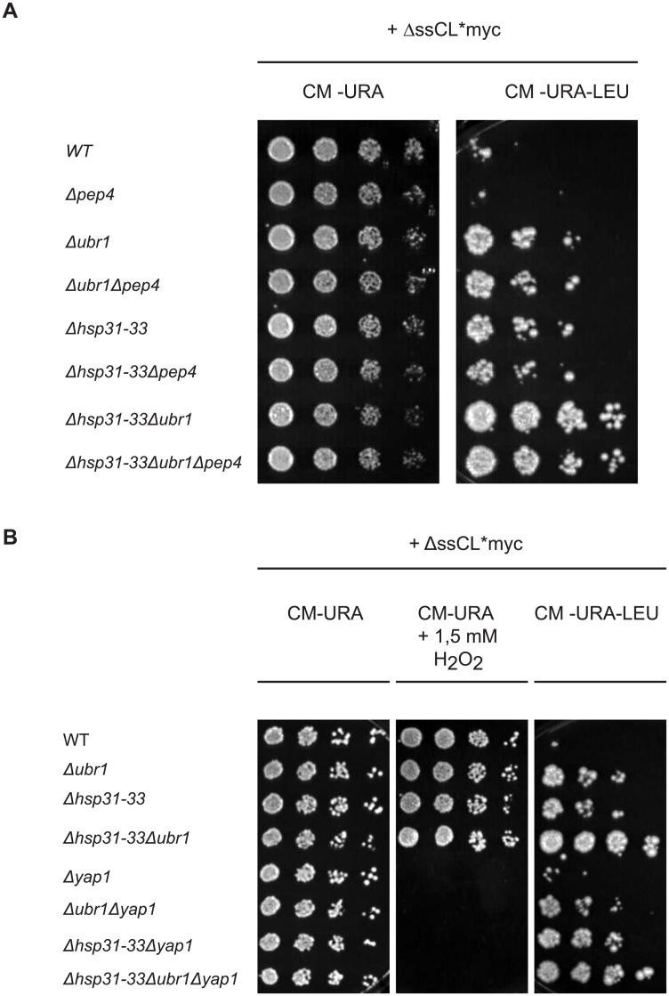
The Saccharomyces cerevisiae heat shock proteins Hsp31, Hsp32, Hsp33 and Hsp34 belong to the DJ-1/ThiJ/PfpI superfamily which includes the human protein DJ-1 (PARK7) as the most prominent member. Mutations in the DJ-1 gene are directly linked to autosomal recessive, early-onset Parkinson's disease. DJ-1 acts as an oxidative stress-induced chaperone preventing aggregation and fibrillation of α-synuclein, a critical factor in the development of the disease. In vivo assays in Saccharomyces cerevisiae using the model substrate ΔssCPY*Leu2myc (ΔssCL*myc) as an aggregation-prone misfolded cytoplasmic protein revealed an influence of the Hsp31 chaperone family on the steady state level of this substrate. In contrast to the ubiquitin ligase of the N-end rule pathway Ubr1, which is known to be prominently involved in the degradation process of misfolded cytoplasmic proteins, the absence of the Hsp31 chaperone family does not impair the degradation of newly synthesized misfolded substrate. Also degradation of substrates with strong affinity to Ubr1 like those containing the type 1 N-degron arginine is not affected by the absence of the Hsp31 chaperone family. Epistasis analysis indicates that one function of the Hsp31 chaperone family resides in a pathway overlapping with the Ubr1-dependent degradation of misfolded cytoplasmic proteins. This pathway gains relevance in late growth phase under conditions of nutrient limitation. Additionally, the Hsp31 chaperones seem to be important for maintaining the cellular Ssa Hsp70 activity which is important for Ubr1-dependent degradation.


Conflict of interest statement

Competing Interests: The authors have declared that no competing interests exist.

Figures

Fig 1
Fig 1. The Hsp31 chaperone family is involved in quality control of misfolded cytoplasmic ΔssCL*myc.
(A) Sequence alignment of the three members of the Hsp31 chaperone family in the yeast W303 strain shows an extraordinary sequence homology of 99% between Hsp32 and Hsp33. When compared with Hsp31 they show a sequence homology of about 70%. (B) Growth tests of deletion strains of the E3-Ligase Ubr1, different chaperones of the Hsp31 family and the corresponding wild type (WT) strain, respectively. All strains defective in the URA3 and LEU2 genes, harbour a centromeric (CEN) plasmid with the URA3 marker and expressing ΔssCL*myc under the control of the PRC1 (CPY) promoter. ΔssCL*myc is composed of the vacuolar protein carboxypeptidase yscY (CPY) harbouring the point mutation G255R [54]. The protein is additionally deleted in the signal sequence required for transport into the endoplasmic reticulum [6]. In order to perform growth tests it is fused to the enzyme β-isopropylmalate dehydrogenase (Leu2) necessary for leucine biosynthesis. For immunodetection it is C-terminally fused to a c-Myc tag. Cells were spotted in a five fold dilution series on solid selection medium lacking uracil and leucine and, as a control, solely uracil, respectively. (C) Growth tests of cells performed as described in the legend to Fig 1B, but expressing Leu2myc (β-isopropylmalate dehydrogenase C-terminally fused to a c-Myc tag) instead of the terminally misfolded substrate ΔssCL*myc. As ΔssCL*myc, the Leu2myc protein is also expressed under control of the PRC1 promoter. (D) Growth tests of cells as described in the legend to Fig 1B, but using yeast strains transformed with a URA3 marker-containing plasmid expressing the ERAD-L model substrate CTL*myc under control of the GAL4 promoter. CTL*myc consists of the misfolded ER-lumenal carboxypeptidase yscY (CPY*) moiety, the last transmembrane domain of Pdr5, and the cytoplasmic β-isopropylmalate dehydrogenase C-terminally fused to a c-Myc tag (Leu2myc). In contrast to ΔssCL*myc, the CPY* moiety is localized in the ER lumen and therefore glycosylated (black hexagons). The Δder3/hrd1 strain defective in ubiquitination of ERAD-L substrates served as control.
Fig 2
Fig 2. The Hsp31 chaperones function in a pathway overlapping with Ubr1-dependent degradation.
(A) Growth tests were performed as described in the legend to Fig 1B using ΔssCL*myc encoded by the plasmid pFE15 as model substrate. Growth of the substrate-expressing Δubr1 and Δhsp31-33 strains on medium lacking leucine was compared with growth of the quadruple deletion strain lacking HSP31, HSP32, HSP33 and UBR1 on the same medium. (B) Growth tests were performed using the substrate ΔssCL*myc as described before. The used yeast strains are additionally transformed either with a high copy plasmid encoding C-terminally HA-tagged Ubr1 (Ubr1HA) or the RING mutant Ubr1HA C1220S (Ubr1HAmut). The corresponding empty plasmid pRS424 served as control. All the three plasmids contain a TRP1 marker for plasmid selection. (C) Growth tests were performed with yeast strains expressing the substrate ΔssCL*myc from a HIS3 marker-containing plasmid (pIA1). In addition, the strains were either transformed with a high-copy plasmid expressing Hsp31 under control of its own promoter (pIA30) or the corresponding URA3 marker-containing empty plasmid pRS426.
Fig 3
Fig 3. N-degrons alter the influence of the Hsp31 family members on the steady state level of cytoplasmic model substrates in cells.
(A) Test of the Arg-βGal levels in cells using a β-galactosidase (βGal) activity assay. The substrate is expressed as a ubiquitin-Arg-βGal fusion protein which is cotranslationally deubiquitinated creating the N-end rule substrate Arg-βGal. βGal activity was measured by incubation of permeabilized cells attached on a filter with X-Gal which is converted into a blue dye by β-galactosidase. (B) Growth tests were performed with strains expressing the type 1 N-end rule substrate Arg-Ura3. Growth of cells on a CM-TRP plate served as control monitoring the presence of the plasmid. (C) Growth tests of cells were performed with strains expressing the misfolded model substrate ΔssCL*myc as in Fig 1B but containing the type 1 N-degron arginine. Initially, the fusion protein ubiquitin-ArgΔssCL*myc is expressed of which ubiquitin is cleaved off. (D) Similar growth tests as shown in Fig 3C were performed with yeast strains expressing ΔssCL*myc containing the type 2 N-degron isoleucine (IleΔssCL*myc), also initially expressed as ubiquitin-IleΔssCL*myc protein.
Fig 4
Fig 4. Function of the Hsp31 family in stationary growth phase of cells.
(A) Pulse-chase analysis was done with exponentially-growing yeast cells expressing ΔssCL*myc. Cells were lysed at the indicated time points. Proteins were immunoprecipitated, separated by SDS-PAGE and analysed using a Phosphorimager (Storm 860; Molecular Dynamics) and the ImageQuant software (Amersham Biosciences). Plotted data represent the mean values of three independent experiments. Error bars represent the standard error of the mean. (B) Steady state analysis of the amount of the ΔssCL*myc substrate in stationary growth phase. Equal amounts of cells were harvested and cell lysates were subjected to SDS-PAGE followed by immunodetection using c-Myc antibody. As reference protein, 3-phosphoglycerate kinase (PGK) was used. (C) Solubility assays of the misfolded protein ΔssCL*myc expressed in the temperature-sensitive Hsp70 (Ssa) mutant strains Δssa2Δssa3Δssa4ssa1-45 ts and Δssa2Δssa3Δssa4ssa1-45 ts Δubr1. Cells were grown at 25°C before splitting into two halves. One half of the yeast culture was shifted to 37°C for 1 h prior to harvesting, lysis and fractionation into supernatant (S) and pellet (P) fractions. The total (T) fraction represents the precleared cell lysate. Exponentially growing cells were harvested at an OD600 value of 1.0 whereas stationary cells were grown 3 days prior to temperature shift, cell lysis and fractionation. The different fractions were subjected to TCA precipitation prior to SDS-PAGE and immunoblotting using c-Myc antibody for substrate detection. PGK served as loading control and reference for a soluble protein. (D) Solubility assays were performed as described for Fig 4C. Strains defective in either Ubr1 or/and the Hsp31 chaperones were used in this assay. The cells were grown at 30°C and harvested either in exponential phase or stationary phase (72h growth), lysed and subjected to fractionation into supernatant (S) and pellet (P) fractions. The samples were subjected to TCA precipitation, SDS-PAGE and immunoblotting using c-Myc antibody. PGK served as loading control and reference for a soluble protein. (E) Growth tests were performed as described earlier. The used strains express the substrate ΔssCL*myc from a HIS3-marker-containing plasmid (pIA1). In addition, the strains were transformed either with the empty plasmid pRS426 containing a URA3 marker or a pRS426-based plasmid expressing functional histidine-tagged Ssa1 under control of the GPD promoter (pAM25).
Fig 5
Fig 5. Diminishing vacuolar function or the oxidative stress response does not alter the dependency of the steady state level of ΔssCL*myc on the Hsp31 family.
(A) Growth tests were performed as described above using yeast strains carrying a PEP4 deletion. All strains were transformed with the plasmid pFE15 encoding the model substrate ΔssCL*myc. Medium lacking uracil served as control selecting only for the presence of the plasmid. (B) Yeast strains transformed with the plasmid pFE15 encoding ΔssCL*myc were used for the growth tests performed as described above. Yeast strains possessing the YAP1 gene were compared with YAP1 deletion strains. Defective growth of cells on plates containing 1.5 mM hydrogen peroxide served as verification for the absence of Yap1. Medium lacking uracil served as control for selection of cells carrying the plasmid pFE15.

References

    1. Parsell DA, Kowal AS, Singer MA, Lindquist S. Protein disaggregation mediated by heat-shock protein Hsp104. Nature. 1994;372(6505):475–8. . - PubMed
    1. Hartl FU, Hayer-Hartl M. Molecular chaperones in the cytosol: from nascent chain to folded protein. Science. 2002;295(5561):1852–8. Epub 2002/03/09. 10.1126/science.1068408 295/5561/1852 [pii]. . - DOI - PubMed
    1. Hartl FU. Molecular chaperones in cellular protein folding. Nature. 1996;381(6583):571–9. Epub 1996/06/13. 10.1038/381571a0 . - DOI - PubMed
    1. Frydman J. Folding of newly translated proteins in vivo: the role of molecular chaperones. Annu Rev Biochem. 2001;70:603–47. Epub 2001/06/08. 70/1/603 [pii] 10.1146/annurev.biochem.70.1.603 . - DOI - PubMed
    1. Mayer MP, Bukau B. Hsp70 chaperones: cellular functions and molecular mechanism. Cell Mol Life Sci. 2005;62(6):670–84. Epub 2005/03/17. 10.1007/s00018-004-4464-6 - DOI - PMC - PubMed

Publication types

MeSH terms

Substances