. 2015 Nov 17;132(20):1898-908.
doi: 10.1161/CIRCULATIONAHA.115.017313. Epub 2015 Oct 14.

Red Blood Cell Dysfunction Induced by High-Fat Diet: Potential Implications for Obesity-Related Atherosclerosis

Affiliations

Red Blood Cell Dysfunction Induced by High-Fat Diet: Potential Implications for Obesity-Related Atherosclerosis

Dusten Unruh et al. Circulation. .

Abstract

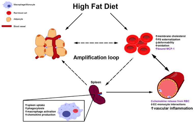
Background: High-fat diet (HFD) promotes endothelial dysfunction and proinflammatory monocyte activation, which contribute to atherosclerosis in obesity. We investigated whether HFD also induces the dysfunction of red blood cells (RBCs), which serve as a reservoir for chemokines via binding to Duffy antigen receptor for chemokines (DARC).

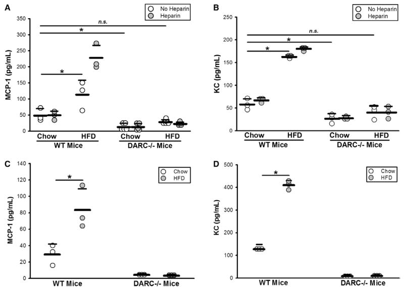
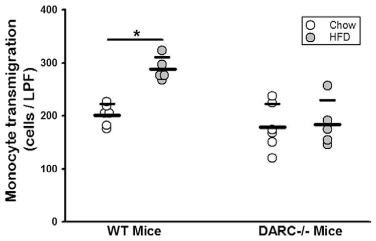
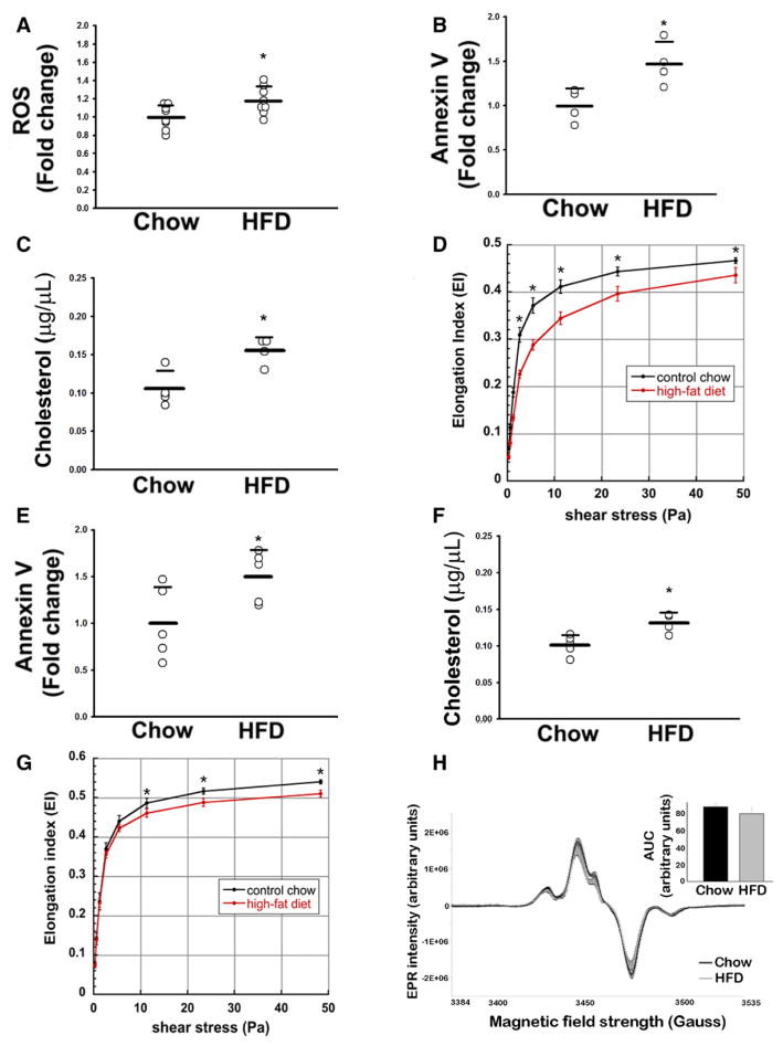
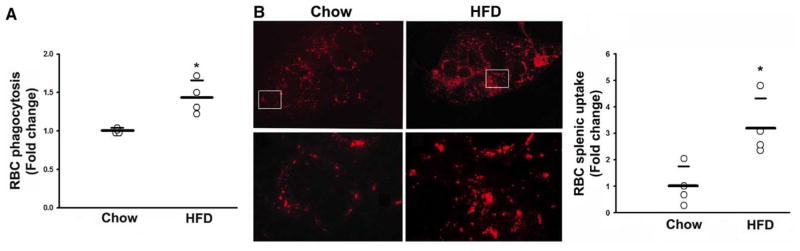
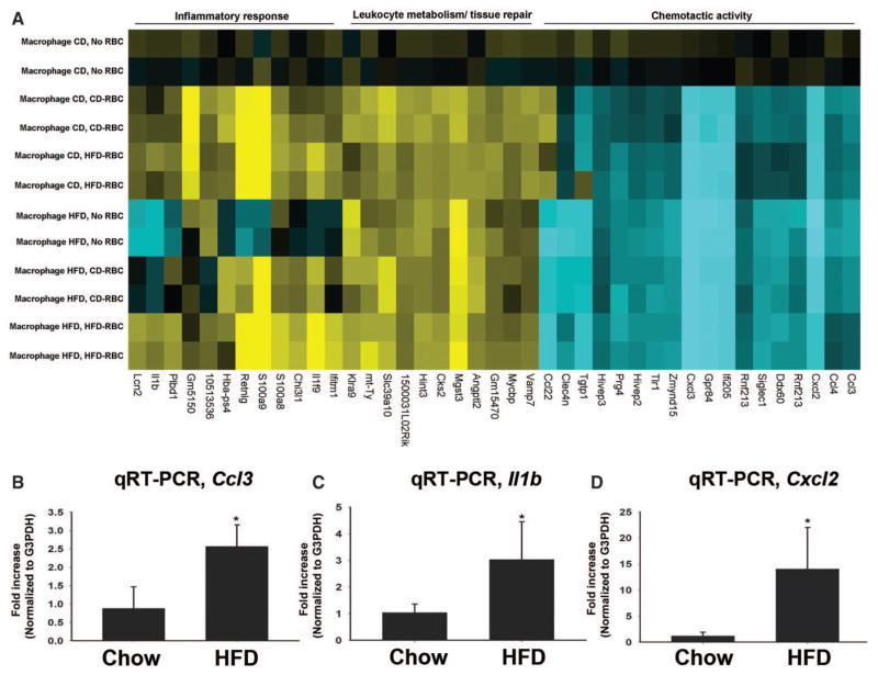
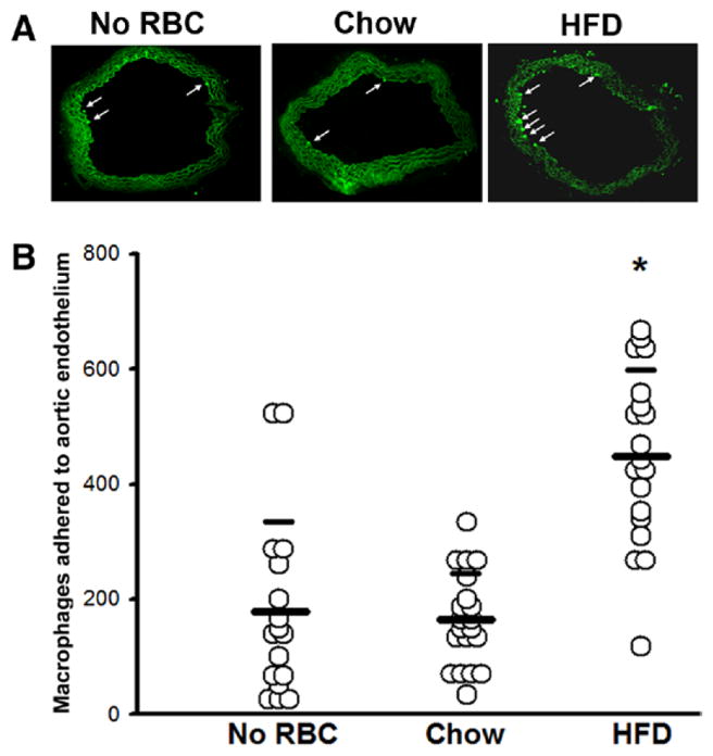
Methods and results: A 60% HFD for 12 weeks, which produced only minor changes in lipid profile in C57/BL6 mice, markedly augmented the levels of monocyte chemoattractant protein-1 bound to RBCs, which in turn stimulated macrophage migration through an endothelial monolayer. Levels of RBC-bound KC were also increased by HFD. These effects of HFD were abolished in DARC(-/-) mice. In RBCs from HFD-fed wild-type and DARC(-/-) mice, levels of membrane cholesterol and phosphatidylserine externalization were increased, fostering RBC-macrophage inflammatory interactions and promoting macrophage phagocytosis in vitro. When labeled ex vivo and injected into wild-type mice, RBCs from HFD-fed mice exhibited ≈3-fold increase in splenic uptake. Finally, RBCs from HFD-fed mice induced increased macrophage adhesion to the endothelium when they were incubated with isolated aortic segments, indicating endothelial activation.

Conclusions: RBC dysfunction, analogous to endothelial dysfunction, occurs early during diet-induced obesity and may serve as a mediator of atherosclerosis. These findings may have implications for the pathogenesis of atherosclerosis in obesity, a worldwide epidemic.

Keywords: atherosclerosis; erythrocytes; leukocytes; obesity.


Figures

Figure 1
Figure 1
HFD-RBCs promote vascular inflammation: increased MCP-1 and KC levels on the surface of the HFD-RBCs in wild-type (WT) mice, but not Duffy antigen receptor for chemokine-deficient (DARC−/−) mice. A and B, ELISA was performed on platelet-poor plasma prepared from whole blood of WT and DARC−/− mice fed CD vs HFD for 12 weeks, with or without heparin treatment. C and D, ELISA was performed on RBC membrane preparations. n=3 to 5 (1 dot = 1 mouse); *P<0.05. CD indicates chow diet; ELISA, enzyme-linked immunosorbent assay; HFD, high-fat diet; MCP-1, monocyte chemoattractant protein-1; n.s., not significant; and RBC, red blood cell.
Figure 2
Figure 2
HFD increases DARC-dependent, RBC-induced monocyte transmigration. Monocyte transmigration assay, with packed RBCs from WT and DARC−/− mice fed CD vs HFD for 12 weeks as the chemoattractant source. n=5 (1 dot = 1 mouse); *P<0.05. CD indicates chow diet; DARC, Duffy antigen receptor for chemokines; HFD, high-fat diet; LPF, low-power field; RBC, red blood cell; and WT, wild type.
Figure 3
Figure 3
HFD alters the biochemical properties of RBCs. Mice were maintained on CD or HFD for ≥12 weeks. A, Intracellular ROS levels (DCFH fluorescence). B, PS externalization as measured by Annexin V staining. C, Cholesterol content of RBC membranes. D, RBC deformability index as a function of various shear rates (black line, CD RBCs; red line, HFD RBCs), WT mice. E through G, Annexin V staining, cholesterol content, and elongation index, respectively, in DARC−/− mice fed CD vs HFD. A through C, E, F, n=3 to 8 (1 dot = 1 mouse); *P<0.05. H, RBC nitrosohemoglobin levels (EPR assay), n=6; *P<0.05. AUC indicates area under the curve; CD, chow diet; DARC−/−, Duffy antigen receptor for chemokine-deficient; DCFH, 2′,7′-dichlorodihydrofluorescein; EPR, electron paramagnetic resonance; HFD, high-fat diet; PS, phosphatidylserine; RBC, red blood cell; ROS, reactive oxygen species; and WT, wild type.
Figure 4
Figure 4
Increased phagocytosis and splenic uptake of HFD-RBCs. A, RBC phagocytosis by mouse macrophages. B, Representative images of splenic sections showing uptake of labeled RBCs from CD- or HFD-fed mice in vivo: Top, low-power images of the spleen sections; Bottom, high-power images of the boxed areas. The graph is a quantification of RBC splenic uptake (see text for details). n=3 to 4 (1 dot = 1 mouse); *P<0.05. CD indicates chow diet; HFD, high-fat diet; and RBC, red blood cell.
Figure 5
Figure 5
HFD induces a proinflammatory phenotype in RBC-exposed macrophages. A, Heat map, changes in expression of 41 genes in macrophages elicited from chow vs HFD mice and incubated under control conditions (no RBC) or with RBC derived from mice maintained for ≥12 weeks on either CD or HFD; see Methods for details. B through D, qRT-PCR validation of the increased expression of Ccl3, Il1b, and Cxcl2, respectively, in macrophages derived from mice maintained on HFD and exposed to HFD-RBCs vs CD-RBCs. n=3; *P<0.05. CD, chow diet; HFD, high-fat diet; qRT-PCR, quantitative real-time polymerase chain reaction; and RBC, red blood cell.
Figure 6
Figure 6
HFD-RBCs increase adhesion of macrophages to the aortic endothelium. Aortic segments were exposed to RBCs from either CD- or HFD-fed mice, washed, and incubated with labeled macrophages; a control group (no RBCs) was also included. The number of macrophages adhered to luminal endothelium was counted in serial sections. A, Representative images (arrows point to macrophages). B, Cumulative data compiled from individual cryosections. *P<0.05. CD indicates chow diet; HFD, high-fat diet; and RBC, red blood cell
Figure 7
Figure 7
Schematic representation of RBC-mediated processes fueling chronic inflammation in the setting of HFD. RBC dysfunction is promoted by several concomitantly acting mechanisms, both dependent on and independent of DARC on RBC surface (text in purple and black, respectively), leading to an increase in the levels of chemokines in the vasculature, enhanced EC-monocyte interactions, and heightened vascular inflammation. Dashed arrows comprising a possible amplification loop are hypothetical. DARC indicates Duffy antigen receptor for chemokines; EC, endothelial cell; HFD, high-fat diet; PS, phosphatidylserine; and RBC, red blood cell.

Comment in

  • Atherothrombosis: Seeing Red?
    Simon DI, Silverstein RL. Simon DI, et al. Circulation. 2015 Nov 17;132(20):1860-2. doi: 10.1161/CIRCULATIONAHA.115.019259. Epub 2015 Oct 14. Circulation. 2015. PMID: 26467255 Free PMC article. No abstract available.

References

    1. Bornfeldt KE, Tabas I. Insulin resistance, hyperglycemia, and atherosclerosis. Cell Metab. 2011;14:575–585. doi: 10.1016/j.cmet.2011.07.015. - DOI - PMC - PubMed
    1. Osborn O, Olefsky JM. The cellular and signaling networks linking the immune system and metabolism in disease. Nat Med. 2012;18:363–374. doi: 10.1038/nm.2627. - DOI - PubMed
    1. Gower RM, Wu H, Foster GA, Devaraj S, Jialal I, Ballantyne CM, Knowlton AA, Simon SI. CD11c/CD18 expression is upregulated on blood monocytes during hypertriglyceridemia and enhances adhesion to vascular cell adhesion molecule-1. Arterioscler Thromb Vasc Biol. 2011;31:160–166. doi: 10.1161/ATVBAHA.110.215434. - DOI - PMC - PubMed
    1. Swirski FK, Libby P, Aikawa E, Alcaide P, Luscinskas FW, Weissleder R, Pittet MJ. Ly-6Chi monocytes dominate hypercholesterolemia-associated monocytosis and give rise to macrophages in atheromata. J Clin Invest. 2007;117:195–205. doi: 10.1172/JCI29950. - DOI - PMC - PubMed
    1. Rothe G, Herr AS, Stöhr J, Abletshauser C, Weidinger G, Schmitz G. A more mature phenotype of blood mononuclear phagocytes is induced by fluvastatin treatment in hypercholesterolemic patients with coronary heart disease. Atherosclerosis. 1999;144:251–261. - PubMed