

Review
doi: 10.14348/molcells.2015.0263.
Epub 2015 Oct 15.
Structural Studies of G Protein-Coupled Receptors
Affiliations
- PMID: 26467290
- PMCID: PMC4625064
- DOI: 10.14348/molcells.2015.0263
Item in Clipboard
Review
Structural Studies of G Protein-Coupled Receptors
Mol Cells.
2015 Oct.
Abstract
G protein-coupled receptors (GPCRs) constitute the largest and the most physiologically important membrane protein family that recognizes a variety of environmental stimuli, and are drug targets in the treatment of numerous diseases. Recent progress on GPCR structural studies shed light on molecular mechanisms of GPCR ligand recognition, activation and allosteric modulation, as well as structural basis of GPCR dimerization. In this review, we will discuss the structural features of GPCRs and structural insights of different aspects of GPCR biological functions.
Keywords: GPCR; activation; allosteric modulation; ligand recognition; structure.
Figures
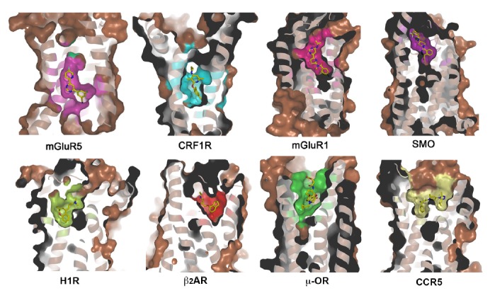
Ligand-binding pockets of mGluR5, CRF1R, mGluR1, SMO, H1R, β2AR, μ-OR and CCR5. Receptors are shown in cartoon and surface representations. Ligands are shown as yellow sticks.
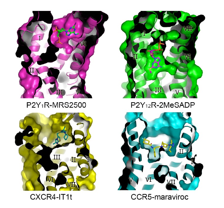
Comparison of the ligand-binding pockets in the structures of P2Y1R-MRS2500, P2Y12R-2MeSADP, CXCR4-IT1t and CCR5-maraviroc complexes. The receptors are shown in cartoon and surface representations, and are colored in magenta, green, yellow and cyan, respectively. The ligands MRS2500, 2MeSADP, IT1t and maraviroc are shown as green, magenta, cyan and yellow sticks, respectively.
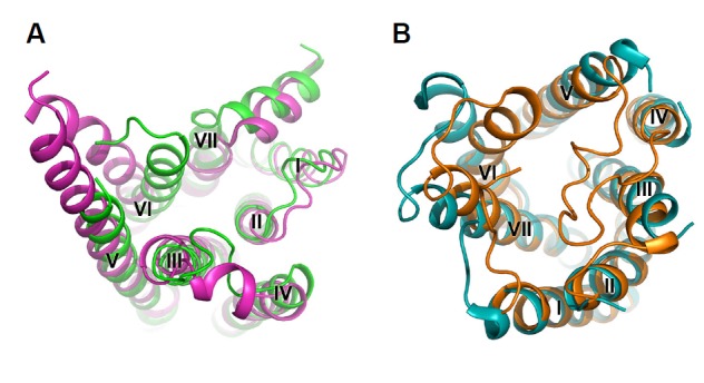
Comparison between active structures and inactive structures of β2AR and P2Y12R. A, Intracellular side of the active structure (magenta) and the inactive structure (green) of β2AR. B, Extracellular side of the active structure (orange) and the inactive structure (cyan) of P2Y12R. The receptors are shown in cartoon representations.
References
-
- Ballesteros J., Weinstein H. Integrated methods for the construction of three-dimensional models and computational probing of structure-function relations in G protein-coupled receptors. Methods Neurosci. 1995;25:366–428.
Publication types
MeSH terms
Substances
LinkOut - more resources
Full Text Sources
Other Literature Sources
