

Essential tremor is associated with disruption of functional connectivity in the ventral intermediate Nucleus--Motor Cortex--Cerebellum circuit
- PMID: 26467643
- PMCID: PMC6867464
- DOI: 10.1002/hbm.23024
Essential tremor is associated with disruption of functional connectivity in the ventral intermediate Nucleus--Motor Cortex--Cerebellum circuit
Abstract
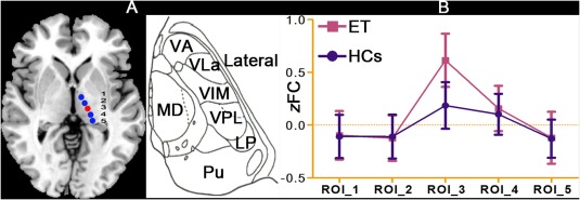
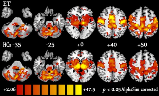
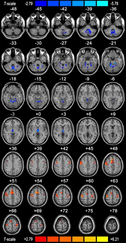
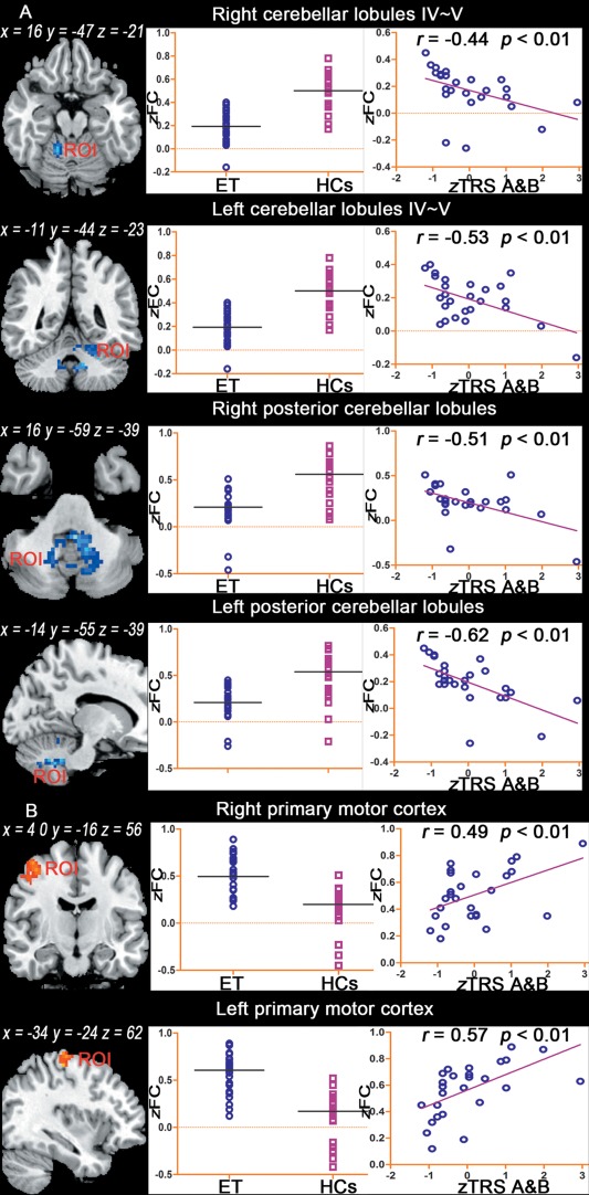
The clinical benefits of targeting the ventral intermediate nucleus (VIM) for the treatment of tremors in essential tremor (ET) patients suggest that the VIM is a key hub in the network of tremor generation and propagation and that the VIM can be considered as a seed region to study the tremor network. However, little is known about the central tremor network in ET patients. Twenty-six ET patients and 26 matched healthy controls (HCs) were included in this study. After considering structural and head-motion factors and establishing the accuracy of our seed region, a VIM seed-based functional connectivity (FC) analysis of resting-state functional magnetic resonance imaging (RS-fMRI) data was performed to characterize the VIM FC network in ET patients. We found that ET patients and HCs shared a similar VIM FC network that was generally consistent with the VIM anatomical connectivity network inferred from normal nonhuman primates and healthy humans. Compared with HCs, ET patients displayed VIM-related FC changes, primarily within the VIM-motor cortex (MC)-cerebellum (CBLM) circuit, which included decreased FC in the CBLM and increased FC in the MC. Importantly, tremor severity correlated with these FC changes. These findings provide the first evidence that the pathological tremors observed in ET patients might be based on a physiologically pre-existing VIM - MC - CBLM network and that disruption of FC in this physiological network is associated with ET. Further, these findings demonstrate a potential approach for elucidating the neural network mechanisms underlying this disease.
Keywords: cerebellum; essential tremor; functional connectivity; functional magnetic resonance imaging; motor cortex; resting state; thalamus; ventral intermediate nucleus.
© 2015 Wiley Periodicals, Inc.
Figures
References
-
- Asanuma C, Thach W, Jones E (1983a): Distribution of cerebellar terminations and their relation to other afferent terminations in the ventral lateral thalamic region of the monkey. Brain Res Rev 5:237–265. - PubMed
-
- Asanuma C, Thach WT, Jones EG (1983b): Cytoarchitectonic delineation of the ventral lateral thalamic region in the monkey. Brain Res 286:219–235. - PubMed
-
- Bagepally BS, Bhatt MD, Chandran V, Saini J, Bharath RD, Vasudev M, Prasad C, Yadav R, Pal PK (2011): Decrease in Cerebral and Cerebellar Gray Matter in Essential Tremor: A Voxel‐Based Morphometric Analysis under 3T MRI. J Neuroimaging 22:275–278. - PubMed
-
- Behrens T, Johansen‐Berg H, Woolrich M, Smith S, Wheeler‐Kingshott C, Boulby P, Barker G, Sillery E, Sheehan K, Ciccarelli O (2003): Non‐invasive mapping of connections between human thalamus and cortex using diffusion imaging. Nat Neurosci 6:750–757. - PubMed
Publication types
MeSH terms
Substances
LinkOut - more resources
Full Text Sources
Other Literature Sources
Miscellaneous
