. 2016 Jan;21(1):792-802.
doi: 10.2119/molmed.2015.00126. Epub 2015 Oct 13.

Differential Sensitivities of Fast- and Slow-Cycling Cancer Cells to Inosine Monophosphate Dehydrogenase 2 Inhibition by Mycophenolic Acid

Affiliations

Differential Sensitivities of Fast- and Slow-Cycling Cancer Cells to Inosine Monophosphate Dehydrogenase 2 Inhibition by Mycophenolic Acid

Kan Chen et al. Mol Med. 2016 Jan.

Abstract

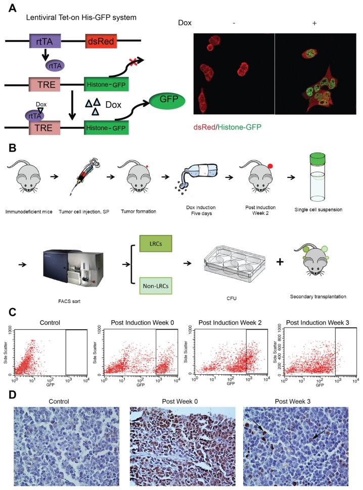
As uncontrolled cell proliferation requires nucleotide biosynthesis, inhibiting enzymes that mediate nucleotide biosynthesis constitutes a rational approach to the management of oncological diseases. In practice, however, results of this strategy are mixed and thus elucidation of the mechanisms by which cancer cells evade the effect of nucleotide biosynthesis restriction is urgently needed. Here we explored the notion that intrinsic differences in cancer cell cycle velocity are important in the resistance toward inhibition of inosine monophosphate dehydrogenase (IMPDH) by mycophenolic acid (MPA). In short-term experiments, MPA treatment of fast-growing cancer cells effectively elicited G0/G1 arrest and provoked apoptosis, thus inhibiting cell proliferation and colony formation. Forced expression of a mutated IMPDH2, lacking a binding site for MPA but retaining enzymatic activity, resulted in complete resistance of cancer cells to MPA. In nude mice subcutaneously engrafted with HeLa cells, MPA moderately delayed tumor formation by inhibiting cell proliferation and inducing apoptosis. Importantly, we developed a lentiviral vector-based Tet-on label-retaining system that enables to identify, isolate and functionally characterize slow-cycling or so-called label-retaining cells (LRCs) in vitro and in vivo. We surprisingly found the presence of LRCs in fast-growing tumors. LRCs were superior in colony formation, tumor initiation and resistance to MPA as compared with fast-cycling cells. Thus, the slow-cycling compartment of cancer seems predominantly responsible for resistance to MPA.


Figures

Figure 1
Figure 1
MPA inhibited cell proliferation and colony formation of different cancer cell lines. (A) Growth curve of seven different cancer cell lines show that HeLa cells grow faster than the other cell lines tested (mean ± SD, n = 6). (B) MPA inhibited cell proliferation of all seven cancer cell lines as determined by MTT; data shown cells were treated by MPA for 72 h (mean ± SD, n = 5). (C) and (D) MPA inhibited single cell colony formation of seven cancer cell lines (mean ± SD, n = 6, ***P < 0.001).
Figure 2
Figure 2
MPA counteracts proliferation of a fast-growing cancer cell line. (A) Clinically-relevant MPA concentrations potently inhibit proliferation of the HeLa cell line as assessed by MTT activity (mean ± SD, n = 5). (B) Clinically-relevant MPA concentrations impair colony formation of HeLa cells (mean ± SD, n = 6). (C) Clinically-relevant MPA concentrations impair colony growth of HeLa cell as determined by image analysis (mean ± SEM, n = 30, **P < 0.01). (D) MPA treatment causes G0/G1 phase cell cycle arrest. The left panel shows cell cycle phase distribution of a vehicle-treated culture, whereas the middle panel shows cell cycle phase distribution in a MPA-treated culture. The right panel shows a quantification of the MPA effects on the cell cycle of HeLa (mean ± SD, n = 3. *P < 0.05; **P < 0.01). (E) FACS analysis of cellular apoptosis through annexin-V positivity and PI incorporation. The left panel provides an example of a vehicle-treated HeLa culture, whereas the middle panel provides an example of the effects seen following MPA treatment. The quantification in the right panel shows statistically significant stimulation of both early and late apoptosis in the 5–25 μg/mL MPA concentration (mean ± SD, n = 3, *P < 0.05; **P < 0.01).
Figure 3
Figure 3
Force expression of a mutated IMPDH2, lacking the MPA-binding site, results in resistance to MPA. (A) An IMPDH2 variant having normal IMP hydrogenase activity but lacking the MPA-binding site was fused to a GFP reporter (mutIMPDH2) and expressed in HeLa cells by a lentiviral vector. FACS analysis showed robust GFP expression in the transduced HeLa cells, but not in mock-transduced cells. Transduced cells appear to be resistant to MPA both in (B) colony formation (mean ± SD, n = 6, **P < 0.01) and (C) cell proliferation assays (mean ± SEM, n = 30, ***P < 0.001). (D) MPA-mediated induction of early apoptotic cells, as well as late apoptotic cells, is reduced significantly in mutIMPDH2 HeLa cells (mean ± SD, n = 3. *P < 0.05; **P < 0.01).
Figure 4
Figure 4
MPA delayed tumor initiation, inhibited cell proliferation and provoked tumor apoptosis in mice. (A) MPA treatment significantly delayed tumor initiation by HeLa cells in nude mice. (B) Following the experiment, animals were killed and tumors were harvested. The photograph illustrates the macroscopic appearance of the tumors in the respective groups. (C) Immunohistochemical staining of harvested tumor tissue sections revealed a significant downregulation of IMPDH2 protein levels following treatment with MPA. (D) Treatment of MPA significantly reduced the percentage of p-histone H3 positive (proliferating) cells in the tumors. (E) MPA treatment of HeLa-grafted nude mice significantly increased the numbers of anti-cleaved caspase-3 immunoreactive (apoptotic) cells (mean ± SEM, PBS, n = 10; 60 mg/kg, n = 11; 240 mg/kg, n = 9, *P < 0.05; **P < 0.01).
Figure 5
Figure 5
Identification of slow-cycling cancer cells using a lentiviral label-retaining system. (A) Illustration of principle; the lentiviral Tet-on histone-GFP strategy employed. Confocal imaging confirms the specific induction of nuclear histone-GFP expression in HeLa cells upon doxycycline treatment. (B) Schematic representation of the experimental approach to identify, isolate and characterize the slow-cycling cells, or termed as label-retaining cells (LRCs) in vivo. (C) FACS characterization of histone-GFP expression in transduced HeLa cells during 3 wks following release from doxycycline induction. (D) Immunohistochemical staining of harvested xenograft tumor tissues confirms the presence of nuclear anti-GFP immunoreactivity in these tissues.
Figure 6
Figure 6
Grafted label-retaining cells are superior in tumor initiation and more resistant to MPA. (A) The macroscopic appearance of the tumors formed following secondary grafting of LRCs and non-LRCs, respectively. (B) Colony formation following grafting LRCs and non-LRCs harvested from xenograft tumor tissues (mean ± SEM, n = 19, ***P < 0.001). (C) Colony formation by LRCs and non-LRCs harvested from xenograft tumor tissues, when treated with MPA at a concentration of 10 μg/mL (mean ± SEM, n = 17, ***P < 0.001). NS, no signification.

References

    1. Hanahan D, Weinberg RA. Hallmarks of cancer: the next generation. Cell. 2011;144:646–74. - PubMed
    1. Natsumeda Y, et al. Two distinct cDNAs for human IMP dehydrogenase. J Biol Chem. 1990;265:5292–5. - PubMed
    1. Carr SF, Papp E, Wu JC, Natsumeda Y. Characterization of human type I and type II IMP dehydrogenases. J Biol Chem. 1993;268:27286–90. - PubMed
    1. Moosavi MA, Yazdanparast R, Sanati MH, Nejad AS. 3-Hydrogenkwadaphnin targets inosine 5′-monophosphate dehydrogenase and triggers post-G1 arrest apoptosis in human leukemia cell lines. Int J Biochem Cell Biol. 2005;37:2366–79. - PubMed
    1. Guidicelli G, et al. The necrotic signal induced by mycophenolic acid overcomes apoptosis-resistance in tumor cells. PLoS One. 2009;4:e5493. - PMC - PubMed

LinkOut - more resources