

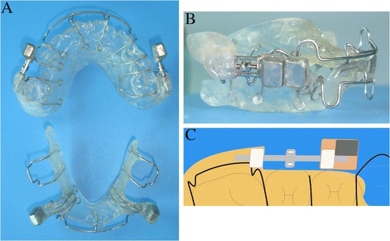
Effects of a novel magnetic orthopedic appliance (MOA-III) on the dentofacial complex in mild to moderate skeletal class III children
- PMID: 26468063
- PMCID: PMC4606506
- DOI: 10.1186/s13005-015-0092-7
Effects of a novel magnetic orthopedic appliance (MOA-III) on the dentofacial complex in mild to moderate skeletal class III children
Abstract
Introduction: The objective of this study was to evaluate the changes of skeletal and dental structures in mild to moderate skeletal Class III children following the use of a new magnetic orthopedic appliance (MOA-III).
Methods: A total of 36 patients (14 boys and 22 girls, mean age 9 years and 5 months) who presented with a mild to moderate skeletal Class III jaw discrepancy were treated with MOA-III. Another group of 20 untreated patients (9 boys and 11 girls, mean age 9 years and 2 months) with the same level of deformity served as the control group. The average treatment time was 6.6 months. Radiographs were taken at the same time intervals for both groups. A paired t test was used to determine the significant differences before and after treatment, and a two-sample t test was used to analyze the differences between the treatment and control groups.
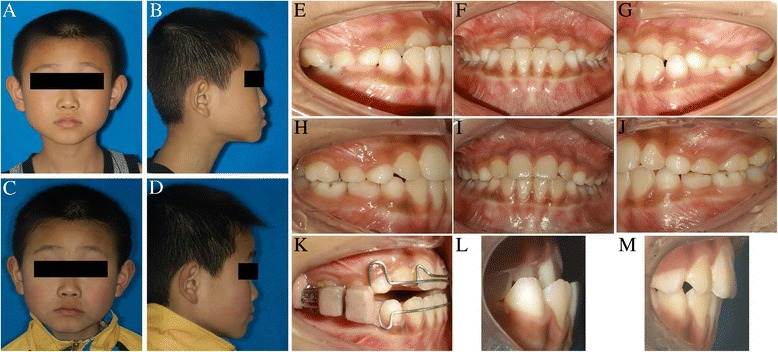
Results: The anterior crossbite in all subjects was corrected after MOA-III therapy. The maxillomandibular relationship showed favorable changes (ANB, Wits, overjet increased significantly, P < 0.001). The maxilla was anteriorly positioned (SNA, ptm-A, ptm-S increased significantly, P < 0.001) with clockwise rotation (PP-FH increased, P < 0.001). The mandible showed a slight downward and backward rotation (SNB decreased, P < 0.05, MP-SN, Y-axis increased, P < 0.05). The length of the mandibular body showed no significant changes (Go-Pg, P > 0.05). Significant upper incisor proclination and lower incisor retroclination were observed (UI-NA increased, P < 0.001, LI-NB, FMIA decreased, P < 0.001). The upper lip moved forward, and the lower lip moved backward (UL-EP increased, P < 0.001, LL-EP decreased, P < 0.05). In the control group, most of the parameters showed normal growth, except for some unfavorable mandibular skeletal and soft tissue changes (Go-Pg, Go-Co, MP-SN, N'-SN-Pg' increased, P < 0.001). Significant positive changes were induced with the MOA-III appliance compared to the untreated group.
Conclusions: The MOA-III was effective for the early treatment of a mild to moderate Class III malocclusion in children.
Figures

References
MeSH terms
LinkOut - more resources
Full Text Sources
Other Literature Sources
Research Materials
Miscellaneous
