. 2016 Jan;126 Suppl 1(Suppl 1):S5-21.
doi: 10.1002/lary.25676. Epub 2015 Oct 15.

Pediatric laryngotracheal reconstruction with tissue-engineered cartilage in a rabbit model

Affiliations

Pediatric laryngotracheal reconstruction with tissue-engineered cartilage in a rabbit model

Ian N Jacobs et al. Laryngoscope. 2016 Jan.

Abstract

Objectives/hypothesis: To develop an effective rabbit model of in vitro- and in vivo-derived tissue-engineered cartilage for laryngotracheal reconstruction (LTR).

Study design: 1) Determination of the optimal scaffold 1% hyaluronic acid (HA), 2% HA, and polyglycolic acid (PGA) and in vitro culture time course using a pilot study of 4 by 4-mm in vitro-derived constructs analyzed on a static culture versus zero-gravity bioreactor for 4, 8, and 12 weeks, with determination of compressive modulus and histology as outcome measures. 2) Three-stage survival rabbit experiment utilizing autologous auricular chondrocytes seeded in scaffolds, either 1% HA or PGA. The constructs were cultured for the determined in vitro time period and then cultured in vivo for 12 weeks. Fifteen LTRs were performed using HA cartilage constructs, and one was performed with a PGA construct. All remaining specimens and the final reconstructed larynx underwent mechanical testing, histology, and glycosaminoglycan (GAG) content determination, and then were compared to cricoid control specimens (n = 13) and control LTR using autologous thyroid cartilage (n = 18).

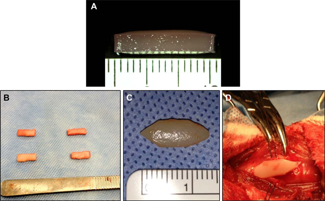
Methods: 1) One rabbit underwent an auricular punch biopsy, and its chondrocytes were isolated and expanded and then encapsulated in eight 4 by 4-mm discs of 1% HA, 2% HA, PGA either in rotary bioreactor or static culture for 4, 8, and 12 weeks, respectively, with determination of compressive modulus, GAG content, and histology. 2) Sixteen rabbits underwent ear punch biopsy; chondrocytes were isolated and expanded. The cells were seeded in 13 by 5 by 2.25-mm UV photopolymerized 1% HA (w/w) or calcium alginate encapsulated synthetic PGA (13 × 5 × 2 mm); the constructs were then incubated in vitro for 12 weeks (the optimal time period determined above in paragraph 1) on a shaker. One HA and one PGA construct from each animal was tested mechanically and histologically, and the remaining eight (4 HA and 4 PGA) were implanted in the neck. After 12 weeks in vivo, the most optimal-appearing HA construct was used as a graft for LTR in 15 rabbits and PGA in one rabbit. The seven remaining specimens underwent hematoxylin and eosin, Safranin O, GAG content determination, and flexural modulus testing. At 12 weeks postoperative, the animals were euthanized and underwent endoscopy. The larynges underwent mechanical and histological testing. All animals that died underwent postmortem examination, including gross and microhistological analysis of the reconstructed airway.

Results: Thirteen of the 15 rabbits that underwent LTR with HA in vitro- and in vivo-derived tissue-engineered cartilage constructs survived. The 1% HA specimens had the highest modulus and GAG after 12 weeks in vitro. The HA constructs became well integrated in the airway, supported respiration for the 12 weeks, and were histologically and mechanically similar to autologous cartilage.

Conclusions: The engineering of in vitro- and in vivo-derived cartilage with HA is a novel approach for laryngotracheal reconstruction. The data suggests that the in vitro- and in vivo-derived tissue-engineered approaches may offer a promising alternative to current strategies used in pediatric airway reconstruction, as well as other head and neck applications.

Level of evidence: NA. Laryngoscope, 126:S5-S21, 2016.

Keywords: Polyglycolic acid (PGA); compressive modulus; flexural modulus; glycosaminoglycan (GAG); hyaluronic acid (HA).


Figures

Fig. 1.
Fig. 1.
Gross morphology of specimens from alginate encapsulated rectangular constructs 6 months after in vivo implantation in the nude mouse..
Fig. 2.
Fig. 2.
Hemotoxylin-Eosin stain of 6-month alginate encapsulated in vivo constructs from nude mouse.
Fig. 3.
Fig. 3.
In vivo-derived 1% hyaluronic acid graft at 24 weeks, revealing severe ossification.
Fig. 4.
Fig. 4.
Timeline of in vitro/in vivo experimental procedure. Time for each rabbit experiments totals 36 weeks.
Fig. 5.
Fig. 5.
(A) 4 × 4-mm pilot construct undergoing compression testing. (B) Three-point bend test for flectoral modulus. (C) Anterior-posterior compression test for anterior-to-posterior compressive modulus.
Fig. 6.
Fig. 6.
Rotating vessel bioreactor with 4 × 4-mm constructs.
Fig. 7.
Fig. 7.
Rabbit with scaffolds in the neck.
Fig. 8.
Fig. 8.
(A) A lateral view of 1% HA construct after 12 weeks in vitro showing gross augmentation in volume due to production of extracellular matrix. The dimensions increase beyond 13 × 5 × 2.25 mm. (B) Four 1% HA after 12 weeks in vivo and 12 weeks in vitro. (C, D) A 1% HA graft carved as a suitable laryngotracheal reconstruction graft. HA = hyaluronic acid.
Fig. 9.
Fig. 9.
(A) TE-LTR using a 1% HA graft. (B) Endoscopic view 12 weeks later. (C) TE-LTR on a different rabbit with 1% HA graft. (D) Endoscopic view showing mucosalized graft 12 weeks later. (E) Control LTR using thyroid alar cartilage. (F) Endoscopic view 12 weeks later. HA = hyaluronic acid; LTR = laryngotracheal reconstruction; TE = tissue-engineered.
Fig. 10.
Fig. 10.
Time course for compressive modulus of 4 × 4-mm discs using 1, 2% hyaluronic acid, and polyglycolic acid-calcium alginate at 4, 8, and 12 weeks in vitro both in a rotary bioreactor (blue-striped columns) and static culture (red columns).
Fig. 11.
Fig. 11.
(A) Postmortem examination of larynx of rabbit s/p PGA-calcium alginate cartilage graft that died of airway obstruction. Lumen reveals a large granuloma that obstructed the airway. (B) Cross section of the airway stained with Safranin O revealing nearly complete obstructive granuloma (20×). (C) Collagen II immune-stain of graft revealing PGA filaments (50×). (D) Safranin O stain of graft revealing decrease in central cells and microfilaments (50×). Death of animal due to brittle bony graft that collapsed into the airway. (E) Bony graft sutured into the airway. (F) Gross postmortem view revealing collapsed graft. (G) Cross section of the airway stained with Safranin O revealing nearly complete obstructive collapsed graft (20×). (H) Safranin O stain of bony graft (cartilage would be red) (50×). PGA 5 polyglycolic acid.
Fig. 12.
Fig. 12.
Flexural modulus of constructs. Individual values are presented by groups. Red bar represents mean with 95% confidence interval. (A) Moduli of HA versus PGA constructs after 12 weeks in vitro. (B) Moduli of HA versus PGA constructs after 12 weeks in vivo compared to control thyroid cartilage. (C) Moduli of HA constructs after 12 weeks in vitro versus HA constructs after 12 weeks in vivo. (D) Moduli of PGA constructs after 12 weeks in vitro versus 12 weeks in vivo. HA = hyaluronic acid; PGA = polyglycolic acid.
Fig. 13.
Fig. 13.
Cricoid compressive moduli. The scatterplots show the range, mean (red bar) and 95% confidence intervals. (A) Anterior-posterior compressive moduli of TE-LTR versus C-LTR (P = 0.111) and cricoid controls (P = 0.004). (B) Medial-lateral compressive moduli of TE-LTR versus C-LTR (P = 0.999) and cricoid dontrols (P = 0.047). C-LTR = control laryngotracheal reconstruction; TE-LTR = tissue-engineered laryngotracheal reconstruction.
Fig. 14.
Fig. 14.
GAG of constructs. Individual values are presented by groups. Bar represents mean with 95% confidence interval. (A) GAG of HA versus PGA 12 in vitro and in vivo compared to control cartilage and negative control. Two high extreme control cartilage values are not shown as they are high on the y-axis. (B) GAG of HA after 12 weeks in vitro versus after 12 weeks in vivo. (C) GAG of PGA constructs after 12 weeks in vitro versus after 12 weeks in vivo. GAG = glycosaminoglycan; PGA = polyglycolic acid.
Fig. 15.
Fig. 15.
GAG determination of the TE grafts versus controls (A) GAG content (% w/w) of constructs of TE-LTR and C-LTR cricoid cartilage versus control cartilage and negative controls (muscle) shown with mean and 95% confidence intervals. Two high extreme outlier values of control cartilage and one of C-LTR is not shown as they are much higher on the y-axis. (B) AP modulus versus GAG content revealed reverse correlation which was statistically significant with (P = 0407) with R-squared = 0.36. (C) ML modulus versus GAG content. This shows trend toward inverse correlation which is not statistically significant (P = 0.1536) and R-squared = 0.19. C-LTR = control laryngotracheal reconstruction; GAG = glycosaminoglycan; ML = GAG = glycosaminoglycan; TE-LTR = tissue-engineered laryngotracheal reconstruction.
Fig. 16.
Fig. 16.
Histology of 1% HA scaffolds after 12 weeks in vitro. (A) Safranin O stain (50×) revealing dense extracellular matrix, which is red. (B) Safranin O stain (100×) showing the same as (C) at closer magnification. (C) Collagen II stain (50×) showing abundant high intensity staining. Histology of 1% HA scaffolds after 12 weeks in vitro and 12 weeks in vivo. (D) Safranin O stain (50×), where the red indicates extracellular matrix of cartilage. (E) Von Kassa/Alcian blue stain (50×) where the blue indicates dense extracellular matrix. (F) Collagen II stain (50×) showing dense staining for collagen II. HA = hyaluronic acid.
Fig. 17.
Fig. 17.
Bony HA construct after 12 weeks in vivo culture. (A, B) Safranin O stain revealing decreased staining of extracellular matrix (50×). (C, D) Safranin O stain of different animal revealing ossification and lack of cartilage (100×). HA = hyaluronic acid.
Fig. 18.
Fig. 18.
Histology of PGA constructs. PGA constructs after 12 weeks in vitro. (A) Safranin O stain revealing extracellular matrix (50×). (B) Safranin O stain revealing decreased intensity of staining and remaining PGA strands (50×). (C) Collagen II immunostain revealing decreased staining intensity (50×). PGA Constructs 12 weeks in vivo. (D) Safranin O stain revealing decreased intensity of staining (50×). (E) Safranin O stain after 12 weeks in vivo revealing sparse center of the PGA construct (50×). (F) Collagen II immunostain revealing decreased intensity in the center (50×). PGA = polyglycolic acid.
Fig. 19.
Fig. 19.
(A) Postmortem view with Safranin O stain of 1% HA tissue-engineered cartilage graft (arrow) at 12 weeks postoperative (20×). (B) Epithelial lining with Safranin O stain (50×). (C) Epithelial lining with Safranin O stain (100×). (D) Center of cartilage graft with Safranin O stain (100×). (E) Collagen II immunostain of construct revealing intense staining (50×). (F) Collagen I immunostain revealing lower intensity staining (50×). (G) Collagen II immunostain of normal cricoid cartilage of the specimen revealing intense staining (50×). (H) Collagen I immunostain revealing minimal intensity staining (50×). HA = hyaluronic acid.
Fig. 20.
Fig. 20.
(A) Control LTR (20×) showing autologous thyroid graft at the top (arrow). (B) Epithelial lining with Safranin O stain (50×). (C) Center of cartilage graft (50×) revealing dense chondrocytes and matrix. (D) Transition zone (50×) revealing graft and fibrous tissue at junction. (E) Collagen II immune stain (50×) revealing intense staining of the graft. (F) Collagen I immune stain (50×) revealing no staining of the graft but some stain of the connective tissue. C-LTR = control laryngotracheal reconstruction.

References

    1. Cotton RT. The problem of pediatric laryngotracheal stenosis: a clinical and experimental study on the efficacy of autologous cartilage grafts placed between the vertically divided halves of the posterior lamina of the cricoid cartilage. Laryngoscope 1991;101(suppl 56):1–34. - PubMed
    1. Hartnick CJ, Hartley BE, Lacy PD, et al. Surgery for pediatric subglottic stenosis-disease specific outcomes. Ann Otol Rhinol Laryngol 2001;110: 1109–1113. - PubMed
    1. Rizzi M, Thorne MC, Zur KB, Jacobs IN. Laryngotracheal reconstruction using posterior costal cartilage grafts: outcomes at a single institution. Otolaryngol Head Neck Surg 2009;140:348–353. - PubMed
    1. Jacobs IN, Podrebarac P, Boden SD, Chen M. Graft-healing in laryngotracheal reconstruction: an experimental rabbit model. Ann Otol Laryngol Rhinol 1999;108:599–605. - PubMed
    1. Langer R, Vacanti JP. Tissue-engineering. Science 1993;260:920–926. - PubMed

Publication types