. 2016 Jan 21;127(3):314-24.
doi: 10.1182/blood-2015-06-651034. Epub 2015 Oct 14.

Gnb isoforms control a signaling pathway comprising Rac1, Plcβ2, and Plcβ3 leading to LFA-1 activation and neutrophil arrest in vivo

Affiliations

Gnb isoforms control a signaling pathway comprising Rac1, Plcβ2, and Plcβ3 leading to LFA-1 activation and neutrophil arrest in vivo

Helena Block et al. Blood. .

Abstract

Chemokines are required for leukocyte recruitment and appropriate host defense and act through G protein-coupled receptors (GPCRs), which induce downstream signaling leading to integrin activation. Although the α and β subunits of the GPCRs are the first intracellular molecules that transduce signals after ligand binding and are therefore indispensable for downstream signaling, relatively little is known about their contribution to lymphocyte function-associated antigen 1 (LFA-1) activation and leukocyte recruitment. We used knockout mice and short hairpin RNA to knock down guanine nucleotide binding protein (GNB) isoforms (GNB1, GNB2, GNB4, and GNB5) in HL60 cells and primary murine hematopoietic cells. Neutrophil function was assessed by using intravital microscopy, flow chamber assays, and chemotaxis and biochemistry studies. We unexpectedly discovered that all expressed GNB isoforms are required for LFA-1 activation. Their downregulation led to a significant impairment of LFA-1 activation, which was demonstrated in vitro and in vivo. Furthermore, we showed that GPCR activation leads to Ras-related C3 botulinum toxin substrate 1 (Rac1)-dependent activation of both phospholipase C β2 (Plcβ2) and Plcβ3. They act nonredundantly to produce inositol triphosphate-mediated intracellular Ca(2+) flux and LFA-1 activation that support chemokine-induced arrest in vivo. In a complex inflammatory disease model, Plcβ2-, Plcβ3-, or Rac1-deficient mice were protected from lipopolysaccharide-induced lung injury. Taken together, we demonstrated that all Gnb isoforms are required for chemokine-induced downstream signaling, and Rac1, Plcβ2, and Plcβ3 are critically involved in integrin activation and leukocyte arrest.


Figures

Figure 1
Figure 1
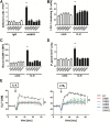
Downregulation of GNB isoforms or GNAI2 strongly affects LFA-1 function. HL60 cells transduced with a nonsilencing scrambled (scr) construct or with shRNAs against GNB1, GNB2, GNB4, GNB5, or GNAI2 were used. (A) Number of adherent cells per field of view (FOV) in the adhesion flow chamber. Chambers were coated with P-selectin, IL-8, and either a control IgG antibody or with the reporter mAb24 recognizing high-affinity LFA-1 (n = 4 experiments). *P < .05 vs all other groups. (B) Transduced HL60 cells were left untreated or were stimulated with IL-8, fixed with paraformaldehyde (4%), and stained with anti-CD11a and AF488-labeled secondary antibody to visualize clustered LFA-1 (n = 3 experiments). *P < .05 vs all other groups. (C) Flow cytometry analyzed allophycocyanin (APC)-labeled ICAM-1 binding in unstimulated (unst.) or IL-8 (100 ng/mL) stimulated cells (n = 4 experiments). *P < .05 vs all other groups. (D) Cells were left unstimulated or were stimulated with IL-8 (100 ng/mL, 3 minutes). The reaction was stopped by adding trichloroacetic acid, which was later removed by adding 1,1,2-trichloro-trifluoroethane-trioctylamine. The aqueous IP3-containing supernatant was used for a competitive radioreceptor assay. Bars indicate IP3 concentration in picomoles per 1 × 10⁷ HL60 cells (n = 3 experiments). *P < .05. (E) Concentration of intracellular calcium measured in Indo-1-labeled HL60 cells before and after chemokine stimulation. Arrow indicates IL-8 or LTB4 stimulation (n = 4 experiments). #P < .05 vs all other groups. MFI, mean fluorescent intensity.
Figure 2
Figure 2
Knockdown of GNB isoforms significantly impairs RAC1-mediated downstream signaling. (A) Representative western blots of scrambled or shRNA-transduced HL60 cells, which were left untreated or stimulated with IL-8 (100 ng/mL), lysed, and used to pull down GTP-bound active RAC1. Total RAC1 served as loading control (n = 3 experiments). (B) Number of adherent cells per field of view in the adhesion flow chamber after pre-incubation with TAT-WT RAC or (C) TAT-CA RAC construct (n = 4 experiments). *P < .05 vs all other groups. (D-E) Chemotactic migration of HL60 cells transduced with a scrambled construct or different shRNAs (n = 3 experiments; >60 cells). *P < .05 vs all groups. (D) Migration velocity and (E) accumulated (accum.) distance toward the applied IL-8 gradient in the ibidi μ-Slide. (F) Transduced HL60 cells were left unstimulated or incubated with IL-8 (100 ng/mL, 3 minutes, 37°C), fixed, and permeabilized to stain intracellular p-p38 MAPK. Staining was analyzed by using flow cytometry (n = 3 experiments). *P < .05 vs all other groups.
Figure 3
Figure 3
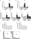
Knockdown of Gnb isoforms in primary neutrophils significantly abolishes chemokine-induced arrest in vivo and neutrophil migration in vitro. Retrovirally transduced hematopoietic stem cells with shRNAs against Gnb1, Gnb2, Gnb4, and Gnb5 or with a scrambled construct were transplanted into lethally irradiated WT recipients. (A) Mixed chimeric mice were exposed to nebulized saline or LPS to induce lung injury. Data indicate number of neutrophils in the BAL 24 hours after inhalation of NaCl (left panel) or LPS (right panel) (n = 3). *P < .05 vs all other groups. (B) Mixed chimeric mice were used to investigate CXCL1-induced arrest in the cremaster muscle (n = 3). *P < .05 vs all other groups. (C) Purified bone marrow–derived neutrophils were used for an ICAM-1-binding assay. During flow cytometry acquisition, cells were gated into GFP-positive (shRNA-containing) and GFP-negative (WT) cell populations and analyzed for CXCL1-induced increase of bound APC-labeled ICAM-1. Data represent MFI of APC in nontransduced and transduced cells, both unstimulated and stimulated (n = 4 experiments). *P < .05 vs all other groups. (D) Isolated PMNs from mixed chimeric mice were left untreated or were stimulated with CXCL1, fixed with paraformaldehyde (4%), and stained with anti-CD11a and AF488-labeled secondary antibody to visualize clustered LFA-1 (n = 3 experiments). *P < .05 vs all other groups. (E) Purified bone marrow–derived neutrophils were left unstimulated or incubated with CXCL1 (100 ng/mL, 3 minutes, 37°C), fixed, and permeabilized to stain intracellular p-p38 MAPK. MFI of bound AF647-labeled p38 MAPK antibody was analyzed by using flow cytometry gating into GFP-positive and GFP-negative populations (n = 3 experiments). *P < .05 vs all other groups. (F-H) Chemotactic migration of purified bone marrow–derived neutrophils of mixed chimeric mice (n = 3 experiments; >40 cells). *P < .05 vs all groups. (F) Migration directness and (G) migration velocity toward the applied CXCL1 gradient in the ibidi μ-Slide. (H) Representative migration plots of cells transduced with a scrambled construct or with an shRNA against 1 Gnb isoform. Arrow indicates applied CXCL1 gradient. no c., no construct; n.s., not significant.
Figure 4
Figure 4
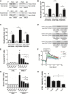
GPCR signaling activates Plcβ2 and Plcβ3 in a Rac1-dependent manner. (A) Co-immunoprecipitation (co-IP) of Plcβ2 or Plcβ3 with Rac1. Bone marrow–derived WT neutrophils were left untreated or were stimulated with CXCL1 (100 ng/mL, 3 minutes, 37°C), lysed, and used for precipitation with Plcβ2 or Plcβ3 antibody and were blotted and incubated with Rac1 antibody (upper panel) or (lower panel) used for precipitation with Rac1 and then blotted and incubated with Plcβ2 or Plcβ3 antibody (n = 3 experiments for both). For densitometric analysis, see supplemental Figure 4A-B. (B-C) Number of arrested cells per square millimeter after injection of 500 ng CXCL1 in cremaster muscles of mice transplanted with retrovirally transduced Plcβ-knockout (KO) bone marrow. Chemokine-induced arrest of (B) Plcβ2-KO bone marrow nontransduced (non-transd.) or transduced with Plcβ2-complementary DNA (cDNA) or Plcβ2-ΔPH and (C) Plcβ3-KO bone marrow nontransduced or transduced with Plcβ3-cDNA or Plcβ3-ΔPH. (D) Representative western blots of WT, Gnai2−/−, Plcβ2−/−, or Plcβ3−/− neutrophils, which were left untreated or stimulated with CXCL1 (100 ng/mL), lysed, and used to pull down GTP-bound active RAC1. Total RAC1 serves as loading control (n = 3 experiments). (E) Intracellular concentrations of IP3 before and after stimulating cells with CXCL1 (100 ng/mL, 3 minutes; n = 3 experiments). #P < .05 WT vs all other groups; *P < .05, Plcβ2−/− vs Gnai2−/− and Rac1−/−, and Plcβ3−/− vs Gnai2−/− and Rac1−/−. (F) Bone marrow–derived Indo-1-labeled neutrophils from WT, Plcb2−/−, Plcb3−/−, and Rac1−/− mice were investigated for intracellular calcium levels before and after stimulation with CXCL1. Arrow indicates CXCL1 treatment (100 ng/mL; n = 4 experiments). #P = .05. (G) Flow cytometry was used to analyze APC-labeled ICAM-1 binding in unstimulated or CXCL1 (100 ng/mL, 3 minutes) stimulated bone marrow–derived neutrophils (n = 3 experiments). #P < .05 WT vs all other groups; *P < .05 Plcβ2−/− vs Gnai2−/− and Rac1−/−, and Plcβ3−/− vs Gnai2−/− and Rac1−/−. (H) Percentage of clustered cells in the inflamed cremaster muscle (tumor necrosis factor-α, 2 hours; n = 4). #P < .05 WT vs all other groups. *P < .05 Plcβ2−/− vs Rac1−/−, and Plcβ3−/− vs Rac1−/−.
Figure 5
Figure 5
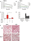
Plcβ and Rac1 deficiency leads to reduced chemokine arrest and protects from LPS-induced lung injury. (A-B) WT mice transplanted with WT, Plcb2−/−, Plcb3−/−, Gnai2−/−, and Rac1−/− bone marrow cells were used to determine chemokine-induced arrest in the cremaster muscle. Arrested cells were counted after injection of 500 ng (A) CXCL1 or (B) LTB4 over 15 minutes. (C) Chemokine-induced arrest of Plcb2−/− mice pretreated with dimethylsulfoxide (DMSO) or with PLC inhibitor U73122. Arrested cells were determined before and after injection of 500 ng CXCL1 (n = 3). *P < .05. (D) WT, Plcb2−/−, Plcb3−/−, or Rac1−/− mice were exposed to nebulized NaCl or LPS; 24 hours later, mice were euthanized and the bronchoalveolar fluid was collected and analyzed for neutrophil recruitment (n = 4). *P < .05 vs all other groups. (E) Representative images of hematoxylin and eosin (H&E)–stained formalin-fixed paraffin-embedded lung sections (slices 5 µm thick) from WT and Rac1−/− mice, which were exposed to nebulized saline or LPS. Images were acquired by using a Zeiss LSM510 (×20 magnification).
Figure 6
Figure 6
Schematic model of downstream signaling after GPCR activation. After chemokine binding, Gα and Gβγ subunits dissociate and GDP is exchanged for GTP. Gβ subunits, Rac1, Plcβ2, and Plcβ3 form a macromolecular complex upon activation, leading to generation of IP3 (black circles), which in turn binds to IP3-gated calcium stores leading to calcium release (black stars). Finally, activated LFA-1 upshifts to the high-affinity conformation. Dotted arrows, question marks, and molecules in light gray circles indicate potential alternative signaling pathways which might also play a role in downstream signaling leading to LFA-1 activation.

Comment in

Similar articles

Cited by

References

    1. Ley K, Laudanna C, Cybulsky MI, Nourshargh S. Getting to the site of inflammation: the leukocyte adhesion cascade updated. Nat Rev Immunol. 2007;7(9):678–689. - PubMed
    1. Herter J, Zarbock A. Integrin Regulation during Leukocyte Recruitment. J Immunol. 2013;190(9):4451–4457. - PubMed
    1. Phillipson M, Heit B, Colarusso P, Liu L, Ballantyne CM, Kubes P. Intraluminal crawling of neutrophils to emigration sites: a molecularly distinct process from adhesion in the recruitment cascade. J Exp Med. 2006;203(12):2569–2575. - PMC - PubMed
    1. Takagi J, Petre BM, Walz T, Springer TA. Global conformational rearrangements in integrin extracellular domains in outside-in and inside-out signaling. Cell. 2002;110(5):599–611. - PubMed
    1. Zarbock A, Kempf T, Wollert KC, Vestweber D. Leukocyte integrin activation and deactivation: novel mechanisms of balancing inflammation. J Mol Med (Berl) 2012;90(4):353–359. - PubMed

Publication types

MeSH terms