. 2015 Dec 4;290(49):29250-8.
doi: 10.1074/jbc.M115.681767. Epub 2015 Oct 14.

cAMP-dependent Protein Kinase (PKA) Signaling Is Impaired in the Diabetic Heart

Affiliations

cAMP-dependent Protein Kinase (PKA) Signaling Is Impaired in the Diabetic Heart

Lee B Bockus et al. J Biol Chem. .

Abstract

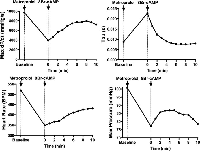
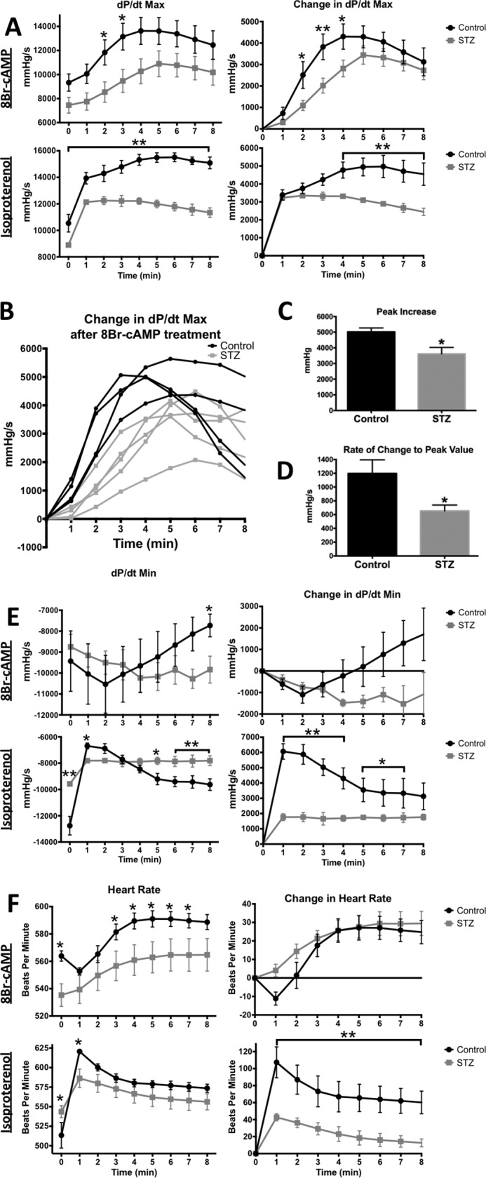
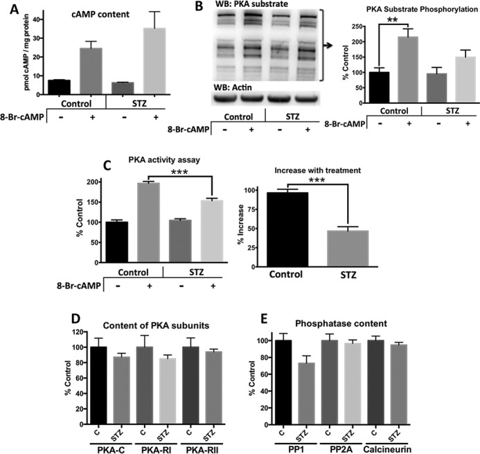
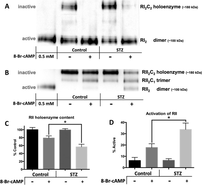
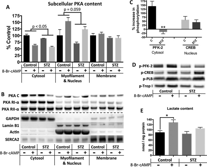
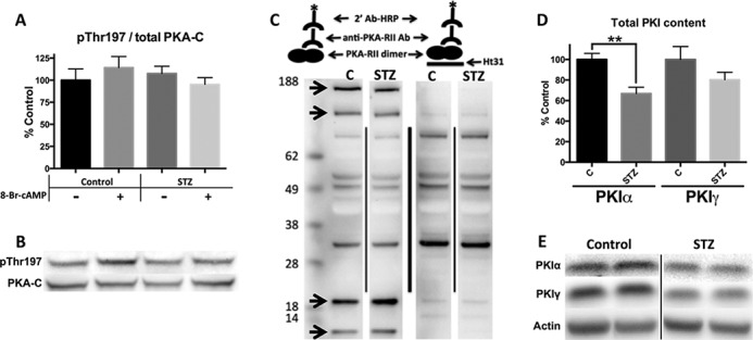
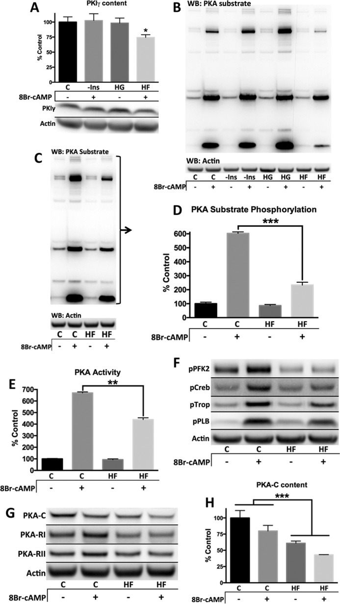
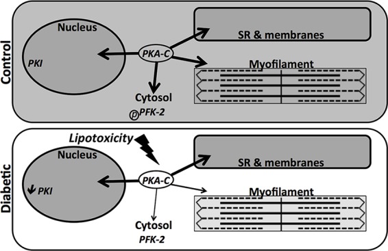
Diabetes mellitus causes cardiac dysfunction and heart failure that is associated with metabolic abnormalities and autonomic impairment. Autonomic control of ventricular function occurs through regulation of cAMP-dependent protein kinase (PKA). The diabetic heart has suppressed β-adrenergic responsiveness, partly attributable to receptor changes, yet little is known about how PKA signaling is directly affected. Control and streptozotocin-induced diabetic mice were therefore administered 8-bromo-cAMP (8Br-cAMP) acutely to activate PKA in a receptor-independent manner, and cardiac hemodynamic function and PKA signaling were evaluated. In response to 8Br-cAMP treatment, diabetic mice had impaired inotropic and lusitropic responses, thus demonstrating postreceptor defects. This impaired signaling was mediated by reduced PKA activity and PKA catalytic subunit content in the cytoplasm and myofilaments. Compartment-specific loss of PKA was reflected by reduced phosphorylation of discrete substrates. In response to 8Br-cAMP treatment, the glycolytic activator PFK-2 was robustly phosphorylated in control animals but not diabetics. Control adult cardiomyocytes cultured in lipid-supplemented media developed similar changes in PKA signaling, suggesting that lipotoxicity is a contributor to diabetes-induced β-adrenergic signaling dysfunction. This work demonstrates that PKA signaling is impaired in diabetes and suggests that treating hyperlipidemia is vital for proper cardiac signaling and function.

Keywords: cardiac metabolism; cardiomyocyte; diabetes; heart; protein kinase A (PKA).


Figures

FIGURE 1.
FIGURE 1.
Acute 8Br-cAMP treatment directly activates cardiac PKA and increases cardiac function. Representative traces of left ventricular hemodynamics from 3-month-old C57BL/6J mice. The data were gathered continuously at 1 k/s and analyzed in LabChart Pro. Data points represent 1-min averages. The mice were pretreated with metoprolol (50 mg/kg, intraperitoneally) 10 min before treatment with 8Br-cAMP (330 mg/kg, intraperitoneally). The effect of metoprolol stabilized prior to 8Br-cAMP treatment.
FIGURE 2.
FIGURE 2.
Diabetic mice show an early, blunted hemodynamic response to 8Br-cAMP and Isoproterenol. Left ventricular hemodynamics from control and diabetic (3 months post-STZ) mice in response to either a 330 mg/kg 8Br-cAMP or a 20 mg/kg isoproterenol intraperitoneal treatment. 1 min average data were obtained as in Fig. 1. Black traces, control; gray traces, STZ. A, systolic performance measured by dP/dt max. Average values (left) and the change with treatment (right). B, individual traces of change in dP/dt max after 8Br-cAMP treatment. C and D, peak increase and rate of change to peak refer to change in dP/dt max. B–D, one control data trace was removed from these figures for an experimental reason but was averaged into all other figures. E, diastolic performance measured by dP/dt min. F, heart rate. *, p < 0.05, unpaired Student's t test (n = 5 for 8Br-cAMP, n = 3 for isoproterenol).
FIGURE 3.
FIGURE 3.
Diabetic mice have less PKA substrate phosphorylation and PKA activity than control mice following 8Br-cAMP treatment. Control and diabetic (4 months post-STZ) mice were treated for 8 min with saline or 330 mg/kg 8Br-cAMP and then euthanized. A, cAMP content was measured in the soluble fraction of cardiac homogenates. The antibody-based system detects both cAMP and 8Br-cAMP. B, phosphorylation of PKA substrates was measured by Western blot (WB) of cardiac homogenates and standardized to actin. Representative blot refers to bands larger than 60 kDa. Densitometric analysis used the summation of all bands >80 kDa (n = 5). C, PKA activity was measured in the soluble fraction of cardiac homogenates in the presence of a phosphatase inhibitor. D and E, PKA subunit content (D) and phosphatases (E) were measured by Western blot analysis. *, p < 0.05; **, p < 0.01 unpaired Student's t test (n = 5 for all four groups).
FIGURE 4.
FIGURE 4.
The RI population of PKA is fully activated by 8Br-cAMP in both control and diabetic mice. RII activation is enhanced in diabetic mice. The soluble fraction of cardiac samples from control and 4 months post-STZ diabetic mice treated with either saline or 330 mg/kg 8Br-cAMP were separated by native gel electrophoresis techniques indicated and then subjected to Western blot analysis. 0.5 mm 8Br-cAMP was added to control homogenates (lanes 1) to differentiate band identities. Representative blots are shown. A, clear native electrophoresis was used to separate the inactive RI holoenzyme from the active RI dimer. B, blue native gel electrophoresis was used to separate the three species of RII complexes. C and D, quantification of RII activation can be shown either by loss of the inactive holoenzyme complex (C) or by calculating the percent active from the percentage in the dimer (fully activated), trimer (half-activated), and holoenzyme (fully inactive) forms (where % active = %RII2C/2 + %RII2) (D). n = 5 for each figure. *, p < 0.05, unpaired Student's t test.
FIGURE 5.
FIGURE 5.
Improper subcellular PKA localization and content is accompanied by deficient PFK-2 phosphorylation and glycolytic flux in diabetic mice. A and B, subcellular fractionation of freshly homogenized cardiac tissue from control and 4 months post-STZ diabetic mice yields cytosol, membrane, and myofilament/nucleus enriched fractions. A, densitometric data showing PKA-catalytic fractional content standardized to PKA-RII as a control, because RII was present in all fractions and neither affected by diabetes status nor treatment. Thus, values can be viewed as the C:RII ratio. p values represent comparison of diabetes status by two-way analysis of variance. B, representative blots of PKA subunits in each fraction, as well as fraction markers. C and D, Western blots of cardiac homogenates using phospho-specific antibodies to PKA targets. C, percent increase in phosphorylation with treatment compared with basal level of phosphorylation. Phospho-antibodies were standardized to total content for each substrate with the ratio [phospho-substrate/actin]/[substrate/actin]. D, representative blots. Total levels of PFK-2, CREB, phospholamban (PLB), and troponin I (Trop I) were not affected by treatment. E, effect of 8Br-cAMP treatment on lactate content was determined using a fluorometric enzyme assay. n = 5 for each figure. *, p < 0.05; **, p < 0.005, unpaired Student's t test.
FIGURE 6.
FIGURE 6.
PKI is decreased in diabetic mice, but PKA phosphorylation and AKAP binding are unchanged. A and B, densitometry and Western blots of cardiac homogenates. The phosphorylated form of PKA standardized to total PKA. C, schematic and representative AKAP screen blot by PKA-RII overlay. PKA-RII bound to HRP via primary and secondary antibodies (left). Binding-deficient PKA-RII construct made by the addition of Ht31 (right) to show nonspecific bands. Arrows represent RII-AKAP interactions. D and E, PKI content shown by densitometry and representative Western blots. Standardized to actin. n = 5 for all data figures. **, p < 0.005, unpaired Student's t test.
FIGURE 7.
FIGURE 7.
Lipid supplemented media causes blunted PKA signaling in primary adult mouse cardiomyocytes by decreasing PKA content. Primary adult mouse cardiomyocytes from control C57B6/J mice were cultured overnight with listed media modifications. All Western blots (WB, excludes E) represent cell pellets lysed in 1× SDS-PAGE sample buffer and were standardized to actin. A and B, primary adult cardiomyocyte culture media was supplemented with 10 mg/liter insulin, excluding the condition lacking insulin (−Ins). The glucose level in the high glucose condition (HG) was increased to 450 mg/dl from 100 mg/dl. The high fat medium (HF) was supplemented with 100 μm oleate/100 μm palmitate conjugated to 0.02% BSA, and all other conditions were supplemented with the appropriate vehicle. C, control. Where applicable, cells were treated with 100 μm 8Br-cAMP for 8 min. C–H, culture medium was supplemented with insulin-transferrin-selenium (10 mg/liter insulin, 5.5 mg/liter transferrin, 6.7 μg/liter sodium selenite) for both conditions, and either 100 μm oleate/100 μm palmitate bound BSA (HF) or vehicle (C). 8Br-cAMP treatment was 100 μm for 8 min. C and D, entire lane was quantified and standardized to actin. E, PKA activity measured in 1% Nonidet P-40 lysed cell fraction without additional cAMP. The values were standardized to protein concentration determined by Bradford protein assay. F–H, representative blots and quantification. n = 3 for all figures, except n = 5 for A. *, p < 0.05; **, p < 0.005; ***, p < 0.0005, unpaired Student's t test except for H, which was analyzed by two-way analysis of variance.
FIGURE 8.
FIGURE 8.
Decreased localized PKA content from lipotoxicity in the diabetic heart leads to deficient phosphorylation. The model represents how altered localized PKA content in diabetic mice may be influenced by lipids and the PKI deficit. Shading represents the extent of phosphorylation in the diabetic heart.

References

    1. Gilbert R. E., and Krum H. (2015) Heart failure in diabetes: effects of anti-hyperglycaemic drug therapy. Lancet 385, 2107–2117 - PubMed
    1. Kannel W. B., Hjortland M., and Castelli W. P. (1974) Role of diabetes in congestive heart failure: the Framingham study. Am. J. Cardiol. 34, 29–34 - PubMed
    1. Aneja A., Tang W. H., Bansilal S., Garcia M. J., and Farkouh M. E. (2008) Diabetic cardiomyopathy: insights into pathogenesis, diagnostic challenges, and therapeutic options. Am. J. Med. 121, 748–757 - PubMed
    1. McMurray J. J., Gerstein H. C., Holman R. R., and Pfeffer M. A. (2014) Heart failure: a cardiovascular outcome in diabetes that can no longer be ignored. Lancet Diabetes Endocrinol. 2, 843–851 - PubMed
    1. Bugger H., and Abel E. D. (2014) Molecular mechanisms of diabetic cardiomyopathy. Diabetologia 57, 660–671 - PMC - PubMed

Publication types

MeSH terms

Substances

LinkOut - more resources