. 2015 Nov 27;290(48):28997-9009.
doi: 10.1074/jbc.M115.668533. Epub 2015 Oct 14.

Subunits of ADA-two-A-containing (ATAC) or Spt-Ada-Gcn5-acetyltrasferase (SAGA) Coactivator Complexes Enhance the Acetyltransferase Activity of GCN5

Affiliations

Subunits of ADA-two-A-containing (ATAC) or Spt-Ada-Gcn5-acetyltrasferase (SAGA) Coactivator Complexes Enhance the Acetyltransferase Activity of GCN5

Anne Riss et al. J Biol Chem. .

Abstract

Histone acetyl transferases (HATs) play a crucial role in eukaryotes by regulating chromatin architecture and locus specific transcription. GCN5 (KAT2A) is a member of the GNAT (Gcn5-related N-acetyltransferase) family of HATs. In metazoans this enzyme is found in two functionally distinct coactivator complexes, SAGA (Spt Ada Gcn5 acetyltransferase) and ATAC (Ada Two A-containing). These two multiprotein complexes comprise complex-specific and shared subunits, which are organized in functional modules. The HAT module of ATAC is composed of GCN5, ADA2a, ADA3, and SGF29, whereas in the SAGA HAT module ADA2b is present instead of ADA2a. To better understand how the activity of human (h) hGCN5 is regulated in the two related, but different, HAT complexes we carried out in vitro HAT assays. We compared the activity of hGCN5 alone with its activity when it was part of purified recombinant hATAC or hSAGA HAT modules or endogenous hATAC or hSAGA complexes using histone tail peptides and full-length histones as substrates. We demonstrated that the subunit environment of the HAT complexes into which GCN5 incorporates determines the enhancement of GCN5 activity. On histone peptides we show that all the tested GCN5-containing complexes acetylate mainly histone H3K14. Our results suggest a stronger influence of ADA2b as compared with ADA2a on the activity of GCN5. However, the lysine acetylation specificity of GCN5 on histone tails or full-length histones was not changed when incorporated in the HAT modules of ATAC or SAGA complexes. Our results thus demonstrate that the catalytic activity of GCN5 is stimulated by subunits of the ADA2a- or ADA2b-containing HAT modules and is further increased by incorporation of the distinct HAT modules in the ATAC or SAGA holo-complexes.

Keywords: KAT2A; chromatin; chromatin modification; chromatin regulation; complex; histone; histone acetylase; histone acetylation; human; mass spectrometry (MS).


Figures

FIGURE 1.
FIGURE 1.
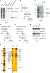
Purification of GCN5, recombinant HAT modules, and endogenous ATAC and SAGA complexes. A, purification of recombinant GCN5. SDS-PAGE analysis of the GCN5 purification procedure showed input and unbound samples (lanes 1 and 2, respectively), the peptide elutions (lanes 4 and 5), and GCN5-bound antibody beads before elution (lane 6). Proteins were visualized by Coomassie Brilliant Blue (CBB) stain. The antibody heavy chains (Ig HC) and light chains (Ig LC) are indicated. MW, molecular mass marker (170, 125, 95, 75, 55, 36, and 24 kDa). B, purification scheme of the recombinant ATAC and SAGA HAT modules (rHAT ATAC and rHAT SAGA). SN, supernatant. Elution, peptide elution with a 1000× excess of peptide against which the indicated antibodies were raised. C, purification of recombinant HAT modules of ATAC and SAGA. SDS-PAGE analysis of the purified complexes following the procedure depicted in B. Proteins were visualized by Coomassie Brilliant Blue stain. Note that the ATAC HAT module contains ADA2a, whereas the SAGA HAT module contains ADA2b. MW, molecular mass marker (as in panel A), and protein identities are indicated on the left and right of the gel, respectively. D and E, schematic representation of the purification of endogenous GCN5-containing ATAC and SAGA complexes from HeLa cell nuclear extracts. SN, supernatant containing the unbound proteins. Elution, peptide elution with a 1000× excess of peptide against which the indicated antibodies were raised. GCN5-ATAC, GCN5-containing ATAC complex; GCN5-SAGA, GCN5-containing SAGA complex. F, Western blot analyses of endogenous ATAC (lane 1) and SAGA (lane 2) complexes following the purification procedures depicted in D and E, respectively. Recombinant GCN5 (lane 3) was also analyzed as the control. Note that the recombinant hGCN5 is FLAG tagged, and thus it migrates slightly slower than the endogenous hGCN5 in ATAC and SAGA. Primary antibodies are indicated on the right. GCN5, ADA3, and SGF29 are common subunits of ATAC and SAGA, whereas ZZZ3 and ADA2b are ATAC- and SAGA-specific subunits, respectively. The GCN5 content of the endogenous complexes was normalized to the amount of recombinant GCN5. G, the subunits of endogenous ATAC (lane 2) and SAGA (lane 3) complexes purified as depicted in D and E, respectively, were visualized by silver nitrate staining. The subunits of the respective complexes are indicated. Molecular mass markers (MW; lane 1) are indicated in kDa. The antibody heavy chains (Ig HC) and light chains (Ig LC) are indicated.
FIGURE 2.
FIGURE 2.
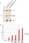
Acetylation activity of the GCN5 enzyme depended on its protein environment. A, normalization of recombinant GCN5 and GCN5-containing HAT complexes. Amounts of purified recombinant ATAC HAT and SAGA HAT modules were normalized to recombinant GCN5 and analyzed by either Coomassie Blue (CB) staining or by WB with two different anti-hGCN5 mouse mAbs 2GC and 4GC. B, purified recombinant GCN5, ATAC HAT, and SAGA HAT modules and endogenous ATAC and SAGA complexes were tested by WB with two different anti-hGCN5 mouse mAbs 2GC and 4GC. C, amounts of purified recombinant GCN5, ATAC HAT, and SAGA HAT modules and endogenous ATAC and SAGA complexes were normalized by WB with the anti-hGCN5 mouse mAbs 2GC. Note that in panels B and C the recombinant hGCN5 is FLAG- and His-tagged alone, or incorporated in the HAT modules (lanes 1, 2, and 4). Thus, it migrates slightly higher than the endogenous non-tagged hGCN5 (lanes 3 and 5). D, acetylation of an H3 tail peptide revealed by a filter-binding assay. H3 tail peptide covering amino acids 5–20 was incubated with radioactive acetyl-coenzyme A and the indicated protein or protein complex preparation. Reactions were carried out in quadruplicate in the presence (H3 amino acids 5–20) or absence of the H3 tail peptide, and acetylation was measured in cpm. Error bars indicate S.D. E, acetylation assay as in D but with an H4 tail peptide encompassing residues 1–19. F, -fold activation of GCN5 activity of the HAT modules or the endogenous ATAC and SAGA complexes compared with the activity of recombinant GCN5 on the H3 and H4 tail peptides used in D and E. Activation was calculated from three biological replicates. Error bars represent S.D.
FIGURE 3.
FIGURE 3.
Acetylation activity of GCN5 was differentially regulated by the subunits ADA2a or ADA2b within recombinant HAT modules. A, Western blot analysis of purified recombinant GCN5 and heterotrimeric and heterotetrameric recombinant HAT modules of ATAC and SAGA. Primary antibodies used to identify subunits are indicated on the right. Amounts of GCN5 within the HAT modules were normalized to recombinant GCN5. B, acetylation of an H3 tail peptide by recombinant GCN5 and heterotrimeric and heterotetrameric HAT modules of ATAC and SAGA. Experimental conditions were as in Fig. 2B. Subunit compositions of the HAT modules tested are indicated.
FIGURE 4.
FIGURE 4.
Subunits of ATAC and SAGA complexes enhanced both the substrate binding and the turnover rate of GCN5-substrate complexes. A, kinetic measurements comparing the activity of GCN5 alone with endogenous purified ATAC and SAGA complexes on different concentration of H3 tail peptide (from amino acid 1–20) using [14C]acetyl CoA in a filter binding assay. Peptide bound radioactivity was quantified in two biological replicates. The activities of the different GCN5-containing preparations are represented in cpm. B, summary of kinetic parameter calculations.
FIGURE 5.
FIGURE 5.
The acetylation activity of GCN5 on recombinant histone assemblies gradually increased with subunit complexity of the HAT modules. A, purification of human H2A/H2B dimers, H3/H4 tetramers, and histone octamers. SDS-PAGE analysis of H2A/H2B dimers and H3/H4 tetramers purified from human HeLa cells and recombinant histone octamers produced in Escherichia coli. Proteins were visualized by Coomassie brilliant blue (CBB) stain. Protein identities are indicated. B, acetylation tests on purified HeLa H2A/H2B dimers with recombinant GCN5, the HAT modules of ATAC and SAGA, and the endogenous ATAC and SAGA complexes. H2A/H2B dimers were incubated with radioactive acetyl-coenzyme A and normalized amounts of GCN5- and GCN5-containing complexes. Histones were resolved by 15% SDS-PAGE and dried, and radioactive signals were detected by radiography using an image intensifier screen. C, acetylation tested were as in B but purified HeLa H3/H4 tetramers were used. D, quantification of acetylation of histones H3 and H4 by GCN5-containing complexes. Quantified signals from two experiments are displayed in arbitrary units with signal deviations indicated as error bars. E, acetylation tests as in B but performed on recombinant histone octamers.
FIGURE 6.
FIGURE 6.
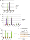
In peptide tail HAT assays the specific acetylation site of GCN5 and the endogenous HAT complexes was H3K14, whereas on full-length histones the acetylation specificity was broader. A, acetylation assays on H3 tail peptides. Histone H3 tail peptides encompassing different regions of the H3 tail (as indicated) were assayed for acetylation by recombinant GCN5 and the purified endogenous ATAC and SAGA complexes as in Fig. 2B. Reactions were carried out in quadruplicate, and acetylation was measured in cpm. B, acetylation assays as in A but using H3 (amino acids 5–20) peptides in which lysine residues Lys-9, Lys-14, and Lys-18 were consecutively substituted with arginines. C, acetylation assays as in A but using the H3 (amino acids 5–20) peptide in which lysine residues Lys-9, Lys-14, and Lys-18 were consecutively substituted with acetylated lysines. D, acetylation tests on recombinant histone octamers with normalized amounts of recombinant GCN5, the HAT modules, and the endogenous complexes (as indicated). Protein preparations were assayed for acetylation activity on recombinant octamers as in Fig. 4 but with cold acetyl-CoA. Histone octamers were resolved by SDS-PAGE and probed for acetylation by Western blot using the indicated antibodies. Successful transfer of histones to the membrane was verified by Ponceau S stain.
FIGURE 7.
FIGURE 7.
GCN5 and the HAT modules of ATAC or SAGA acetylated the same lysine residues on histone octamers. A and B, acetylation reactions were set up with GCN5 and the HAT modules of ATAC or SAGA using recombinant histone octamers. Acetylated lysine residues were identified on histone H3 (A) or H4 (B) by LC-MS/MS analysis using Orbitrap. The numbers of specific acetylated peptide-spectrum matches (PSM) containing the indicated different individual lysine (K) residues or their identified combination (as indicated) on H3 or H4 are represented. Buffer, the reaction in which no enzyme was included (control).

References

    1. Kornberg R. D., and Lorch Y. (1999) Twenty-five years of the nucleosome, fundamental particle of the eukaryote chromosome. Cell 98, 285–294 - PubMed
    1. Kouzarides T. (2007) Chromatin modifications and their function. Cell 128, 693–705 - PubMed
    1. Hong L., Schroth G. P., Matthews H. R., Yau P., and Bradbury E. M. (1993) Studies of the DNA binding properties of histone H4 amino terminus. Thermal denaturation studies reveal that acetylation markedly reduces the binding constant of the H4 “tail” to DNA. J. Biol. Chem. 268, 305–314 - PubMed
    1. Li R., Yang L., Fouts E., and Botchan M. R. (1993) Site-specific DNA-binding proteins important for replication and transcription have multiple activities. Cold Spring Harb. Symp. Quant. Biol. 58, 403–413 - PubMed
    1. Nagy Z., and Tora L. (2007) Distinct GCN5/PCAF-containing complexes function as co-activators and are involved in transcription factor and global histone acetylation. Oncogene 26, 5341–5357 - PubMed

Publication types

MeSH terms