. 2015 Dec 4;290(49):29217-30.
doi: 10.1074/jbc.M115.687731. Epub 2015 Oct 14.

The Inflammasome Adaptor ASC Induces Procaspase-8 Death Effector Domain Filaments

Affiliations

The Inflammasome Adaptor ASC Induces Procaspase-8 Death Effector Domain Filaments

Parimala R Vajjhala et al. J Biol Chem. .

Abstract

Inflammasomes mediate inflammatory and cell death responses to pathogens and cellular stress signals via activation of procaspases-1 and -8. During inflammasome assembly, activated receptors of the NLR or PYHIN family recruit the adaptor protein ASC and initiate polymerization of its pyrin domain (PYD) into filaments. We show that ASC filaments in turn nucleate procaspase-8 death effector domain (DED) filaments in vitro and in vivo. Interaction between ASC PYD and procaspase-8 tandem DEDs optimally required both DEDs and represents an unusual heterotypic interaction between domains of the death fold superfamily. Analysis of ASC PYD mutants showed that interaction surfaces that mediate procaspase-8 interaction overlap with those required for ASC self-association and interaction with the PYDs of inflammasome initiators. Our data indicate that multiple types of death fold domain filaments form at inflammasomes and that PYD/DED and homotypic PYD interaction modes are similar. Interestingly, we observed condensation of procaspase-8 filaments containing the catalytic domain, suggesting that procaspase-8 interactions within and/or between filaments may be involved in caspase-8 activation. Procaspase-8 filaments may also be relevant to apoptosis induced by death receptors.

Keywords: apoptosis; caspase; cell death; death domain; death domain filaments; inflammasome; pattern recognition receptor (PRR).


Figures

FIGURE 1.
FIGURE 1.
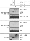
Interaction of procaspase-8 DEDs with ASC PYD. Procaspase-8 DED1, DED2, or DED1-DED2 was in vitro translated with [35S]methionine and incubated with purified bead-bound GST-ASC PYD or with GST alone. Volumes of in vitro translated protein used are indicated, and an amount representing 10% of the input used for binding studies is shown. Bead-bound proteins were resolved by SDS-PAGE. GST and GST-ASC PYD were detected by Coomassie stain, whereas 35S-labeled proteins were detected by phosphorimaging. The data are representative of two independent experiments.
FIGURE 2.
FIGURE 2.
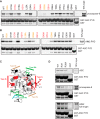
Identification of residues on ASC PYD that are important for interaction with procaspase-8 and AIM2. A and B, wild-type or mutant GST-ASC PYD or GST alone were purified on glutathione-agarose beads and then used for binding assays with in vitro translated [35S]methionine-labeled procaspase-8 (A) or AIM2 PYD (B). Bead-bound proteins were resolved by SDS-PAGE. GST-ASC PYD was detected by Coomassie stain, whereas 35S-labeled proteins were detected by phosphorimaging. The minor band below GST-ASC PYD appears to be a truncated product. An amount representing 10% of the input of in vitro translated protein used for binding studies is shown. Data are representative of three independent experiments. C, model of ASC PYD (one monomer from the EM-derived structure of the ASC PYD filament; Protein Data Bank entry 3J63) (20) showing the location of residues that affect interactions to the type I, II, and III interaction surfaces. The type I, II, and III interaction surfaces are indicated with bars colored red, light brown, and green, respectively. Residues that reduced interactions when mutated are colored to match the surface they belong to. Arg-38, which is proximal to the type Ib interaction surface, is shown in blue. Residues in black increased binding of procaspase-8. D, binding of in vitro translated [35S]methionine-labeled ASC, procaspase-8, and AIM2 to WT or mutant GST-ASC PYD or GST alone. Full-length AIM2 was used in D, whereas the PYD alone was used in B.
FIGURE 3.
FIGURE 3.
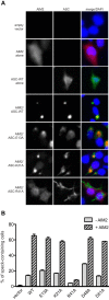
AIM2 induces and co-localizes with speck-like structures formed by ASC mutants but does not induce or co-localize with filaments formed by the R41A mutant. HEK293 cells were either transfected with empty vector alone or co-transfected with plasmids expressing WT or mutant ASC and either empty vector or a plasmid expressing AIM2. A, cells were immunostained with anti-ASC (green) and anti-AIM2 (red), and the nuclei were counterstained with DAPI (blue). The brightness of cells expressing ASC-WT alone, AIM2 alone, or ASC-R41A mutant with AIM2 was increased to enhance visibility. B, cells were immunostained with anti-ASC and subjected to flow cytometry on an Accuri C6. The proportion of cells with the diffuse and “speck” form of ASC can be determined from the fluorescence peak height/peak area ratio. The percentage of ASC speck-containing cells (high peak height/area ratio) determined for a population falling within the same window of moderate ASC expression under each transfection condition is shown. Error bars, range of duplicates in the same experiment. Results were similar in two independent experiments.
FIGURE 4.
FIGURE 4.
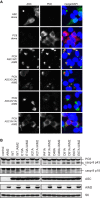
Procaspase-8 is recruited to the AIM2-induced speck-like structures formed by ASC mutants and is weakly activated in cells expressing these mutants but not in cells expressing the R41A mutant that forms filaments. A, HEK293 cells were transfected with plasmids expressing WT ASC alone, V5-tagged procaspase-8 C360S inactive mutant (PC8) alone, or AIM2 with PC8 and either WT or mutant ASC. Cells were immunostained with anti-ASC (green) or anti-V5 (red), and the nuclei were counterstained with DAPI (blue). The brightness of cells expressing ASC-WT alone or procaspase-8 alone or ASC-R41A mutant with AIM2 and procaspase-8 was increased to improve visibility. B, plasmids expressing WT or mutant ASC were co-transfected either with a plasmid expressing AIM2 or with empty vector into HEK293 cells. After 24 h, processing of endogenous procaspase-8 to the p43 and p18 forms was detected by Western blotting. A band below the p43 form marked with an asterisk is constitutively present and has not been identified. The level of S6 ribosomal protein was used as a loading control. The data are representative of two independent experiments.
FIGURE 5.
FIGURE 5.
Procaspase-8 DEDs can substitute for AIM2 PYD to induce ASC specks with lower efficiency. A, procaspase-8 DEDs or PYDs of ASC and NLRP3 were fused to the AIM2 HIN domain and mCherry-tagged. B, HEK293 cells were transfected with plasmids expressing ASC-GFP and increasing amounts of plasmids expressing either mCherry-tagged AIM2 or one of the mCherry-tagged AIM2 HIN fusion proteins. At 16 h post-transfection, cells were analyzed by flow cytometry to quantify the number of cells containing GFP-ASC specks. For each sample, the percentage of speck-containing cells in the same expression window of ASC-GFP expression is shown and is plotted against the expression levels of the mCherry-tagged proteins, which were also determined by flow cytometry and are expressed as mean fluorescence intensity (MFI). Data from two replicates are plotted separately. The data are representative of two independent experiments. Notably, some mCherry-tagged proteins were expressed at higher levels with equivalent amounts of plasmid transfected.
FIGURE 6.
FIGURE 6.
ASC specks induced by PC8 DED fused to AIM2 HIN are morphologically similar to those induced by AIM2. HEK293 cells were co-transfected with plasmids expressing ASC and either mCherry-tagged AIM2 or one of the mCherry-tagged AIM2 HIN fusion proteins (described in the legend to Fig. 5) or untagged AIM2. Cells were immunostained with anti-ASC (green), and the nuclei were counterstained with DAPI (blue).
FIGURE 7.
FIGURE 7.
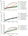
Full-length ASC or GFP-ASC PYD oligomers promote formation of procaspase-8 DED filaments. Fluorescence polarization was used to assess oligomerization of TAMRA-labeled procaspase-8 tandem DEDs with the Y8A mutation and C-terminal SUMO tag (His6-MBP-procaspase-8 DEDs-Y8A-SUMO). Oligomerization was initiated by the addition of TEV protease to cleave off a His6-MBP-solubilizing tag present on both the DED construct and ASC constructs that prevented spontaneous association. A, 2.4-μm TAMRA-labeled His6-MBP-procaspase-8 DEDs-Y8A-SUMO was incubated with full-length (FL) ASC, ASC PYD, or ASC CARD at various ratios. B, 4 μm TAMRA-labeled His6-MBP-procaspase-8 DEDs-Y8A-SUMO was incubated with full-length ASC with mutations in the PYD. C, 2.2 μm TAMRA-labeled His6-MBP-procaspase-8 DEDs-Y8A-SUMO was incubated with full-length ASC or preformed GFP-ASC PYD aggregates at various ratios.
FIGURE 8.
FIGURE 8.
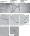
Association of ASC with one terminus of procaspase-8 DED filaments. Electron micrographs of procaspase-8 DEDs-Y8A-biotin·ASC binary complexes. Monomeric biotinylated His6-MBP-procaspase-8 DEDs-Y8A was incubated in the absence or presence of substoichiometric amounts of monomeric His6-MBP-ASC (1:0.1 and 1:0.01). TEV protease was added to cleave the solubilizing His6-MBP tags. After co-polymerization, the samples were analyzed by negative stain EM analysis or in the bottom two rows stained with either streptavidin-6-nm gold or anti-ASC-6-nm gold.
FIGURE 9.
FIGURE 9.
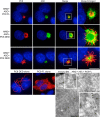
Procaspase-8 DED filaments are initiated focally by AIM2 inflammasomes. HEK293 cells were transfected with plasmids expressing either Myc-tagged full-length procaspase-8 (PC8 FL) or V5-tagged procaspase-8 DEDs (PC8 DEDs) either alone or with plasmids expressing ASC and AIM2. Cells were immunostained with anti-ASC (green) and either anti-Myc (red) or anti-V5 (red), and the nuclei were counterstained with DAPI (blue). Images were acquired using a Personal DeltaVision Olympus IX71 inverted wide field deconvolution microscope. Two different images of inflammasomes with procaspase-8 DEDs are shown, with the bottom panel obtained by maximum projection image processing. Scale bars, 10 and 5 μm on normal and enlarged panels respectively. In the immuno-EM image in the bottom right, HEK293 cells were co-transfected with plasmids expressing ASC, AIM2, and HA-tagged procaspase-8. Ultrathin cryosections were immunogold-labeled with an anti-HA antibody followed by 10-nm protein A-gold. Sections were viewed on a JEOL 1011 electron microscope. A section of a speck is shown, and regions at the periphery of the speck show labeled filamentous projections (arrows). Scale bar (EM image), 500 nm. *, a central region of the speck. Arrowheads, 10-nm gold labeling along procaspase-8 filaments.
FIGURE 10.
FIGURE 10.
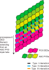
Model to illustrate interaction of ASC PYDs and procaspase-8 DEDs. Shown is a flattened view of a proposed ASC PYD·procaspase-8 DED filament to show the different types of death domain interactions. ASC PYDs belonging to each strand of the triple helix are colored yellow, light green, or dark green, whereas procaspase-8 tandem DEDs are colored pink. Our data indicate that procaspase-8 DEDs are recruited to ASC PYD via the same type I, II, and III interactions that mediate assembly of the ASC PYD triple helix, and we hypothesize that the same interactions mediate assembly of the DED filament. Type I, II, and III interactions are indicated on domains at/near the interface as thick black, blue, and orange lines, respectively. The model shows how both DEDs of procaspase-8 can interact with ASC PYDs at the interface between the filaments.

References

    1. Lamkanfi M., and Dixit V. M. (2014) Mechanisms and functions of inflammasomes. Cell 157, 1013–1022 - PubMed
    1. Aachoui Y., Sagulenko V., Miao E. A., and Stacey K. J. (2013) Inflammasome-mediated pyroptotic and apoptotic cell death, and defense against infection. Curr. Opin. Microbiol. 16, 319–326 - PMC - PubMed
    1. Guo H., Callaway J. B., and Ting J. P. (2015) Inflammasomes: mechanism of action, role in disease, and therapeutics. Nat. Med. 21, 677–687 - PMC - PubMed
    1. Li P., Allen H., Banerjee S., Franklin S., Herzog L., Johnston C., McDowell J., Paskind M., Rodman L., and Salfeld J. (1995) Mice deficient in IL-1 β-converting enzyme are defective in production of mature IL-1 β and resistant to endotoxic shock. Cell 80, 401–411 - PubMed
    1. Gu Y., Kuida K., Tsutsui H., Ku G., Hsiao K., Fleming M. A., Hayashi N., Higashino K., Okamura H., Nakanishi K., Kurimoto M., Tanimoto T., Flavell R. A., Sato V., Harding M. W., Livingston D. J., and Su M. S. (1997) Activation of interferon-γ inducing factor mediated by interleukin-1β converting enzyme. Science 275, 206–209 - PubMed

Publication types

Associated data