Review
. 2016 Jan;69(1):1-5.
doi: 10.1136/jclinpath-2015-203258. Epub 2015 Oct 14.

Traditional serrated adenomas of the upper digestive tract

Review

Traditional serrated adenomas of the upper digestive tract

C A Rubio. J Clin Pathol. 2016 Jan.

Abstract

For many years, it was generally accepted that the vast majority of the colorectal carcinomas (CRCs) evolved from conventional adenomas, via the adenoma-carcinoma sequence. More recently, serrated colorectal polyps (hyperplastic polyps, sessile serrated polyps and traditional serrated adenomas (TSAs)) have emerged as an alternative pathway of colorectal carcinogenesis. It has been estimated that about 30% of the CRC progress via the serrated pathway. Recently, TSAs were also detected in the upper digestive tract. In this work, we review the literature on TSA in the oesophagus, the stomach, the duodenum, the pancreatic main duct and the gallbladder. The review indicated that 53.4% (n=39) out of the 73 TSA of the upper digestive tract now in record showed a simultaneously growing invasive carcinoma. As a corollary, TSAs of the upper digestive tract are aggressive adenomas that should be radically excised, either endoscopically or surgically, to rule out the possibility of a synchronously growing invasive adenocarcinoma or to prevent cancer progression. The present findings substantiate a TSA pathway of carcinogenesis in the upper digestive tract.

Keywords: GALL BLADDER CANCER; GASTRIC CANCER; OESOPHAGUS; PANCREATIC CANCER.


Figures

Figure 1
Figure 1
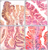
Traditional serrated adenoma (TSA) of Barrett’s oesophagus. (A) Low-power view of the TSA (H&E, ×4). (B) Closer view showing unlocked serrated configurations with low-grade dysplasia (H&E, ×10). (C) Basal aspect of the TSA with high-grade dysplasia (H&E ×20). (D) Detail from another area of the TSA of the oesophagus showing a tripolar mitosis (periodic acid Schiff stain, ×40).
Figure 2
Figure 2
Traditional serrated adenoma (TSA) of the stomach. (A) Section from the resected specimen (H&E, ×1). (B) ‘Christmas-tree’-like serrated configuration (H&E, ×10). (C) Another area with ‘Christmas-tree’-like serrated configuration with high-grade dysplasia (H&E, ×10). (D) Detail from TSA showing unlocked serrated configurations with high-grade dysplasia (H&E, ×20). (E) ‘Christmas-tree’-like serrated configuration showing cell proliferation (Ki67, batch MIB1, ×10). (F) Invasive carcinoma arising in a gastric TSA (periodic acid Schiff stain (PAS), ×4).
Figure 3
Figure 3
Traditional serrated adenoma (TSA) of the duodenum. (A) Detail of the TSA of the duodenum to show unlocked serrated crypts with high-grade dysplasia (H&E, ×10). (B) Another area of the TSA showing unlocked serrated configurations (H&E, ×20).
Figure 4
Figure 4
Traditional serrated adenoma (TSA) of the main pancreatic duct. (A) TSA of the main pancreatic duct (section from the resected specimen (H&E, ×1). (B) TSA showing unlocked serrated configurations lined with low-grade dysplasia (H&E, ×10). (C) Another area of the TSA showing eosinophilic cytoplasm (H&E, ×10. (D) Detail from the TSA of the main pancreatic duct, showing unlocked serrated configurations lined with low-grade dysplasia. Note the eosinophic cytoplasm (H&E, ×20). (E) Invasive carcinoma, arising in a TSA of the main pancreatic duct (H&E, 20×).
Figure 5
Figure 5
Traditional serrated adenoma (TSA) of the gall bladder. (A) TSA of the gallbladder (section from the resected specimen (H&E, ×1). (B) Low-power view of the TSA showing unlocked serrated configurations (H&E, ×4). (C) Closer view of the TSA showing unlocked serrated configurations with high-grade dysplasia (H&E, ×20). (D) Detail from another area of the TSA showing unlocked serrated configurations (H&E, ×20).

References

    1. O'Brien MJ, Gibbons D. The adenoma–carcinoma sequence in colorectal neoplasia. Surg Oncol Clin N Am 1996;5:513–22. - PubMed
    1. Longacre TA, Fenoglio-Preiser CM. Mixed hyperplastic adenomatous polyps/serrated adenomas. A distinct form of colorectal neoplasia. Am J Surg Pathol 1990;14:524–37. 10.1097/00000478-199006000-00003 - DOI - PubMed
    1. Jass JR. Serrated route to colorectal cancer: back street or super highway?. J Pathol 2001;193:283–5. 10.1002/1096-9896(200103)193:3<283::AID-PATH799>3.0.CO;2-9 - DOI - PubMed
    1. Torlakovic E, Snover DC. Serrated adenomatous polyposis in humans. Gastroenterology 1996;110:748–55. 10.1053/gast.1996.v110.pm8608884 - DOI - PubMed
    1. Snover DC, Jass JR, Fenoglio-Preiser C, et al. . Serrated polyps of the large intestine: a morphologic and molecular review of an evolving concept. Am J Surg Pathol 2005;124:380–91. - PubMed

MeSH terms

Substances