. 2015 Oct 14;90(1):117-28.
doi: 10.1128/JVI.01756-15. Print 2016 Jan 1.

Comparative Efficacy of Monoclonal Antibodies That Bind to Different Epitopes of the 2009 Pandemic H1N1 Influenza Virus Neuraminidase

Affiliations

Comparative Efficacy of Monoclonal Antibodies That Bind to Different Epitopes of the 2009 Pandemic H1N1 Influenza Virus Neuraminidase

Lianlian Jiang et al. J Virol. .

Abstract

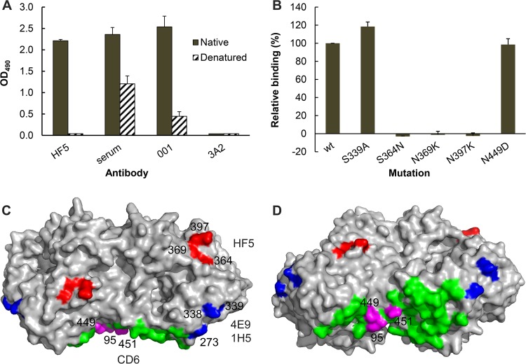
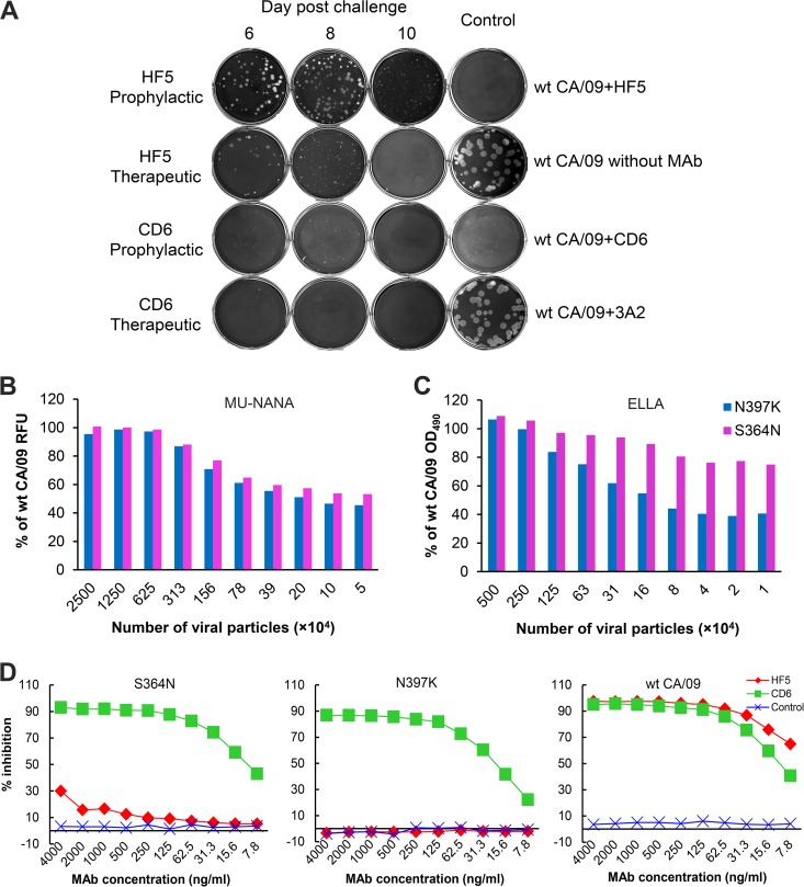
Antibodies against the neuraminidase (NA) of influenza virus correlate with resistance against disease, but the effectiveness of antibodies against different NA epitopes has not been compared. In the present study, we evaluated the in vitro and in vivo efficacies of four monoclonal antibodies (MAbs): HF5 and CD6, which are specific to two different epitopes in the NA of 2009 pandemic H1N1 (pH1N1) virus, and 4E9 and 1H5, which are specific to a conserved epitope in the NA of both H1N1 and H5N1 viruses. In the in vitro assays, HF5 and CD6 inhibited virus spread and growth more effectively than 4E9 and 1H5, with HF5 being the most effective inhibitor. When administered prophylactically at 5 mg/kg of body weight, HF5 and CD6 protected ~90 to 100% of DBA/2 mice against lethal wild-type pH1N1 virus challenge; however, at a lower dose (1 mg/kg), HF5 protected ~90% of mice, whereas CD6 protected only 25% of mice. 4E9 and 1H5 were less effective than HF5 and CD6, as indicated by the partial protection achieved even at doses as high as 15 mg/kg. When administered therapeutically, HF5 protected a greater proportion of mice against lethal pH1N1 challenge than CD6. However, HF5 quickly selected pH1N1 virus escape mutants in both prophylactic and therapeutic treatments, while CD6 did not. Our findings confirm the important role of NA-specific antibodies in immunity to influenza virus and provide insight into the properties of NA antibodies that may serve as good candidates for therapeutics against influenza.

Importance: Neuraminidase (NA) is one of the major surface proteins of influenza virus, serving as an important target for antivirals and therapeutic antibodies. The impact of NA-specific antibodies on NA activity and virus replication is likely to depend on where the antibody binds. Using in vitro assays and the mouse model, we compared the inhibitory/protective efficacy of four mouse monoclonal antibodies (MAbs) that bind to different sites within the 2009 pandemic H1N1 (pH1N1) virus NA. The ability of each MAb to protect mice against lethal pH1N1 infection corresponded to its ability to inhibit NA activity in vitro; however, the MAb that was the most effective inhibitor of NA activity selected pH1N1 escape variants in vivo. One of the tested MAbs, which binds to a conserved region in the NA of pH1N1 virus, inhibited NA activity but did not result in escape variants, highlighting its suitability for development as a therapeutic agent.


Figures

FIG 1
FIG 1
MAbs HF5, CD6, 4E9, and 1H5 bind different epitopes in the NA of pH1N1 virus. (A) MAb HF5 binds to native but not denatured CA/09-X179A. Data from ELISA show the binding of HF5 to purified whole virus (solid bars) but not CA/09-X179A that has been dissociated and heat denatured (hatched bars). Assay controls are labeled serum (mouse serum against CA/09 virus), 001 (rabbit MAb against CA/09 NA), and 3A2 (MAb specific to the NA of BR/07-like H1N1 virus). Shown are mean OD490 values from two independent assays run in duplicate wells; standard deviations (SDs) are shown with error bars. (B) Binding of MAb HF5 to a panel of mutant CA/09 NAs expressed on 293T cells. The binding was measured by a cell-based ELISA, with the signals being normalized to those obtained with mouse serum against CA/09, which are therefore expressed as relative binding. Shown are the data from two independent assays run in duplicate wells. (C) A model that depicts the residues in an NA dimer (Protein Data Bank accession number 3NSS) that are critical for the binding of MAbs HF5 (red), CD6 (magenta), as well as 4E9 and 1H5 (blue). The remaining 27 residues within the CD6 epitope (Protein Data Bank accession number 4QNP) are highlighted in green. The image was generated with PyMOL software (Delano Scientific). (D) The same model in panel C shown from a different view.
FIG 2
FIG 2
Different functional properties of MAbs HF5, CD6, 4E9, and 1H5 in vitro. (A) Inhibition of CA/09 NA activity by each MAb measured by ELLA using H6N1CA/09 and wt CA/09 viruses. (B) CA/09 virus plaques formed in the presence of various concentrations of MAbs HF5, CD6, 4E9, and 1H5 (0.1 to 10 μg/ml) or the control MAb, 3A2 (10 μg/ml), in the overlay agar. (C) Diameters of the CA/09 virus plaques shown in panel B. Plaques from each treatment were randomly chosen, and the diameters were measured and compared to the diameter of the control. Shown are mean diameters (n = 20); SDs are shown with error bars. Diameters that were significantly different from those of the control (P < 0.05) are indicated by lines and asterisks. (D) Growth kinetics of CA/09 in MDCK cells in the presence of MAb HF5, CD6, 4E9, 1H5, or 3A2 (1, 5, or 10 μg/ml). Cells growing in 12-well plates were infected with CA/09 at an MOI of 0.001, and the viral titers in the supernatant at the indicated time points were measured by plaque assay. Shown are the average titers of duplicate wells; SDs are shown with error bars. *, P < 0.05. The asterisks above the green line or below the red line indicate a significant difference between the viral titers generated in the presence of the tested MAbs (CD6 or HF5) and the control MAb 3A2, asterisks between the green and red lines indicate significant differences between the viral titers generated in the presence of MAbs CD6 and HF5, and asterisks below the blue line in the right panel indicate significant differences between groups receiving 4E9 and 1H5 and the control group (which received 3A2).
FIG 3
FIG 3
Prophylactic efficacy of MAbs HF5, CD6, 4E9, and 1H5 against lethal pH1N1 virus challenge in mice. DBA/2 mice (n = 14 or 20 per group) were treated i.p. with HF5, CD6, 4E9, or 1H5 at the indicated doses, followed by challenge i.n. with 10 MLD50 CA/09 12 h later. MAb 3A2, which is specific to the NA of seasonal H1N1 virus BR/07, was used as a negative control. Survival (A, D, G, J, and M) and weight loss (B, E, H, K, and N) (n = 8 per group) were monitored for up to 14 days. Lungs were collected on different days, and viral titers (C, F, I, L, and O) were determined by titration in MDCK cells. Titers are expressed as the log10 50% tissue culture infective dose (TCID50) per milliliter (n = 3); SDs are shown with error bars. The dotted lines denote the detection limit of 2.2 log10 TCID50/ml. A titer of 0.7 log10 TCID50/ml was arbitrarily assigned to samples with titers below the detection limit. The viral titers of the control groups at day 8 p.c. were used for analysis of the titers measured on days 10 and 12 p.c. because none of the mice in the control groups survived at these time points. Significant differences between the titers measured in each group and the MAb 3A2-treated control groups are shown (*, P < 0.05).
FIG 4
FIG 4
Therapeutic efficacy of MAbs HF5, CD6, and 1H5 against lethal pH1N1 virus challenge in mice. DBA/2 mice (n = 14 or 20 per group) were infected i.n. with 10 MLD50 of CA/09, followed by injection i.p. with a single dose of each MAb on day 1 p.c., two doses on days 1 and 5 p.c., or three doses (5 mg/kg per dose) on days 1, 3, and 5 p.c (shown as treatments 1, 2, and 3, respectively). MAb 3A2, which is specific to the seasonal H1N1 BR/07 virus, was used as a negative control. Survival (A, D, and G) and weight loss (B, E, and H) (n = 8 per group) were monitored for up to 14 days. Lungs were collected on different days, and viral titers (C, F, and I) were determined by titration in MDCK cells. Titers are expressed as the log10 50% tissue culture infective dose (TCID50) per milliliter; SDs are shown with error bars. The dotted line denotes the detection limit of 2.2 log10 TCID50/ml. A titer of 0.7 log10 TCID50/ml was assigned to samples with titers below the detection limit. The viral titers of the control groups at day 8 p.c. were used for analysis of the titers measured on days 10 and 12 p.c. because none of the mice in the control groups survived at these time points. Significant differences between titers measured in each group and the MAb 3A2-treated control groups are shown (*, P < 0.05).
FIG 5
FIG 5
MAb HF5 selected escape mutants of pH1N1 virus in mice. (A) Lung samples collected on days 6, 8, and 10 p.c. from mice in the prophylactic study (5 mg/kg of MAb before CA/09 challenge) and the therapeutic study (three doses of 5 mg/kg MAb after CA/09 challenge) were homogenized, and the supernatant was tested in a plaque assay, in which MAb HF5 or CD6 was supplemented in the agar overlay. (B and C) Enzyme activity of CA/09 escape variants selected with HF5. The NA activities of CA/09 variants with the S364N or N397K mutations in the NA were compared with the NA activity of the wt parent virus in an MU-NANA assay, which uses a small substrate, MU-NANA (B), and ELLA, which uses a large substrate, fetuin (C). The assay mixtures were adjusted so that they contained the same number of viral particles per milliliter, and the three viruses were tested in duplicate. The relative fluorescence units (RFUs) and OD values generated with the mutants are expressed as a percentage of the signals obtained with wt CA/09. (D) CA/09 mutants with the S364N mutation (left) or the N397K mutation (middle) in the NA were examined for the sensitivities of their NAs to MAbs HF5, CD6, and 3A2 in an ELLA. (Right) Results obtained with wt CA/09 NA were included for comparison.

References

    1. Kilbourne ED, Johansson BE, Grajower B. 1990. Independent and disparate evolution in nature of influenza A virus hemagglutinin and neuraminidase glycoproteins. Proc Natl Acad Sci U S A 87:786–790. doi:10.1073/pnas.87.2.786. - DOI - PMC - PubMed
    1. Air GM. 2012. Influenza neuraminidase. Influenza Other Respir Viruses 6:245–256. doi:10.1111/j.1750-2659.2011.00304.x. - DOI - PMC - PubMed
    1. Matrosovich MN, Matrosovich TY, Gray T, Roberts NA, Klenk HD. 2004. Neuraminidase is important for the initiation of influenza virus infection in human airway epithelium. J Virol 78:12665–12667. doi:10.1128/JVI.78.22.12665-12667.2004. - DOI - PMC - PubMed
    1. Wagner R, Wolff T, Herwig A, Pleschka S, Klenk HD. 2000. Interdependence of hemagglutinin glycosylation and neuraminidase as regulators of influenza virus growth: a study by reverse genetics. J Virol 74:6316–6323. doi:10.1128/JVI.74.14.6316-6323.2000. - DOI - PMC - PubMed
    1. Palese P, Compans RW. 1976. Inhibition of influenza virus replication in tissue culture by 2-deoxy-2,3-dehydro-N-trifluoroacetylneuraminic acid (FANA): mechanism of action. J Gen Virol 33:159–163. doi:10.1099/0022-1317-33-1-159. - DOI - PubMed

Publication types

MeSH terms

LinkOut - more resources