. 2015 Oct 14;18(4):463-70.
doi: 10.1016/j.chom.2015.09.010.

Blood-Derived CD4 T Cells Naturally Resist Pyroptosis during Abortive HIV-1 Infection

Affiliations

Blood-Derived CD4 T Cells Naturally Resist Pyroptosis during Abortive HIV-1 Infection

Isa Muñoz-Arias et al. Cell Host Microbe. .

Abstract

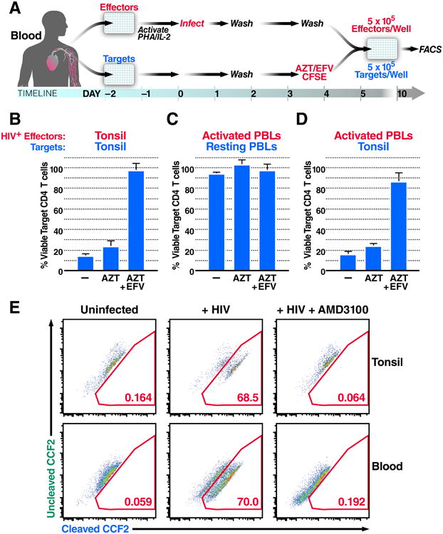
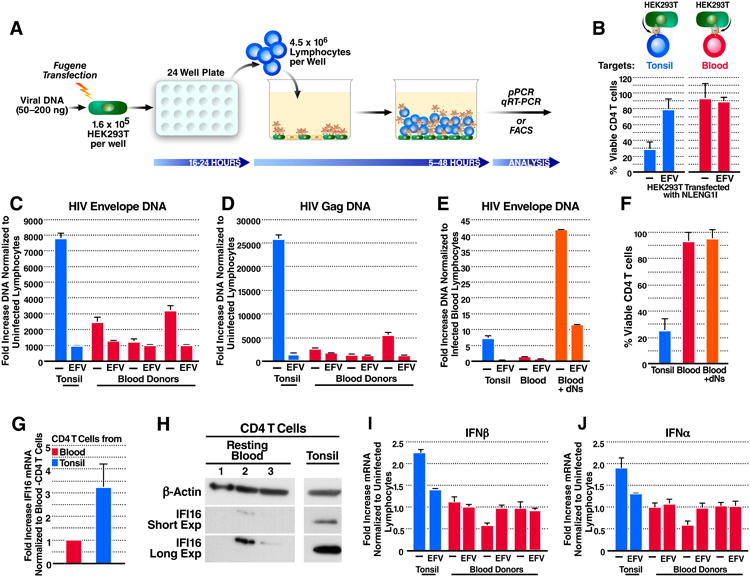
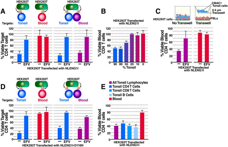
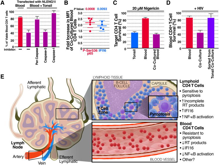
Progression to AIDS is driven by CD4 T cell depletion, mostly involving pyroptosis elicited by abortive HIV infection of CD4 T cells in lymphoid tissues. Inefficient reverse transcription in these cells leads to cytoplasmic accumulation of viral DNAs that are detected by the DNA sensor IFI16, resulting in inflammasome assembly, caspase-1 activation, and pyroptosis. Unexpectedly, we found that peripheral blood-derived CD4 T cells naturally resist pyroptosis. This resistance is partly due to their deeper resting state, resulting in fewer HIV-1 reverse transcripts and lower IFI16 expression. However, when co-cultured with lymphoid-derived cells, blood-derived CD4 T cells become sensitized to pyroptosis, likely recapitulating interactions occurring within lymphoid tissues. Sensitization correlates with higher levels of activated NF-κB, IFI16 expression, and reverse transcription. Blood-derived lymphocytes purified from co-cultures lose sensitivity to pyroptosis. These differences highlight how the lymphoid tissue microenvironment encountered by trafficking CD4 T lymphocytes dynamically shapes their biological response to HIV.


Figures

Figure 1
Figure 1. Blood-Derived CD4 T Cells Are Naturally Resistant to HIV-Mediated Depletion
(A) The HLAC system. Uninfected cells were labeled with CFSE (target cells) and treated with medium, azidothymidine (AZT), or AZT and efavirenz (EFV), and then co-cultured with NL4-3 productively infected (effector) cells for 5 days. Cells were harvested and analyzed by flow cytometry. (B) Percent viable target tonsil CD4 T cells co-cultured with infected tonsil cells. (C) Percent viable target blood CD4 T cells co-cultured with infected PBLs. (D) Percent viable target tonsil CD4 T cells co-cultured with infected PBLs. (E)Virion based fusion assays were performed with BLAM-Vpr-NL4-3-infected tonsil lymphocytes or PBLs. Cells were then loaded with the CCF2-AM dye. Gated populations represent the percentage of fused CD4 T cells scoring positive for BLAM-dependent CCF2-AM cleavage. Data presented in B-D reflect cumulative results from three experiments; data in E are representative of a single experiment performed three times with similar results. Error bars, SEM. See also Figure S1.
Figure 2
Figure 2. Blood-Derived CD4 T Cells Accumulate Fewer Reverse Transcripts and Express Less IFI16 DNA Sensor than Tonsil CD4 T Cells
(A) The HEK293T overlay culture system. Lymphocytes were overlaid on NLENG1I-transfected HEK293T cells for 5-48 h. Samples were then immunostained and analysed by flow cytometry, or lysed for DNA or mRNA purification and analyzed by qPCR or RT-qPCR. (B) Percentages of viable tonsil- or blood-derived CD4 T cells 48 h after overlay. (C and D) Fold-increase in HIV DNA versus the corresponding uninfected lymphocytes 5 h after overlay. (C) Envelope or (D) Gag HIV DNA. (E) Virus-producing HEK293T cells were overlaid with dNs-treated PBLs for 5 h. Shown is the fold-increase in HIV Envelope DNA versus the infected untreated PBLs. (F) Percentages of viable CD4 T cells 48 h post overlay on virus-producing HEK293T. (G) Fold-increase IFI16 mRNA in purified CD4 T cells from three tonsil versus three blood donors. (H) Immuno-blot of IFI16 expression in resting blood- or tonsil-purified CD4 T cells. IFI16 expression varied among different cellular compartments (25-99% less IFI16 in blood than tonsil CD4 T cells). Tonsil- and blood-purified CD4 T samples were analyzed on the same gel exposed for the same length of time, but are shown separately for graphical clarity. (I and J) Fold increase type I interferon mRNA versus corresponding uninfected lymphocytes 24 h post overlay on virus-producing HEK293T cells (I) interferon-β or (J) interferon-α mRNA. Data shown in B and F-G represent cumulative results from at least three experiments, while representative results from a single experiment repeated three times with similar results are shown in C-E and H-J. Error bars, SEM.
Figure 3
Figure 3. Blood-Derived CD4 T Cells Co-Cultured with Lymphoid Tissue-Derived Cells Are Susceptible to HIV-Mediated Depletion
(A) Percent viable target CD4 T cells 48 h post overlay on pNLENG1I-transfected HEK293T cells. The data presented for isolated cultures of tonsil or blood cells are the same as presented in Figure 2B, which corresponded to a portion of the larger experiment shown here. (See also Figure S2). (B) Percent viable blood CD4 T cells 48 h post co-culture with different percentages of CMAC+ tonsil cells and overlay on virus producing HEK293T cells. (C) Percent viable blood CD4 T cells 48 h post overlay on virus-producing HEK293T in direct contact (left panel) with or separated by a transwell insert (right panel) from CMAC+ tonsil cells. (D) Percent viable target-CD4 T cells 48 h post overlay on D116N-NLENG1I-transfected HEK293T. (E) Percent viable blood CD4 T cells 48 h post co-culture with enriched CMAC+ tonsil-derived-B cells, -CD8 T cells, or -CD4 T cells and overlay on virus-producing HEK293T cells. Data shown in A-E represent cumulative results from at least three experiments. Error bars, SEM. See also Figure S2.
Figure 4
Figure 4. Co-culture with Lymphoid Tissue-Derived Cells Sensitizes Blood CD4 T Cells to Pyroptosis Induced by Multiple Inflammasomes
(A) Co-cultured PBLs were treated with media, or inhibitors of caspase-1, caspase-3, or a pan-caspase inhibitor and then overlaid on NLENG1I-transfected HEK293T for 48 h. Of note, the caspase-1 inhibitor also weakly inhibits caspase-4 and -5. Shown is the percentage of viable blood-derived CD4 T cells in the presence of these different inhibitors. (B) PBLs were co-cultured with CMAC+ tonsil cells for 48 h and stained intracellularly for P-Ser536 RelA or IFI16. Shown is the fold increase median fluorescence intensity (MFI) for each protein in co-cultured PBLs versus PBLs cultured alone. (C) Percent viable CD4 T cells 6 h post nigericin treatment (20 μM). (D) PBLs cultured alone, co-cultured with tonsil cells, or sorted from tonsil co-cultures were overlaid on pNLENG1I-transfected HEK293T cells for 48. Shown is the percentage of viable blood CD4 T cells. Cumulative results from (A, C-D) three, or (B) four experiments are presented. Error bars, SEM. See also Figure S3. (E) Blood-trafficking CD4 T cells are resistant to HIV mediated pyroptosis due at least in part to less reverse transcription and IFI16 expression. When these cells return to the lymphoid tissues, cell-cell interactions sensitize them to this mechanism of HIV induced depletion.

References

    1. Berg RK, Rahbek SH, Kofod-Olsen E, Holm CK, Melchjorsen J, Jensen DG, Hansen AL, Jorgensen LB, Ostergaard L, Tolstrup M, et al. T cells detect intracellular DNA but fail to induce type I IFN responses: implications for restriction of HIV replication. PLoS One. 2014;9:e84513. - PMC - PubMed
    1. Brenchley JM, Schacker TW, Ruff LE, Price DA, Taylor JH, Beilman GJ, Nguyen PL, Khoruts A, Larson M, Haase AT, et al. CD4+ T cell depletion during all stages of HIV disease occurs predominantly in the gastrointestinal tract. J Exp Med. 2004;200:749–759. - PMC - PubMed
    1. Cavrois M, De Noronha C, Greene W. A sensitive and specific enzyme-based assay detecting HIV-1 virion fusion in primary T lymphocytes. Nat Biotechnol. 2002;20:1151–1154. - PubMed
    1. Chen LF, Greene WC. Shaping the nuclear action of NF-kappaB. Nat Rev Mol Cell Biol. 2004;5:392–401. - PubMed
    1. Cyster JG. Chemokines, sphingosine-1-phosphate, and cell migration in secondary lymphoid organs. Annu Rev Immunol. 2005;23:127–159. - PubMed

Publication types