. 2015 Oct 15;10(10):e0140445.
doi: 10.1371/journal.pone.0140445. eCollection 2015.

Small RNA Sequencing Uncovers New miRNAs and moRNAs Differentially Expressed in Normal and Primary Myelofibrosis CD34+ Cells

Affiliations

Small RNA Sequencing Uncovers New miRNAs and moRNAs Differentially Expressed in Normal and Primary Myelofibrosis CD34+ Cells

Paola Guglielmelli et al. PLoS One. .

Abstract

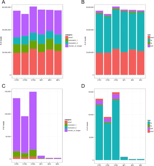
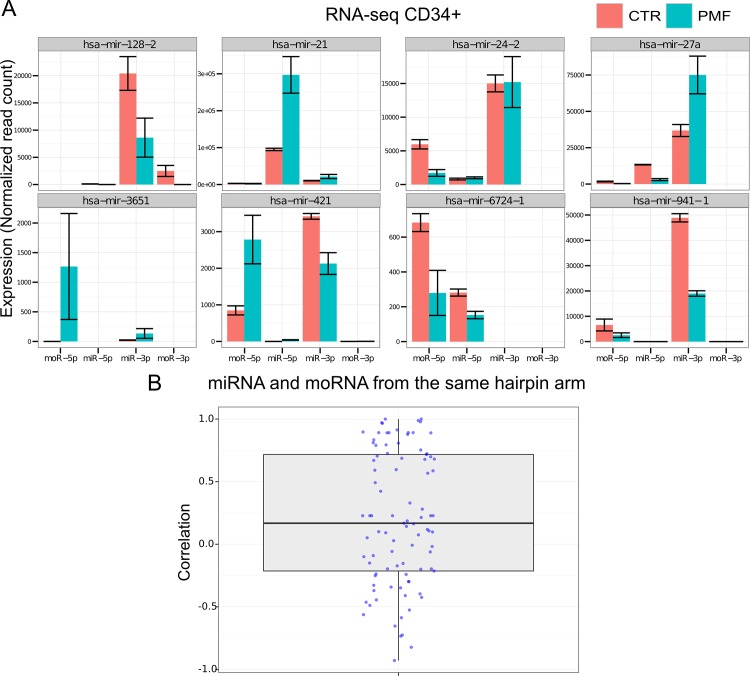
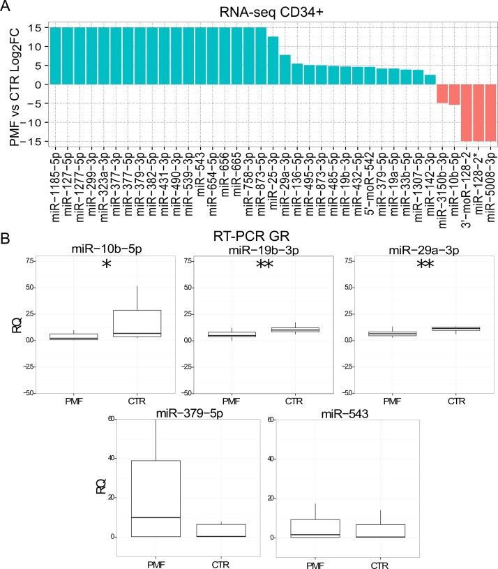
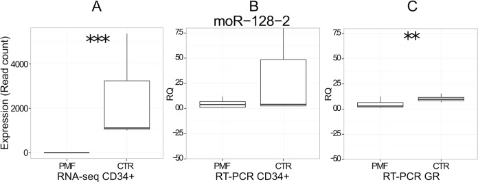
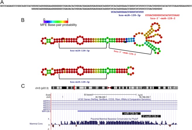
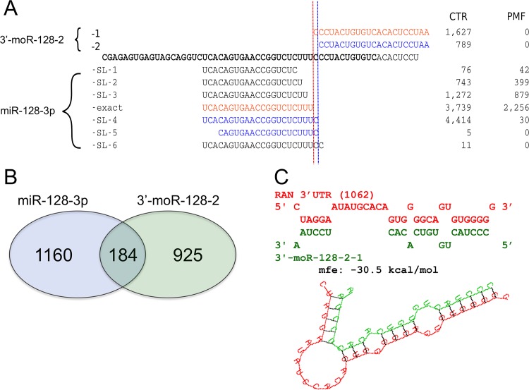
Myeloproliferative neoplasms (MPN) are chronic myeloid cancers thought to arise at the level of CD34+ hematopoietic stem/progenitor cells. They include essential thrombocythemia (ET), polycythemia vera (PV) and primary myelofibrosis (PMF). All can progress to acute leukemia, but PMF carries the worst prognosis. Increasing evidences indicate that deregulation of microRNAs (miRNAs) might plays an important role in hematologic malignancies, including MPN. To attain deeper knowledge of short RNAs (sRNAs) expression pattern in CD34+ cells and of their possible role in mediating post-transcriptional regulation in PMF, we sequenced with Illumina HiSeq2000 technology CD34+ cells from healthy subjects and PMF patients. We detected the expression of 784 known miRNAs, with a prevalence of miRNA up-regulation in PMF samples, and discovered 34 new miRNAs and 99 new miRNA-offset RNAs (moRNAs), in CD34+ cells. Thirty-seven small RNAs were differentially expressed in PMF patients compared with healthy subjects, according to microRNA sequencing data. Five miRNAs (miR-10b-5p, miR-19b-3p, miR-29a-3p, miR-379-5p, and miR-543) were deregulated also in PMF granulocytes. Moreover, 3'-moR-128-2 resulted consistently downregulated in PMF according to RNA-seq and qRT-PCR data both in CD34+ cells and granulocytes. Target predictions of these validated small RNAs de-regulated in PMF and functional enrichment analyses highlighted many interesting pathways involved in tumor development and progression, such as signaling by FGFR and DAP12 and Oncogene Induced Senescence. As a whole, data obtained in this study deepened the knowledge of miRNAs and moRNAs altered expression in PMF CD34+ cells and allowed to identify and validate a specific small RNA profile that distinguishes PMF granulocytes from those of normal subjects. We thus provided new information regarding the possible role of miRNAs and, specifically, of new moRNAs in this disease.


Conflict of interest statement

Competing Interests: The authors have declared that no competing interests exist.

Figures

Fig 1
Fig 1. Distribution of reads per isomiR category (A) and per type (B) across samples, considering all 818 expressed miRNAs.
“Exact” reads are identical to the mature miRNA sequence annotated in miRBase, whereas “mismatch” reads present respectively one or two nucleotides different from the annotated sequence but identical length; the last category includes reads perfecly matching the miRNA precursor (and genomic) sequence but shorter or longer than the annotated mature miRNA. IsomiR types indicate if the sequence difference fall in the 5’ region of the miRNA, in the 3’ region, or in both regions. Abundance of reads falling in different isomiR categories (C) and types (D) for miR-10b-5p, showing that isomiRs different from the annotated mature miRNA sequence are very abundant.
Fig 2
Fig 2. A) Expression, in CD34+ cells of PMF patients and controls, of miRNAs and moRNAs produced from the same hairpin, considering the hairpins expressing most abundant moRNAs; the boxplot in panel (B) shows the distribution of Pearson correlation values calculated pairwise between expression profiles of moRNAs and miRNAs produced from the same hairpin arm, considering all moRNAs detected in CD34+ cells.
Fig 3
Fig 3. Differential expression of small RNAs in PMF vs CTR CD34+.
A) Log2 FC of 37 small RNA differentially expressed considering PMF vs CTR CD34+, according to RNA-seq data (FDR<0.05). When a small RNA was not expressed in one sample category, the ratio was infinite and we represent it as the arbitrary maximum value of 15. B) RT-PCR expression calculation of five selected miRNAs in granulocytes collected from an independent cohort of normal controls (n = 10) and of PMF (50) samples; ***, ** and * indicate respectively a p-value <0.001, <0.01 or <0.05.
Fig 4
Fig 4. Differential expression of 3’-moR-128-2 in PMF (n = 3) vs CTR (3) cells.
A) moR expression in PMF and CTR CD34+ according to RNA-seq data. B) RT-PCR expression (RQ) in CD34+ cells from independent cohort of normal controls (n = 8) and of PMF (20) samples. C) RT-PCR expression (RQ) in granulocytes from independent cohort of normal controls (n = 10) and of PMF (50) samples; ***, ** and * indicate respectively a p-value <0.001, <0.01 or <0.05.
Fig 5
Fig 5. The 3’-moR-128-2 is produced by the precursor sequence of miR-128-3p.
A) The moRNA is derived from a region of the primary miRNA sequence exceeding the canonical hairpin precursor sequence, and it is not exaclty adjactent to the annotated miRNA. B) Minimum free energy (MFE) folding structure, predicted by RNAfold, for the canonical hairpin sequence and for the longer one, from which the moRNA is probably derived. C) Both the considered small RNAs are conserved in evolution through vertebrates.
Fig 6
Fig 6. Origin, sequence variability, and relations beween 3’-moR-128-2 and the adjacent miR-128-3p.
A) 3’-moR-128-2 and miR-128-3p map to the same locus and both shows sequence variability (isomiRs and isomoRs). Both the major and the minor isomoRs are found in normal CD34+ cells and not in PMF samples. Red and blue colors indicate isomiR and isomoR groups that can be produced with an unique sequence cutting sites. The most expressed isomoR is not associated to the corresponding most expressed isomiR. Moreover, expression levels, in CTR and PMF samples, of isomiRs and isomoRs are poorly correlated intragroup. These observations, point against the moRNA being simply a by-product of the miRNA biogenesis. A similar indication is given by the fact that some abundant isomiRs are not associated to detected isomoR sequences. B) 3’-moR-128-2 and miR-128-3p have different, poorly overlapping, sets of predicted targets. C) 3’-moR-128-2 sequence can stably bind a target site in the 3’UTR of the RAN mRNA, causing post transcriptional silencing.

References

    1. Tefferi A, Vardiman JW. Classification and diagnosis of myeloproliferative neoplasms: The 2008 World Health Organization criteria and point-of-care diagnostic algorithms. Leukemia. 2007;22: 14–22. - PubMed
    1. Vannucchi AM, Guglielmelli P, Tefferi A. Advances in Understanding and Management of Myeloproliferative Neoplasms. 2009;59: 171–191. - PubMed
    1. Baxter EJ, Scott LM, Campbell PJ, East C, Fourouclas N, Swanton S, et al. Acquired mutation of the tyrosine kinase JAK2 in human myeloproliferative disorders. 2005;365: 1054–1061. - PubMed
    1. Jones AV, Kreil S, Zoi K, Waghorn K, Curtis C, Zhang L, et al. Widespread occurrence of the JAK2 V617F mutation in chronic myeloproliferative disorders. Blood. 2005;106: 2162–2168. - PubMed
    1. James C, Ugo V, Le Couédic J, Staerk J, Delhommeau F, Lacout C, et al. A unique clonal JAK2 mutation leading to constitutive signalling causes polycythaemia vera. Nature. 2005;434: 1144–1148. - PubMed

Publication types

MeSH terms