. 2015 Dec;185(12):3189-201.
doi: 10.1016/j.ajpath.2015.08.008. Epub 2015 Oct 24.

Resolvin D1 Reduces Emphysema and Chronic Inflammation

Affiliations

Resolvin D1 Reduces Emphysema and Chronic Inflammation

Hsi-Min Hsiao et al. Am J Pathol. 2015 Dec.

Abstract

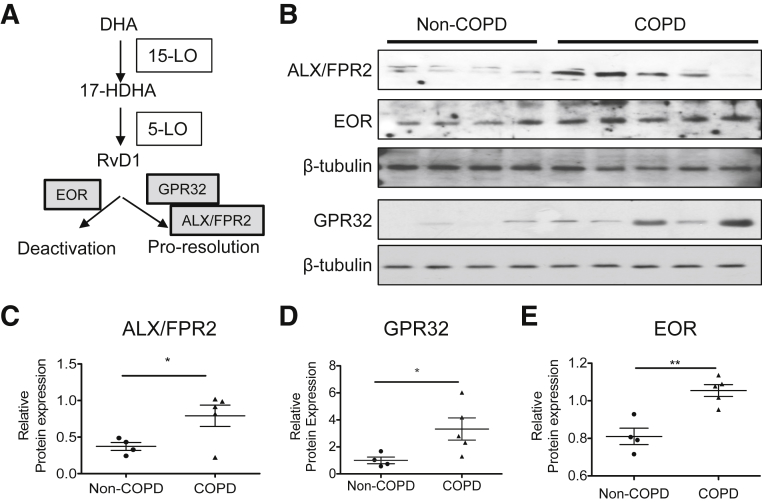
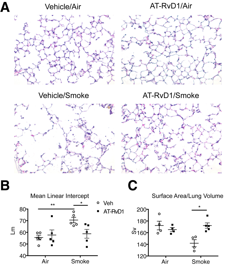
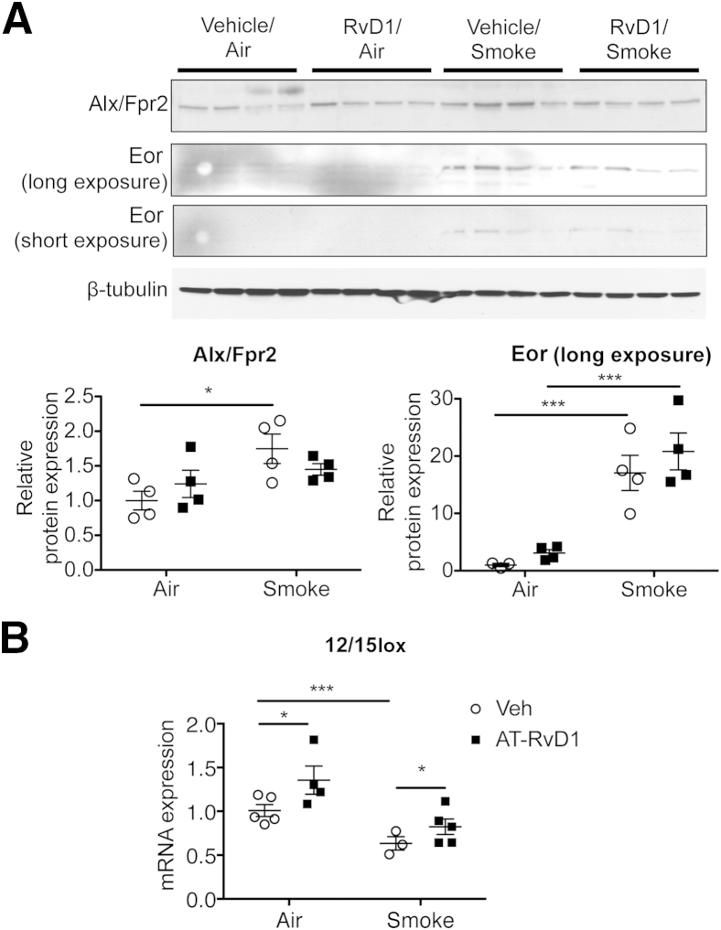
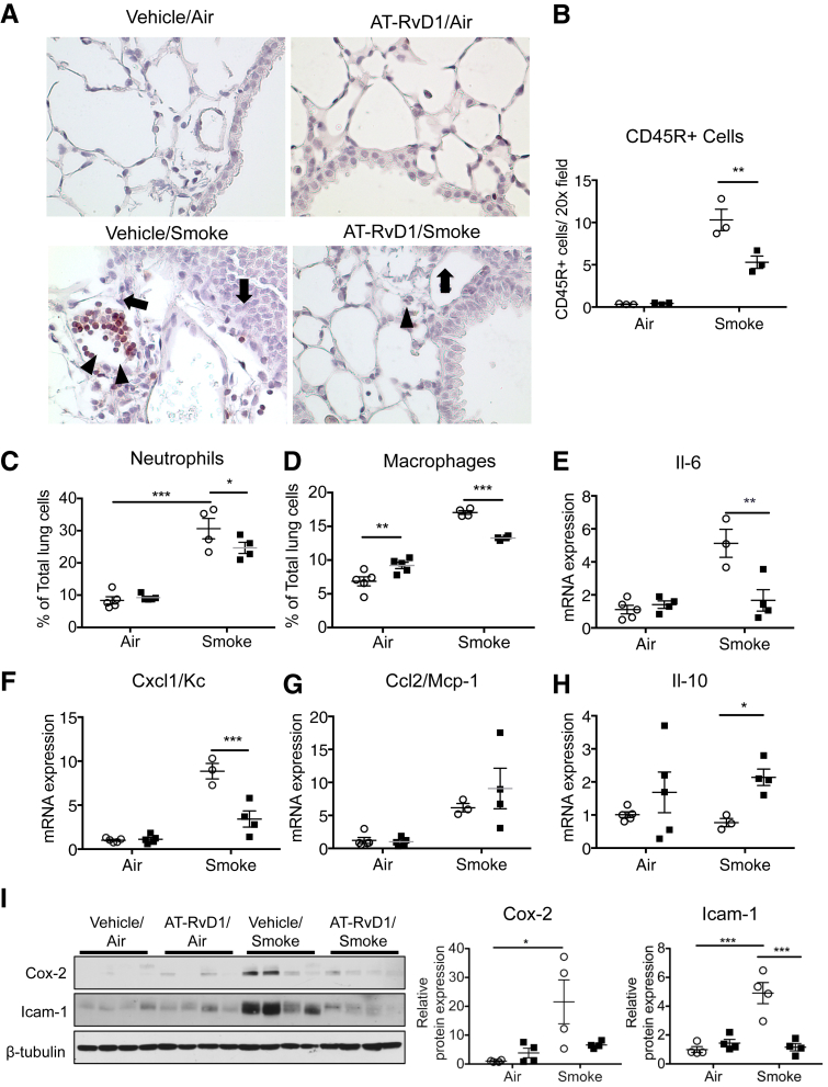
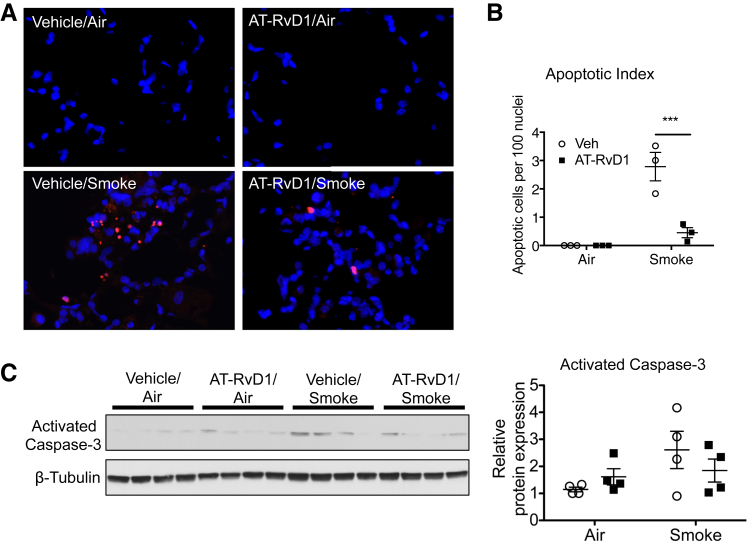
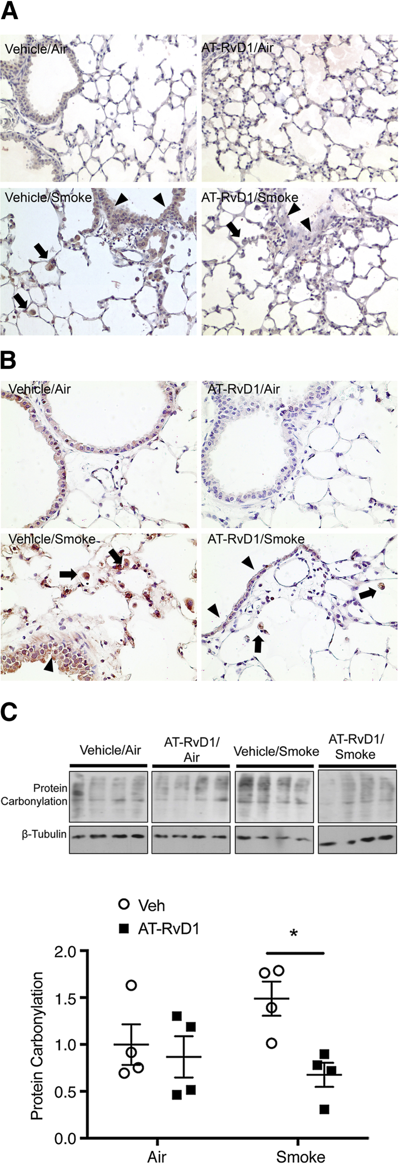
Chronic obstructive pulmonary disease is characterized, in part, by chronic inflammation that persists even after smoking cessation, suggesting that a failure to resolve inflammation plays an important role in the pathogenesis of the disease. It is widely recognized that the resolution of inflammation is an active process, governed by specialized proresolving lipid mediators, including lipoxins, resolvins, maresins, and protectins. Here, we report that proresolving signaling and metabolic pathways are disrupted in lung tissue from patients with chronic obstructive pulmonary disease, suggesting that supplementation with proresolving lipid mediators might reduce the development of emphysema by controlling chronic inflammation. Groups of mice were exposed long-term to cigarette smoke and treated with the proresolving mediator resolvin D1. Resolvin D1 was associated with a reduced development of cigarette smoke-induced emphysema and airspace enlargement, with concurrent reductions in inflammation, oxidative stress, and cell death. Interestingly, resolvin D1 did not promote the differentiation of M2 macrophages and did not promote tissue fibrosis. Taken together, our results suggest that cigarette smoking disrupts endogenous proresolving pathways and that supplementation with specialized proresolving lipid mediators is an important therapeutic strategy in chronic lung disease, especially if endogenous specialized proresolving lipid mediator signaling is impaired.


Figures

Figure 1
Figure 1
Proresolving signaling involving resolvin D1 (RvD1) is altered in lungs from patients with chronic obstructive pulmonary disease (COPD). A: RvD1 signaling pathway, showing synthetic and degrading enzymes and receptors. B: Levels of lipoxin A4 receptor/formyl peptide receptor 2 (ALX/FPR2), G-protein receptor (GPR)32, and eicosanoid oxidoreductase (EOR) in human lung lysates were examined by Western blot analysis. Each lane represents a lung sample from an individual donor. ALX/FPR2 and EOR were from the same blot, whereas GPR32 was from a second blot. Each individual blot was stripped and reprobed with β-tubulin as a loading control. C–E: Levels of ALX/FPR2 (C) GPR32 (D), and EOR (E) were quantitated by densitometry and normalized to β-tubulin. Data are expressed as means ± SEM. n = 4 (control, non-COPD lungs); n = 5 (COPD lungs). P < 0.05, ∗∗P < 0.01 by nonparametric, two-tailed t-test. 5-LO, 5-lipoxygenase; 15-LO, 15-lipoxygenase; 17-HDHA, 17-hydroxy docosahexaenoic acid; DHA, docosahexaenoic acid.
Figure 2
Figure 2
Aspirin-triggered resolvin D1 (AT-RvD1) attenuates long-term cigarette smoke–induced destructive airspace enlargement. A: Representative hematoxylin and eosin–stained photomicrographs of lung sections from mice subjected to 12 weeks of cigarette smoke exposure with AT-RvD1 treatment or without (Veh). B and C: Mean linear intercept (Lm) (B) and alveolar surface-to-volume ratio (Sv) (C) were determined as described in Materials and Methods. n = 5 mice per treatment group. P < 0.05, ∗∗P < 0.01 by t-tests with Welch's correction.
Figure 3
Figure 3
Cigarette smoke exposure dysregulates proresolving signaling in mouse lung. A: Whole-lung lysates were analyzed by Western blot analysis for the expression of lipoxin A4 receptor/formyl peptide receptor 2 (Alx/Fpr2) and eicosanoid oxidoreductase (Eor). β-Tubulin was used as a loading control. Original blots were cropped for illustration purposes. The results were quantitated by densitometry and normalized to β-tubulin. B: 12/15-Lipoxygenase (12/15Lox) mRNA was determined in whole-lung RNA by real-time PCR. Data are expressed as means ± SEM. n = 3 to 5 mice per group. P < 0.05, ∗∗∗P < 0.001 by two-way analysis of variance. RvD1, resolvin D1; Veh, vehicle.
Figure 4
Figure 4
Aspirin-triggered resolvin D1 (AT-RvD1) reduces chronic inflammation elicited by chronic cigarette smoke. A: Representative photomicrographs of CD45-stained lung sections after exposure to air or cigarette smoke, treated with vehicle (closed squares) or AT-RvD1 (open circles). The arrow indicates CD45+ cells in peribronchial and perivascular spaces; arrowhead, accumulated CD45+ cells in the interstitial spaces. B: CD45+ cells were counted in 5 high-power fields per mouse. C and D: Percentages of Gr-1+ neutrophils (C) and F4/80+ macrophages (D) were determined by flow cytometry. E–H: mRNA levels of Il-6 (E), chemokine (C-X-C motif) ligand 1/keratinocyte chemoattractant (Cxcl1/Kc) (F), monocyte chemotactic protein 1 (MCP1/Ccl2) (G), and Il-10 (H) were quantified in whole-lung homogenates by real-time PCR. I: Levels of cyclooxygenase (Cox)-2 and intercellular adhesion molecule (Icam)-1 in mouse lung were determined by Western blot analysis. β-Tubulin was used as a loading control. The results were quantitated by densitometry and normalized to β-tubulin. Data are expressed as means ± SEM. n = 4 mice per group (F, G, and I); n = 5 mice per group (B–E and H). P < 0.05, ∗∗P < 0.01, and ∗∗∗P < 0.001 by two-way analysis of variance with Bonferroni post-test.
Figure 5
Figure 5
Aspirin-triggered resolvin D1 (AT-RvD1) reduces cigarette smoke–induced cell death. A: Representative images of lung sections stained using terminal deoxynucleotidyl transferase-mediated dUTP nick-end labeling (TUNEL). The nuclei of TUNEL-positive cells were labeled in red. Tissue sections were counterstained with DAPI to visualize cell nuclei (blue nuclei). B: TUNEL-positive cells and DAPI-stained nuclei were counted in five high-power fields per mouse. The apoptotic index was defined as the number of TUNEL-positive cells per 100 DAPI-stained nuclei. C: Level of cleaved caspase-3 was examined by Western blot analysis. The results were quantitated by densitometry and normalized to β-tubulin. Data are expressed as means ± SEM. n = 5 mice per group (B). ∗∗∗P < 0.001 for smoke/vehicle versus smoke/AT-RvD1 by two-way analysis of variance with Bonferroni post-test (B). Veh, vehicle.
Figure 6
Figure 6
Aspirin-triggered resolvin D1 (AT-RvD1)-treated lung shows a reduction in oxidative stress. A: Mice were exposed to air or cigarette smoke and treated with vehicle or AT-RvD1, and lung sections were stained for anti–8-hydroxydeoxyguanosine (OHdG). 8-OHdG staining (brown) is evident in smoke-exposed mice in both epithelium (arrowheads) and macrophages (arrows), and is reduced with treatment with AT-RvD1. B: Lung sections from smoke-exposed and AT-RvD1–treated mice were stained for nitrotyrosine (red). Nitrotyrosine staining is broadly distributed in lung sections from smoke-exposed and vehicle-treated mice (Veh), especially in the epithelium (arrowheads) and macrophages (arrows), and is reduced with treatment with AT-RvD1. C: Protein carbonylation was measured in lung homogenates using an OxyBlot kit following the manufacturer's instructions (EMD Millipore, Billerica, MA). The same blot was stripped and reprobed with β-tubulin as a loading control. Each lane represents an individual mouse. Protein carbonylation was quantified by densitometry normalized to β-tubulin. Data are expressed as means ± SEM. P < 0.05 versus vehicle/smoke.
Figure 7
Figure 7
Aspirin-triggered resolvin D1 (AT-RvD1) neither affects macrophage phenotype nor induces fibrosis. A–D: Total RNA was isolated from lung F4/80+ macrophages and subjected to quantitative PCR to determine the levels of Arg1 (A), mannose receptor (Mrc)-1 (B), inducible nitric oxide synthase (iNos; C), and tumor necrosis factor (Tnf)-α (D). 18sRNA was used as an internal control. E: Representative photomicrographs of lungs exposed to chronic smoking, with or without AT-RvD1 treatment, and stained for Gomori's trichrome stain. Collagen deposition was stained blue. Sections were counterstained with hematoxylin for nuclei. Data are expressed as means ± SEM. n = 4 or 5 mice per group. P < 0.05, ∗∗∗P < 0.001 versus vehicle/air by two-way analysis of variance with Bonferroni post-test.

References

    1. Yoshida T., Tuder R.M. Pathobiology of cigarette smoke-induced chronic obstructive pulmonary disease. Physiol Rev. 2007;87:1047–1082. - PubMed
    1. Salvi S., Barnes P.J. Is exposure to biomass smoke the biggest risk factor for COPD globally? Chest. 2010;138:3–6. - PubMed
    1. Barnes P.J. New therapies for chronic obstructive pulmonary disease. Med Princ Pract. 2010;19:330–338. - PubMed
    1. Tuder R.M., Petrache I. Pathogenesis of chronic obstructive pulmonary disease. J Clin Invest. 2012;122:2749–2755. - PMC - PubMed
    1. Barnes P.J. Mediators of chronic obstructive pulmonary disease. Pharmacol Rev. 2004;56:515–548. - PubMed

Publication types

MeSH terms

Substances