. 2015 Oct 15;526(7573):385-390.
doi: 10.1038/nature15700.

Glia-derived neurons are required for sex-specific learning in C. elegans

Affiliations

Glia-derived neurons are required for sex-specific learning in C. elegans

Michele Sammut et al. Nature. .

Abstract

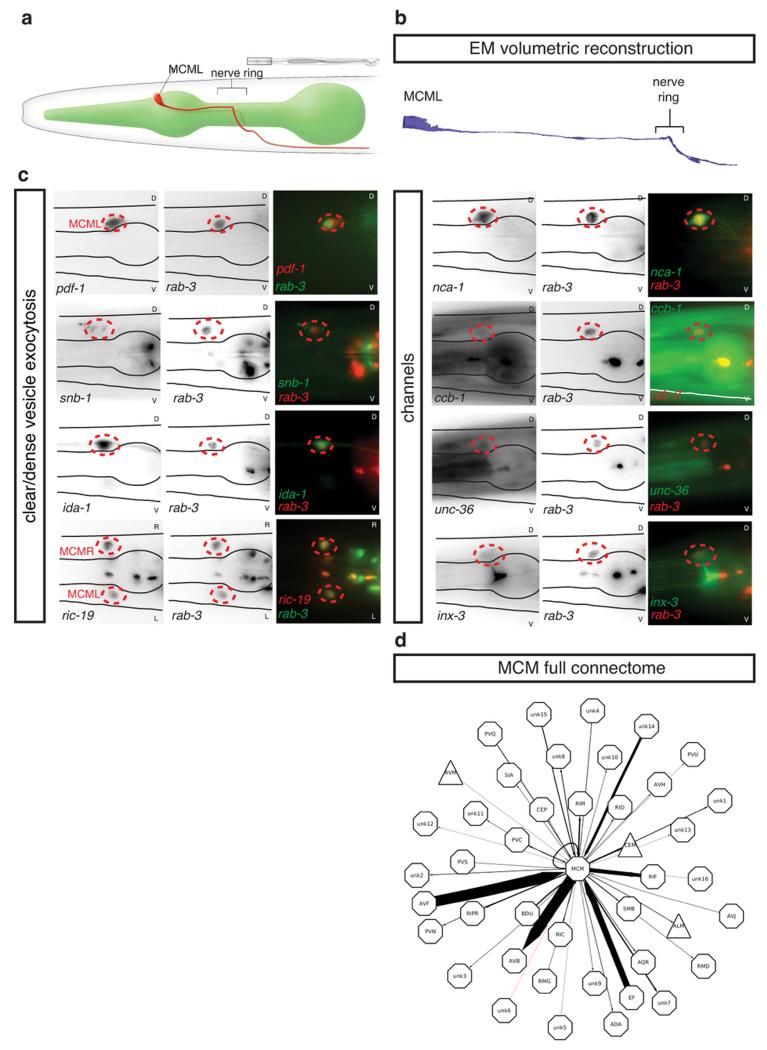
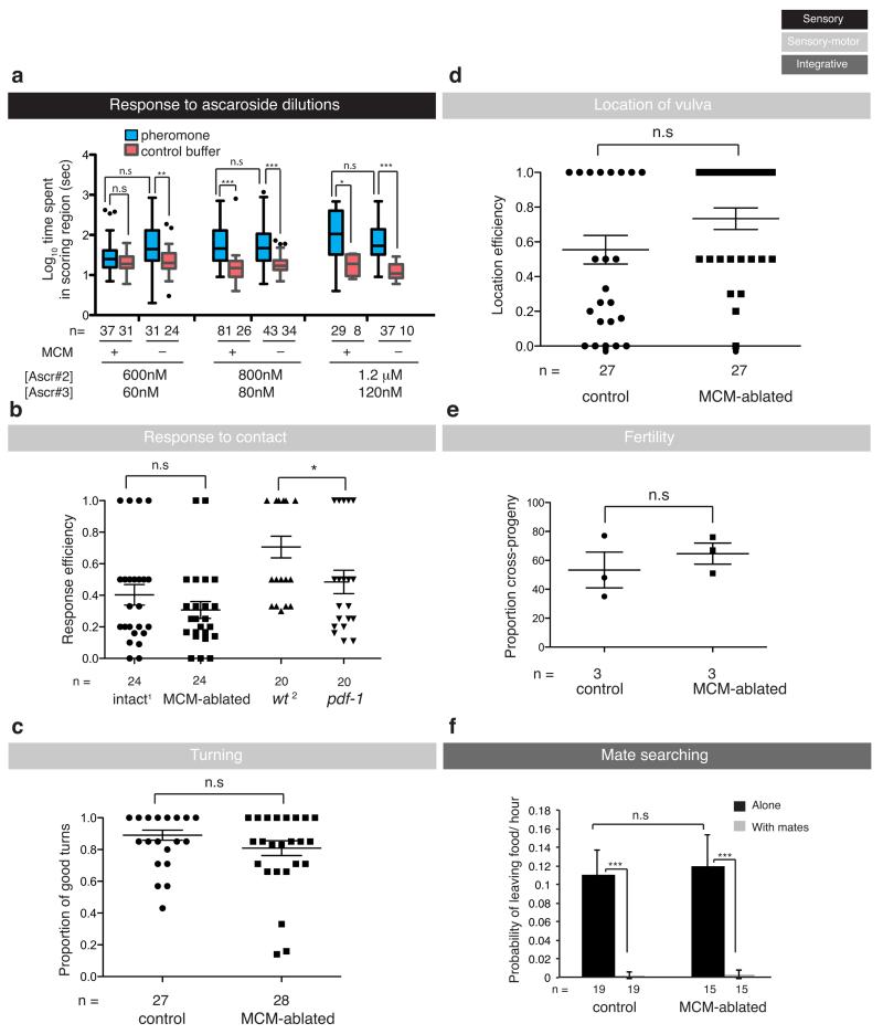
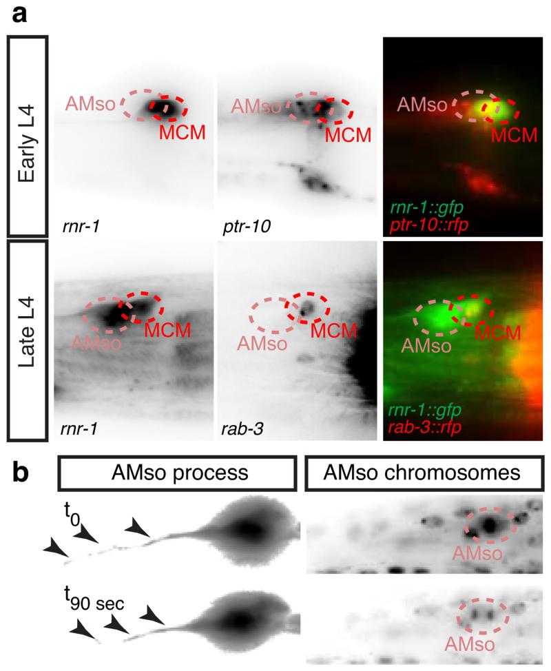
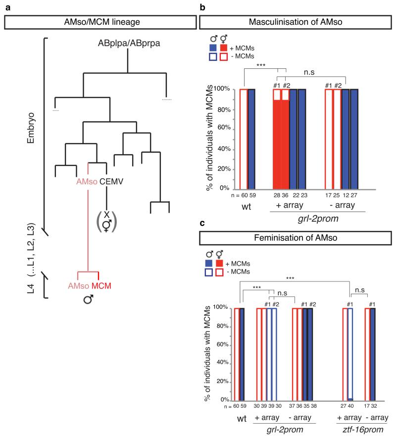
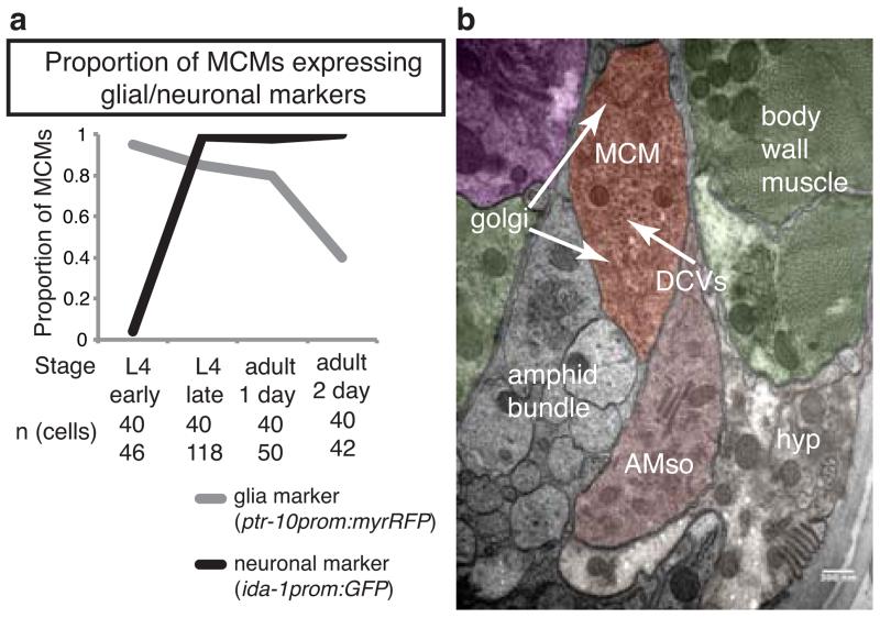
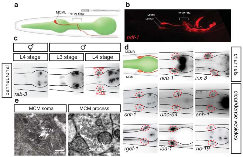
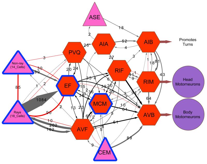
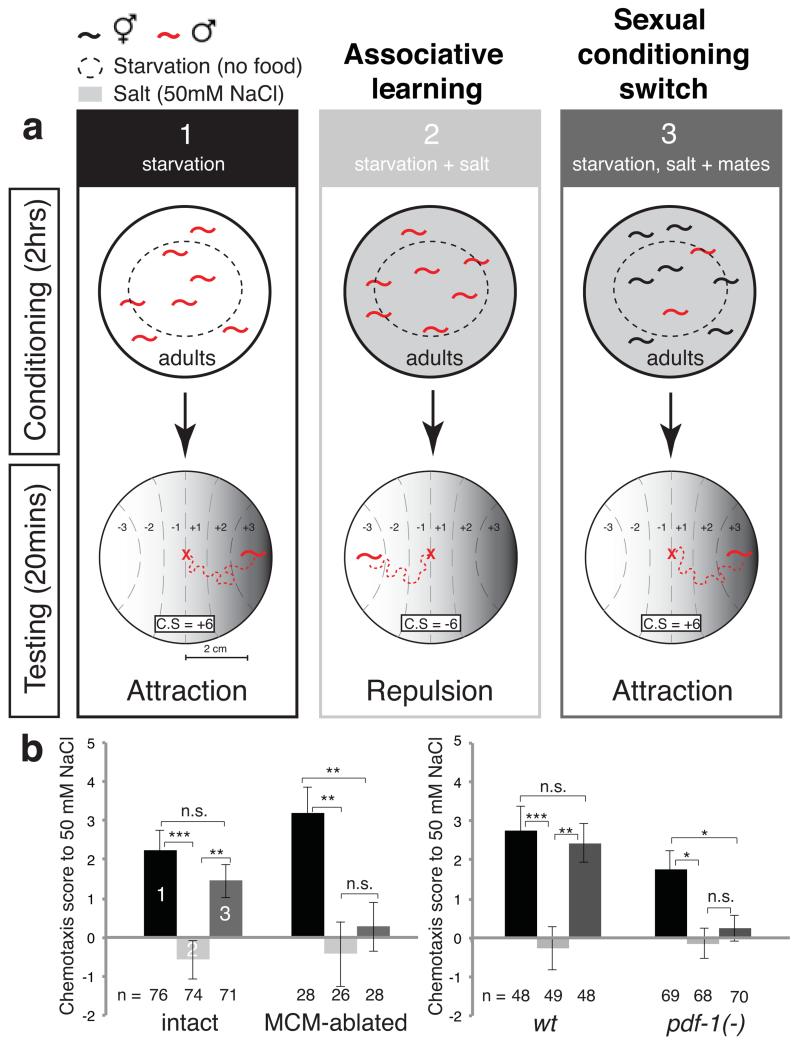
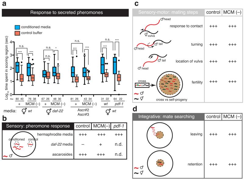
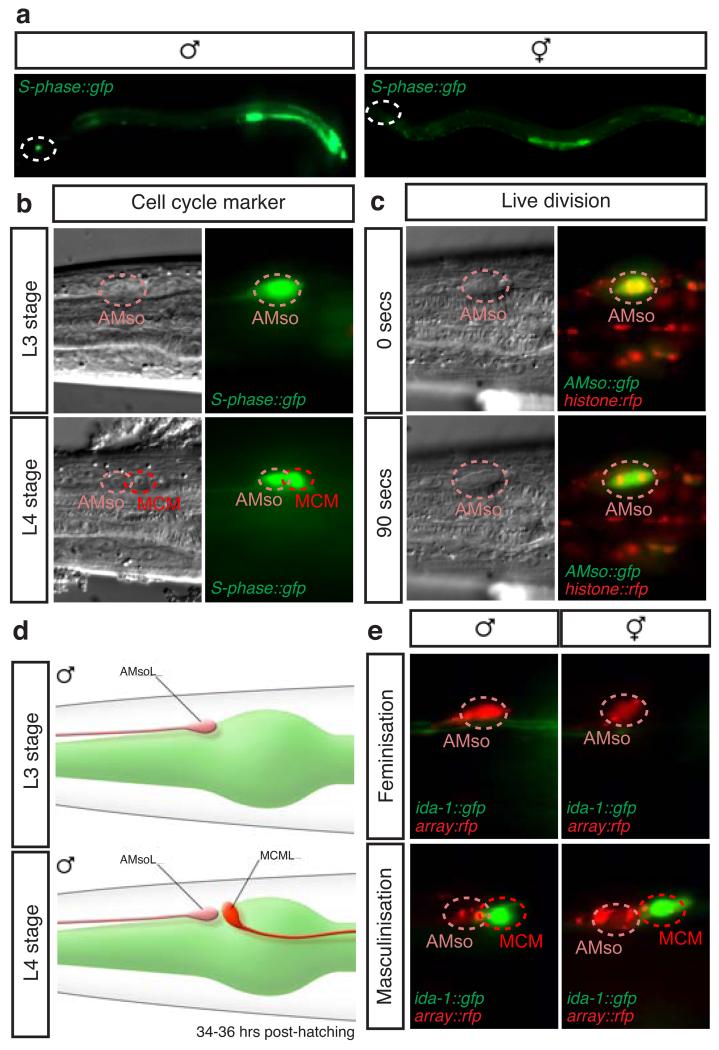
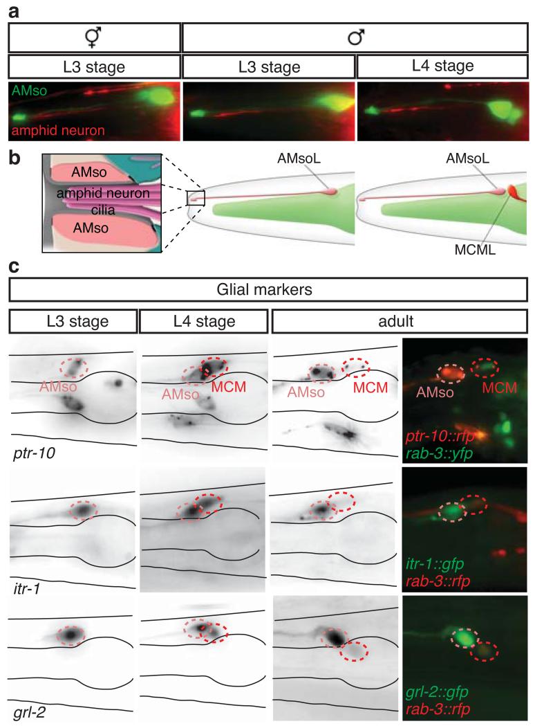
Sex differences in behaviour extend to cognitive-like processes such as learning, but the underlying dimorphisms in neural circuit development and organization that generate these behavioural differences are largely unknown. Here we define at the single-cell level-from development, through neural circuit connectivity, to function-the neural basis of a sex-specific learning in the nematode Caenorhabditis elegans. We show that sexual conditioning, a form of associative learning, requires a pair of male-specific interneurons whose progenitors are fully differentiated glia. These neurons are generated during sexual maturation and incorporated into pre-exisiting sex-shared circuits to couple chemotactic responses to reproductive priorities. Our findings reveal a general role for glia as neural progenitors across metazoan taxa and demonstrate that the addition of sex-specific neuron types to brain circuits during sexual maturation is an important mechanism for the generation of sexually dimorphic plasticity in learning.


Figures

Extended Data Figure 1
Extended Data Figure 1. The MCMs are newly identified male-specific neurons
a, WormAtlas-style diagram depicting the morphology and position of one of the bilateral pair of MCM neurons in the head of a male worm and its projection within the nerve ring and along the ventral cord. b, Volumetric reconstruction of the MCML cell body and projection based on tracing of serial EM sections. c, Co-expression of transgenes for neuronal markers in the rab-3-positive cells identified as MCMs (indicated with dashed red circles). All photographs are lateral views of animals oriented anterior to the left and dorsal to the top except for ric-19, which are dorsal views. Transgenes are listed in table S1. pdf-1 (neuropeptide pigment dispersing factor); snb-1 (synaptobrevin); ida-1 (tyrosine phosphatse-like receptor, ortholog of mammalian phogrin); ric-19 (rab-2 effector); nca-1 (NALCN Na+ channel subunit); ccb-1 (voltage-gated Ca2+ channel subunit); unc-36 (voltage-gated Ca2+ channel subunit); inx-3 (gap junction innexin). R/L=Right/Left and D/V=Dorsal/Ventral. d, Diagram of the neurons that directly connect to and from the MCMs. Triangles, sensory neurons; octagons, interneurons and unidentified neurons. The thickness of the arrows is proportional to the anatomical strength of the connections (Extended Data Table 2).
Extended Data Figure 2
Extended Data Figure 2. The MCMs are not required for other male-specific behaviours
a, Response of intact and MCM-ablated males (inIs179(ida-1prom::gfp);him-8(e1489) and otIs356(rab-3prom::rfp)him-5(e1490)) to dilutions of ascaroside pheromones (Ascr). Graphs represent Tukey box plots of logarithmic transformations of the data; n= number of independent events (i.e. entry in scoring region). T-test with Bonferroni correction was used for statistical analysis. ***P<0.001; **P<0.01; *P<0.05; n.s.= no statistically significant difference (p≥0.05). b, Response efficiency to mate contact of intact, MCM-ablated and pdf-1(tm1996) mutant males measured as the proportion of responses out of total contacts with an hermaphrodite. 1 Intact animals were inIs179(ida-1prom::gfp);him-8(e1489). 2 Control animals were him-5(e1490). A response indicates that the male placed its tail ventral down on the mate’s body and backed along it to make a turn. c and d, Proportion of good turns (c) and location of vulva efficiency (d) of intact and MCM-ablated males (inIs179(ida-1prom::gfp);him-8(e1489) and otIs356(rab-3prom::rfp)him-5(e1490)). e, Fertility (measured as proportion of cross-progeny) of intact and MCM-ablated males (otIs356(rab-3prom::rfp)him-5(e1490)). For be, n= number of individual animals tested. Error bars indicate SEM. Mann-Whitney test was used for statistical analysis. *P<0.05; n.s.= no statistically significant difference (p≥0.05). f, Mate-searching behavior, measured as PL values (probability of leaving food per hour) in the absence or presence of mates, of intact and MCM-ablated males (otIs356(rab-3prom::rfp)him-5(e1490)). n= number of individual animals tested. Two independent population assays were performed on different days. Maximum likelihood statistical analysis was used to compare PL values. Error bars indicate SEM. ***P<0.001; n.s.= no statistically significant difference (p≥0.05).
Extended Data Figure 3
Extended Data Figure 3. The MCMs arise from a division of the AMso glial cell
All photographs are lateral views of animals oriented anterior to the left and dorsal to the top. a, Fluorescent photographs showing the two cells expressing rnr-1prom::gfp co-labeled with the glial marker ptr-10prom::rfp and the neuronal marker rab-3prom::rfp in the head of males at the early and late L4 stages. The AMso and MCM cell bodies are indicated with dashed lines. b, Fluorescent images of the AMso cell body and its projection at two time points during cell division. Photos are overexposed for visualization of the projection, indicated by arrows. The chromosomes are labeled with a histone:rfp transgene, and the AMso cell body is indicated by dashed lines.
Extended Data Figure 4
Extended Data Figure 4. AMso plasticity is regulated by AMso genetic sex
a, Diagram of the left AMso and MCM lineage. b and c, proportion of individuals with MCMs in control animals and animals expressing sex-reversing transgenes in AMso. b, AMso masculinization with grl-2prom::fem-3::SL2::mCherry transgenes (oleEx18 and oleEx24). c, AMso feminization with grl-2prom::tra-2IC::SL2::mCherry transgenes (oleEx19 and oleEx23) and ztf-16enhancer::tra-2IC::SL2::mCherry transgene oleEx22. MCM cell fate was identified with ida-1prom::gfp or rab-3prom::yfp reporter transgenes. In the head, the grl-2 promoter drives expression in AMso and the excretory duct and pore cells, and the ztf-16 glial enhancer drives expression in the AMso and amphid sheath glia. # indicates an independent transgenic array line for each manipulation. χ2 test was used for statistical analysis; ***P<0.001; n.s.= no statistical significant difference (p≥0.05); n= number of animals scored.
Extended Data Figure 5
Extended Data Figure 5. The MCMs lose molecular and structural characteristics of glia after birth
a, Proportion of MCMs with presence of the glial marker ptr-10prom::myrRfp or the neuronal marker ida-1prom::gfp at different stages after MCM birth. b, Electron micrograph of a cross section of an adult male head showing the MCM and AMso cell body ultrastructure. Neighbouring tissues are color-coded according to WormAtlas (http://www.wormatlas.org/colorcode.htm). Purple (pharynx), muscle (green), hypodermis (light cream), AMso (amphid socket, pink). The dendrites of the amphid neurons (amphid bundle) are not colored.
Figure 1
Figure 1. The MCMs are newly identified male-specific neurons
Lateral views (a and b) and dorsal views (c and d) of animals oriented anterior to the left. a, WormAtlas-style diagram depicting the morphology and position, adjacent to the pharynx, of one of the bilateral pair of MCM neurons in the head of a male. b, Confocal projection of pdf-1prom::rfp expression in the head of an adult male (same region as in a). c, Expression of the pan-neuronal reporter transgene rab-3prom:rfp (Ras GTPase) in the head of an hermaphrodite and males at the third (L3) and fourth (L4) larval stages. The position of the MCMs is indicated with dashed red circles. R/L=Right/Left. d, Expression in the MCMs in adult males of reporter transgenes for neuronal markers. snt-1 (synaptotagmin); unc-64 (syntaxin); snb-1 (synaptobrevin); rgef-1 (Ras exchange factor); nca-1 (NALCN Na+ channel subunit); inx-3 (gap junction innexin); ida-1 (tyrosine phosphatase-like receptor, phogrin); pdf-1 (neuropeptide pigment dispersing factor). e, Electron micrographs of MCM showing dense-core vesicles (DCV) (left) and a synapse (right).
Figure 2
Figure 2. MCM connectivity
a, Connectivity diagram of the MCMs showing the main inputs and outputs. The connections to the neurons known to regulate sexual conditioning (rays and CEMs), chemosensory plasticity (AIA, RIF), and salt sensation (ASE) are included. Grey and red connections indicate chemical and electrical synapses, respectively. The triplet motifs created by the MCMs are highlighted in black. The thickness of the lines is proportional to the anatomical strength of the connections, indicated in numbers (the number of EM serial sections of scored synaptic connectivity). Pink triangles, sensory neurons; red octagons, interneurons; purple circles, motor neurons; blue outline, male-specific.
Figure 3
Figure 3. The MCMs are required for male-specific associative learning
a, Diagram depicts the salt-avoidance learning and sexual conditioning assay. C.S, chemotaxis score. b, Salt chemotaxis scores of previously conditioned intact and MCM-ablated males [inIs179(ida-1prom::gfp);him-8(e1489)], and wild-type [him-5(e1490)] and pdf-1(tm1996) mutant males. n= number of individual animals tested. Error bars indicate SEM. Mann-Whitney test was used for statistical analysis. ***P<0.001; **P<0.01; *P<0.05; n.s.= no statistically significant difference (p≥0.05);
Figure 4
Figure 4. MCM ablation does not affect other male-specific behaviours
Efficiency of intact and MCM-ablated males [inIs179(ida-1prom::gfp);him-8(e1489) or otIs356(rab-3prom::rfp)him-5(e1490)], and wild-type [him-5(e1490)] and pdf-1(tm1996) mutant males in male-specific behaviours. a, Response to wild-type or daf-22 (m130) hermaphrodite-conditioned media and purified ascaroside pheromones (80nM Ascr#3, 800nM Ascr#2). Graphs represent Tukey box plots of logarithmic transformations of the data; n= number of independent events (i.e. entry in scoring region); T-test with Bonferroni correction was used for statistical analysis. ***P<0.001; *P<0.05; n.s.= no statistically significant difference (p≥0.05). b, Summary diagram of the data plotted in a. Statistical significance compared to control buffer is indicated by +++, P<0.001; +, P<0.05; − = no statistically significant difference. n.d., not determined. c and d, Execution of mating sub-steps (c) and exploration in search of mates (d). +++ indicates performance level of intact, control males.
Figure 5
Figure 5. The MCMs originate from a male-specific cell division of the AMso glial cells
Lateral views of animals oriented anterior to the left and dorsal to the top. a and b, Expression of the S-phase reporter transgene rnr-1prom:gfp in whole animals at the L4 stage (a) and in the head of L3 and L4 males (b). The male-specific division in the head is indicated by dashed circles in a. The rest of expression corresponds to developing reproductive structures. c, DIC and fluorescent images of the AMso cell body with RFP-labeled histones during cell division. d, WormAtlas diagram depicting the morphology and position of the AMso and the MCM in the male head at the L3 and L4 stages. e, Head of animals with sex-reversal genetic manipulations of AMso. Feminisation by expression of the grl-2prom:tra-2IC::SL2:mCherry transgene oleEx23. Masculinisation by expression of grl-2prom:fem-3::SL2:mCherry transgenes oleEx18 and oleEx24.
Figure 6
Figure 6. The male AMso cells are fully differentiated glia before and after the division that generates the MCM neuron
Lateral views of animals oriented anterior to the left and dorsal to the top. a, AMso glial cell (green, grl-2prom:gfp) and amphid sensory neuron dendrites (red, pdf-1prom:rfp) in an L3 hermaphrodite and L3 and L4 males. b, Diagram of the AMso glial cell and the distal end of its projection to the nose, where it ensheaths the cilia of sensory dendrites. c, Expression in AMso and perdurance in MCM of reporter transgenes for glial markers in L3, L4 and adult males. ptr-10 (Patch related receptor); itr-1 (IP3 receptor); grl-2 (hedgehog-like/Ground related).

References

    1. Kimura K-I, Ote M, Tazawa T, Yamamoto D. Fruitless specifies sexually dimorphic neural circuitry in the Drosophila brain. Nature. 2005;438:229–233. - PubMed
    1. Ruta V, et al. A dimorphic pheromone circuit in Drosophila from sensory input to descending output. Nature. 2010;468:686–690. - PubMed
    1. Yang CF, et al. Sexually dimorphic neurons in the ventromedial hypothalamus govern mating in both sexes and aggression in males. Cell. 2013;153:896–909. - PMC - PubMed
    1. Rideout EJ, Dornan AJ, Neville MC, Eadie S, Goodwin SF. Control of sexual differentiation and behavior by the doublesex gene in Drosophila melanogaster. Nature Neuroscience. 2010;13:458–466. - PMC - PubMed
    1. Stowers L, Logan DW. Sexual dimorphism in olfactory signaling. Curr. Opin. Neurobiol. 2010;20:770–775. - PMC - PubMed

Publication types

MeSH terms