. 2015 Oct 15:5:14649.
doi: 10.1038/srep14649.

Quantification of Interfibrillar Shear Stress in Aligned Soft Collagenous Tissues via Notch Tension Testing

Affiliations

Quantification of Interfibrillar Shear Stress in Aligned Soft Collagenous Tissues via Notch Tension Testing

Spencer E Szczesny et al. Sci Rep. .

Abstract

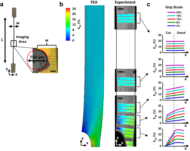
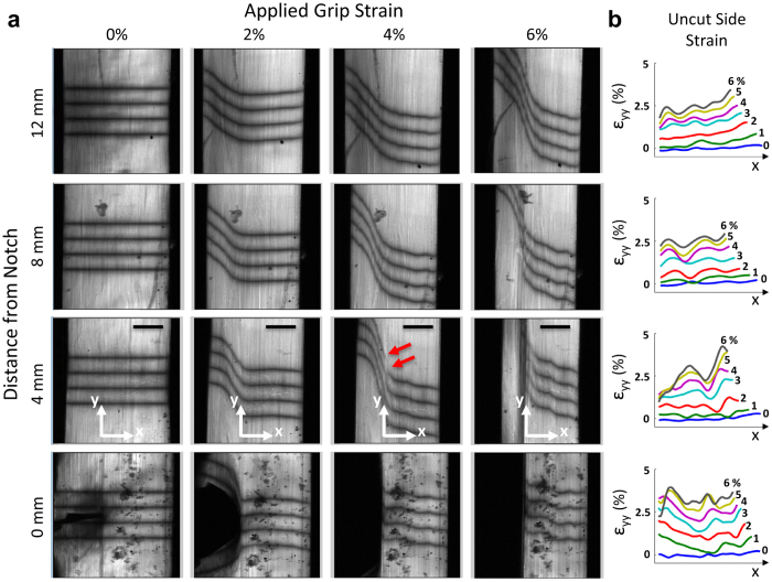
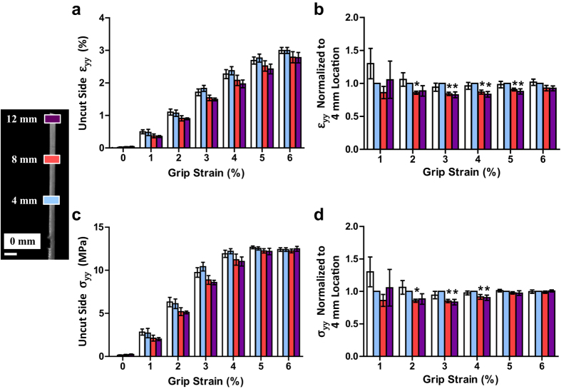
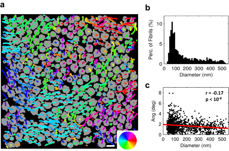
The mechanical function of soft collagenous tissues is largely determined by their hierarchical organization of collagen molecules. While collagen fibrils are believed to be discontinuous and transfer load through shearing of the interfibrillar matrix, interfibrillar shear stresses have never been quantified. Scaling traditional shear testing procedures down to the fibrillar length scale is impractical and would introduce substantial artifacts. Here, through the use of a novel microscopic variation of notch tension testing, we explicitly demonstrate the existence of interfibrillar shear stresses within tendon fascicles and provide the first measurement of their magnitude. Axial stress gradients along the sample length generated by notch tension testing were measured and used to calculate a value of 32 kPa for the interfibrillar shear stress. This estimate is comparable to the interfibrillar shear stress predicted by previous multiscale modeling of tendon fascicles, which supports the hypothesis that fibrils are discontinuous and transmit load through interfibrillar shear. This information regarding the structure-function relationships of tendon and other soft collagenous tissues is necessary to identify potential causes for tissue impairment with degeneration and provide the foundation for developing regenerative repair strategies or engineering biomaterials for tissue replacement.


Conflict of interest statement

The authors declare no competing financial interests.

Figures

Figure 1
Figure 1. Demonstration of notch tension technique using a linear elastic isotropic gelatin gel.
(a) Uniaxial tension was applied to a 20% (w/v) gel formed with similar dimensions as a tendon fascicle (h: 540 μm, w: 680 μm, L: 33.0 mm) and with a semi-circular notch. (b) At a grip-to-grip strain of 8%, the axial strain field (εyy) predicted by finite element analysis closely matched the experimentally measured strains. (c) Close to the notch, the axial strains are concentrated on the uncut (right) side of the gel, while the strains are uniform far from the notch. These gradients in the axial strain (and hence stress) across the gel width and length are classic features of notch tension testing and are produced by shear stresses that transmit load from the uncut to the cut side of the sample. Scale bars, 200 μm.
Figure 2
Figure 2. Notch tension testing of tendon fascicles.
(a) With increasing grip-to-grip strain, the notch widened and the photobleached lines became steeply angled, representing shear strains developing within the tissue. Consistently at 4% grip-to-grip strain, discontinuities in the photobleached lines first appeared at the location 4 mm from notch (red arrows). These discontinuities propagated parallel to the fascicle axis demarcating the interface between the cut and uncut portions of the tissue. (b) At all non-zero distances from the notch, the axial strains (εyy) on the uncut side of the tissue exhibit gradients similar to those seen in the gel (Fig. 1c). Scale bars, 200 μm.
Figure 3
Figure 3. Axial strain and stress gradients produced in tendon fascicles.
(a,b) The axial strains (εyy) averaged over the uncut side of the tissue decreased with distance away from the notch. This axial strain gradient along the sample length is similar to that observed on the uncut side of the gelatin gel and demonstrates the existence of shear stress within the tissue. (c,d) Conversion of these strains to stress values shows that the axial stress (σyy) on the uncut side of the tissue decreased with distance away from the notch until 4% grip-to-grip strain. This drop in axial stress corresponded to an interfibrillar shear stress of 32 ± 33 kPa (mean ± s.d.). *p ≤ 0.05. Error bars, s.e.m. Scale bar, 1 mm.
Figure 4
Figure 4. Trajectories of individual collagen fibrils.
(a) Fibrils (shown in gray) from first image with overlay of lateral trajectories along 8.7 μm of fascicle length. Color wheel indicates direction of lateral trajectory. (b) Histogram of fibril diameters. (c) While there was a weak correlation between angle and fibril diameter (r = −0.17, p < 10−8), on average the fibrils were oriented 1.7 ± 1.0 deg (mean ± s.d.) with respect to the fascicle axis, demonstrating that the fibrils are highly aligned within the fascicles and suggesting that few fibrils cross the cut/uncut tissue interface. Scale bar, 500 nm.

References

    1. Orgel J. P. R. O., Irving T. C., Miller A. & Wess T. J. Microfibrillar structure of type I collagen in situ. Proc. Natl. Acad. Sci. USA 103, 9001–9005 (2006). - PMC - PubMed
    1. Hulmes D. J., Wess T. J., Prockop D. J. & Fratzl P. Radial packing, order, and disorder in collagen fibrils. Biophys. J. 68, 1661–1670 (1995). - PMC - PubMed
    1. Bailey A. J., Paul R. G. & Knott L. Mechanisms of maturation and ageing of collagen. Mech. Ageing Dev. 106, 1–56 (1998). - PubMed
    1. Silver F. H., Kato Y. P., Ohno M. & Wasserman A. J. Analysis of mammalian connective tissue: relationship between hierarchical structures and mechanical properties. J. Long. Term Eff. Med. Implants 2, 165–198 (1992). - PubMed
    1. Szczesny S. E. & Elliott D. M. Interfibrillar shear stress is the loading mechanism of collagen fibrils in tendon. Acta Biomater. 10, 2582–2590 (2014). - PMC - PubMed

Publication types

LinkOut - more resources