

Low-Cost High-Performance MRI
- PMID: 26469756
- PMCID: PMC4606787
- DOI: 10.1038/srep15177
Low-Cost High-Performance MRI
Abstract
Magnetic Resonance Imaging (MRI) is unparalleled in its ability to visualize anatomical structure and function non-invasively with high spatial and temporal resolution. Yet to overcome the low sensitivity inherent in inductive detection of weakly polarized nuclear spins, the vast majority of clinical MRI scanners employ superconducting magnets producing very high magnetic fields. Commonly found at 1.5-3 tesla (T), these powerful magnets are massive and have very strict infrastructure demands that preclude operation in many environments. MRI scanners are costly to purchase, site, and maintain, with the purchase price approaching $1 M per tesla (T) of magnetic field. We present here a remarkably simple, non-cryogenic approach to high-performance human MRI at ultra-low magnetic field, whereby modern under-sampling strategies are combined with fully-refocused dynamic spin control using steady-state free precession techniques. At 6.5 mT (more than 450 times lower than clinical MRI scanners) we demonstrate (2.5 × 3.5 × 8.5) mm(3) imaging resolution in the living human brain using a simple, open-geometry electromagnet, with 3D image acquisition over the entire brain in 6 minutes. We contend that these practical ultra-low magnetic field implementations of MRI (<10 mT) will complement traditional MRI, providing clinically relevant images and setting new standards for affordable (<$50,000) and robust portable devices.
Conflict of interest statement
The authors declare no competing financial interests.
Figures
 , and
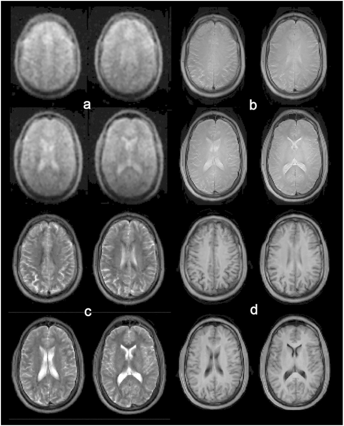
, and  weighted contrast at 3 T, respectively. Most of the anatomic features seen at higher magnetic field can be identified on the ultra-low field scans. At low field,
  approaches
  , and the resulting image contrast in (a) is very similar to PD-weighting (b).
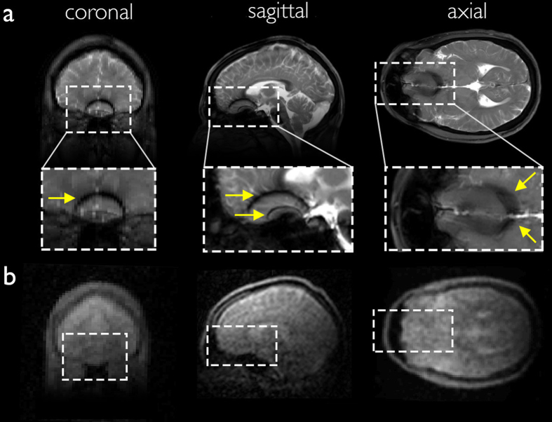
References
-
- Sepponen R. E., Sipponen J. T. & Sivula A. Low Field (0.02 T) Nuclear Magnetic Resonance Imaging of the Brain. Journal of Computer Assisted Tomography 9, 237 (1985). - PubMed
-
- Macovski A. & Conolly S. Novel approaches to low-cost MRI. Magnetic Resonance in Medicine 30, 221–230 (1993). - PubMed
-
- Venook R. D. et al. Prepolarized magnetic resonance imaging around metal orthopedic implants. Magnetic Resonance in Medicine 56, 177–186 (2006). - PubMed
-
- Robert Kraus J., Espy M., Magnelind P. & Volegov P. Ultra-Low Field Nuclear Magnetic Resonance. (Oxford University Press, 2014). 10.1093/med/9780199796434.001.0001/med-9780199796434. - DOI
-
- Clarke J. & Braginski A. I. The SQUID Handbook: Fundamentals and Technology of SQUIDs and SQUID Systems, Volume I. (Wiley-VCH Verlag GmbH & Co. KGaA, 2004).
Publication types
Grants and funding
LinkOut - more resources
Full Text Sources
Other Literature Sources
