. 2015 Oct 15;10(10):e0140824.
doi: 10.1371/journal.pone.0140824. eCollection 2015.

Dengue-3 Virus Entry into Vero Cells: Role of Clathrin-Mediated Endocytosis in the Outcome of Infection

Affiliations

Dengue-3 Virus Entry into Vero Cells: Role of Clathrin-Mediated Endocytosis in the Outcome of Infection

Luana E Piccini et al. PLoS One. .

Abstract

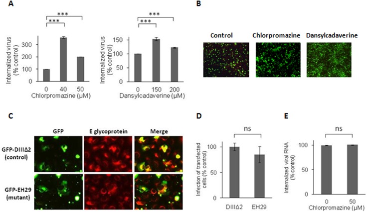
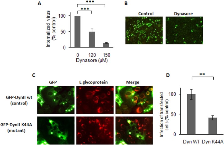
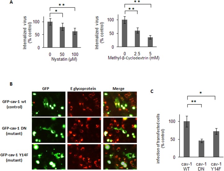
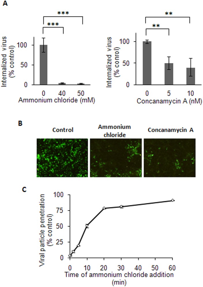
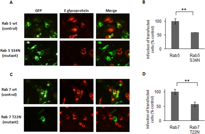
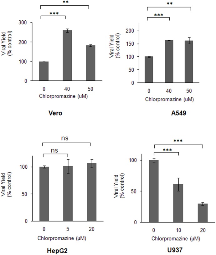
The endocytic uptake and intracellular trafficking for penetration of DENV-3 strain H-87 into Vero cells was analyzed by using several biochemical inhibitors and dominant negative mutants of cellular proteins. The results presented show that the infective entry of DENV-3 into Vero cells occurs through a non-classical endocytosis pathway dependent on low pH and dynamin, but non-mediated by clathrin. After uptake, DENV-3 transits through early endosomes to reach Rab 7-regulated late endosomes, and according with the half-time for ammonium chloride resistance viral nucleocapsid is released into the cytosol approximately at 12 min post-infection. Furthermore, the influence of the clathrin pathway in DENV-3 infective entry in other mammalian cell lines of human origin, such as A549, HepG2 and U937 cells, was evaluated demonstrating that variable entry pathways are employed depending on the host cell. Results show for the first time the simultaneous coexistence of infective and non -infective routes for DENV entry into the host cell, depending on the usage of clathrin-mediated endocytosis.


Conflict of interest statement

Competing Interests: The authors have declared that no competing interests exist.

Figures

Fig 1
Fig 1. Clathrin is not required for DENV-3 infective internalization into Vero cells.
(A) Cells were treated with chlorpromazine or dansylcadaverine and infected with DENV-3. After 1h of internalization in presence of the drugs, monolayers were treated with proteinase K and the cell pellets were plated onto Vero cells to determine internalized virus by an infectious centre assay. (B) Cells treated with 40 μM chlorpromazine, 150 μM dansylcadaverine or untreated (control) were infected with DENV-3. At 48 h p.i., immunofluorescence staining was carried out using mouse anti-E glycoprotein antibody. (C) Cells transiently transfected with GFP-DIII∆2 or GFP-EH29 were infected with DENV-3. After 24 h cells were fixed and viral antigen expression was visualized by immunofluorescence staining using mouse anti-E glycoprotein antibody and TRITC-labelled anti-mouse IgG. (D) For quantification of samples shown in C, 250 transfected cells with similar levels of GFP expression were screened and cells positive for viral antigen were scored. (E) Vero cells were infected and processed as in (A) to obtain cell pellets, then total RNA was extracted and real-time RT-PCR was performed to determine the amount of internalized viral RNA molecules. In (A), (D) and (E) results are expressed as the mean of three independent experiments ± SD. Asterisks indicate statistical significance (*** p < 0.001), ns: non-significant difference between treated sample and control.
Fig 2
Fig 2. DENV-3 entry into Vero cells is dependent on dynamin.
(A) Cells were treated with dynasore and infected with DENV-3. After 1h of internalization in presence of the drug, monolayers were treated with proteinase K and the cell pellets were plated onto Vero cells to determine internalized virus by an infectious centre assay. (B) Cells treated with 150 μM dynasore or untreated (control) were infected with DENV-3. At 48 h p.i., immunofluorescence staining was carried out using mouse anti-E glycoprotein antibody. (C) Cells transiently transfected with GFP-Dyn II wt or GFP-Dyn II K44A were infected with DENV-3. After 24 h, cells were fixed and viral antigen expression was visualized by immunofluorescence staining using mouse anti-E glycoprotein antibody and TRITC-labelled anti-mouse IgG. (D) For quantification of samples shown in C, 250 transfected cells with similar levels of GFP expression were screened and cells positive for viral antigen were scored. In (A) and (D) results are expressed as the mean of three independent experiments ± SD. Asterisks indicate statistical significance (** p < 0.01, *** p < 0.001).
Fig 3
Fig 3. Caveola-dependence for DENV-3 entry.
(A) Cells were pretreated with nystatin or methyl-β-cyclodextrin. Then monolayers were washed and infected with DENV-3. After 1h of internalization, cells were treated with proteinase K and the cell pellets were plated onto Vero cells to determine internalized virus by an infectious centre assay. (B) Cells transiently transfected with GFP-cav-1 wt, GFP-cav-1 DN or GFP-cav-1 Y14F were infected with DENV-3. After 24 h, cells were fixed and viral antigen expression was visualized by immunofluorescence staining using mouse anti-E glycoprotein antibody and TRITC-labelled anti-mouse IgG. (C) For quantification of samples shown in B, 250 transfected cells with similar levels of GFP expression were screened and cells positive for viral antigen were scored. In (A) and (C) results are expressed as the mean of three independent experiments ± SD. Asterisks indicate statistical significance (* p < 0.05, ** p < 0.01).
Fig 4
Fig 4. Low pH-dependence of DENV-3 entry into Vero cells.
(A) Cells were treated with ammonium chloride or concanamycin A and infected with DENV-3. After 1h of internalization in presence of the drugs, monolayers were treated with proteinase K and the cell pellets were plated onto Vero cells to determine internalized virus by an infectious centre assay. (B) Cells treated with 50 mM ammonium chloride, 10 nM concanamycin A or untreated (control) were infected with DENV-3. At 48 h p.i., immunofluorescence staining was carried out using mouse anti-E glycoprotein antibody. (C) 100–200 PFU/well of DENV-3 were bound to cells at 4°C and then allowed to internalize by warming at 37°C. Ammonium chloride was added at different times post-temperature shift. After 3h of incubation at 37°C, extracellular virus was inactivated with citrate buffer and cells were overlaid with plaquing medium. Plaque number was normalized to the values in control cultures without ammonium chloride. In (A) and (C) results are expressed as the mean of three independent experiments ± SD. Asterisks indicate statistical significance (** p < 0.01, *** p < 0.001).
Fig 5
Fig 5. Transport of DENV-3 particles to early and late endosomes.
Cells transiently transfected with the GFP-tagged versions of Rab5 wt and S34N (A, B) or Rab7 wt and T22N (C, D) were infected with DENV-3. After 24 h cells were fixed and viral antigen expression was visualized by immunofluorescence staining using mouse anti-E glycoprotein antibody and TRITC-labelled anti-mouse IgG. (B)(D)For quantification of samples, 250 transfected cells with similar levels of GFP expression were screened and cells positive for viral antigen were scored. Results are expressed as the mean of three independent experiments ± SD. Asterisks indicate statistical significance (** p < 0.01).
Fig 6
Fig 6. Effect of chlorpromazine on DENV-3 infection of different host cells.
Vero, A549, HepG2 and U937 cells were treated with chlorpromazine and infected with DENV-3. At 48 h p.i., virus yields were determined by plaque formation in Vero cells. Results are expressed as the mean of three independent experiments ± SD. Asterisks indicate statistical significance (** p < 0.01, *** p < 0.001). ns: non-significant difference between treated sample and control.

References

    1. Halstead SB. Dengue. Lancet. 2007; 370: 1644–1652. - PubMed
    1. Kyle JL, Harris E. Global spread and persistence of dengue. Annu Rev Microbiol. 2008; 62: 71–92. 10.1146/annurev.micro.62.081307.163005 - DOI - PubMed
    1. Guzmán MG, Halstead SB, Artsob H, Buchy P, Farrar J, Gubler DJ, et al. Dengue: a continuing global threat. Nat Rev Microbiol. 2010; 8: S7–S16. 10.1038/nrmicro2460 - DOI - PMC - PubMed
    1. Sampath A, Padmanabhan R. Molecular targets for flavivirus drug discovery. Antiviral Res. 2009; 81: 6–15. 10.1016/j.antiviral.2008.08.004 - DOI - PMC - PubMed
    1. Noble CG, Chen Y-L, Dong H, Gu F, Lim SP, Schul W, et al. Strategies for development of dengue virus inhibitors. Antiviral Res. 2010; 85: 450–462. 10.1016/j.antiviral.2009.12.011 - DOI - PubMed

Publication types