

Development of Snake Fungal Disease after Experimental Challenge with Ophidiomyces ophiodiicola in Cottonmouths (Agkistrodon piscivorous)
- PMID: 26469977
- PMCID: PMC4607413
- DOI: 10.1371/journal.pone.0140193
Development of Snake Fungal Disease after Experimental Challenge with Ophidiomyces ophiodiicola in Cottonmouths (Agkistrodon piscivorous)
Abstract
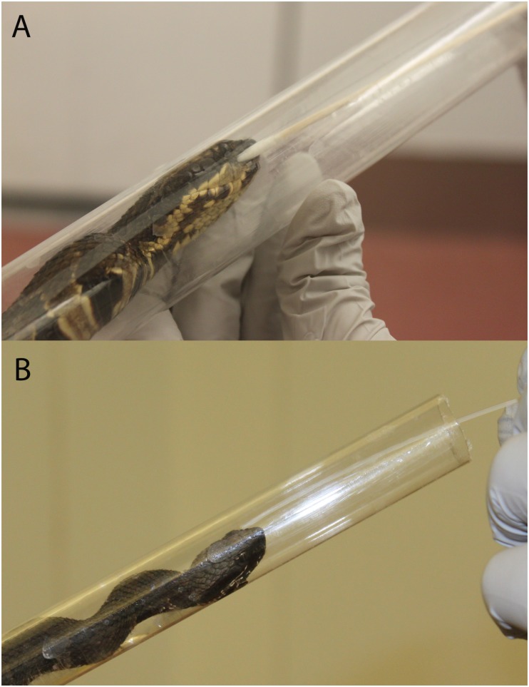
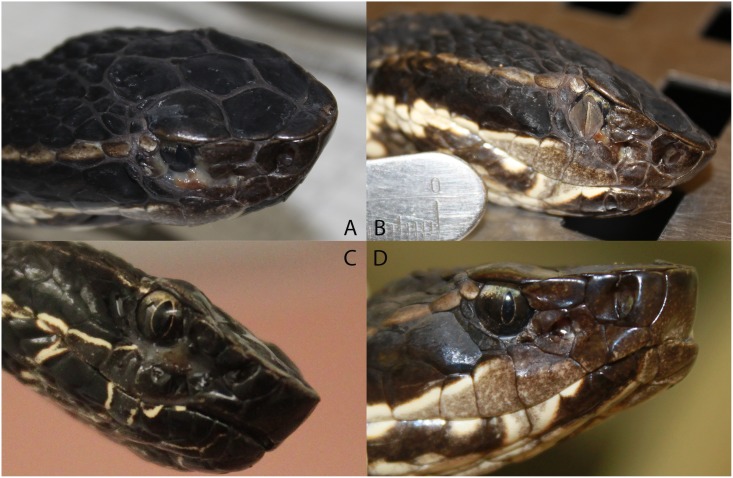
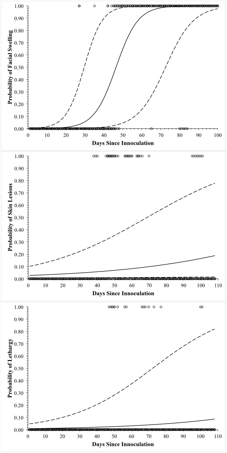
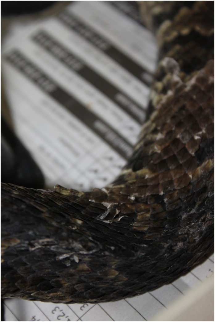
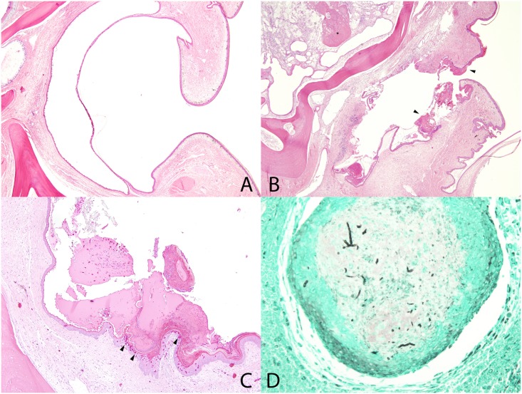
Snake fungal disease (SFD) is a clinical syndrome associated with dermatitis, myositis, osteomyelitis, and pneumonia in several species of free-ranging snakes in the US. The causative agent has been suggested as Ophidiomyces ophiodiicola, but other agents may contribute to the syndrome and the pathogenesis is not understood. To understand the role of O. ophiodiicola in SFD, a cottonmouth snake model of SFD was designed. Five cottonmouths (Agkistrodon piscivorous) were experimentally challenged by nasolabial pit inoculation with a pure culture of O. ophiodiicola. Development of skin lesions or facial swelling at the site of inoculation was observed in all snakes. Twice weekly swabs of the inoculation site revealed variable presence of O. ophiodiicola DNA by qPCR in all five inoculated snakes for 3 to 58 days post-inoculation; nasolabial flushes were not a useful sampling method for detection. Inoculated snakes had a 40% mortality rate. All inoculated snakes had microscopic lesions unilaterally on the side of the swabbed nasolabial pit, including erosions to ulcerations and heterophilic dermatitis. All signs were consistent with SFD; however, the severity of lesions varied in individual snakes, and fungal hyphae were only observed in 3 of 5 inoculated snakes. These three snakes correlated with post-mortem tissue qPCR evidence of O. ophiodiicola. The findings of this study conclude that O. ophiodiicola inoculation in a cottonmouth snake model leads to disease similar to SFD, although lesion severity and the fungal load are quite variable within the model. Future studies may utilize this model to further understand the pathogenesis of this disease and develop management strategies that mitigate disease effects, but investigation of other models with less variability may be warranted.
Conflict of interest statement
Figures
References
-
- Daszak P, Cunningham AA, Hyatt AD. Emerging infectious diseases of wildlife—threats to biodiversity and human health. Science 2000; 287: 43–449. - PubMed
-
- Allender MC, Dreslik MJ, Wylie DB, Wylie SJ, Scott JW, Phillips CA. Ongoing health assessment and prevalence of Chrysosporium in the Eastern Massasauga (Sistrurus catenatus catenatus). Copeia 2013: 97–102.
-
- Clark RW, Marchand MN, Clifford BJ, Stechert R, Stephens S. Decline of an isolated timber rattlesnake (Crotalus horridus) population: interactions between climate change, disease, and loss of genetic diversity. Biolog Conserv 2011; 144:886–891.
-
- McBride MP, Wojick KB, Georoff TA, Kimbro J, Garner MM, Wang X, et al. Ophidiomyces ophiodiicola dermatitis in eight free-ranging timber rattlesnakes (Crotalus horridus) from Massachusetts. J Zoo Wildl Med 2015; 46:86–94. - PubMed
Publication types
MeSH terms
Substances
LinkOut - more resources
Full Text Sources
Other Literature Sources
Medical
