. 2016 Feb;64(2):300-16.
doi: 10.1002/glia.22930. Epub 2015 Oct 15.

Sequential activation of microglia and astrocyte cytokine expression precedes increased Iba-1 or GFAP immunoreactivity following systemic immune challenge

Affiliations

Sequential activation of microglia and astrocyte cytokine expression precedes increased Iba-1 or GFAP immunoreactivity following systemic immune challenge

Diana M Norden et al. Glia. 2016 Feb.

Abstract

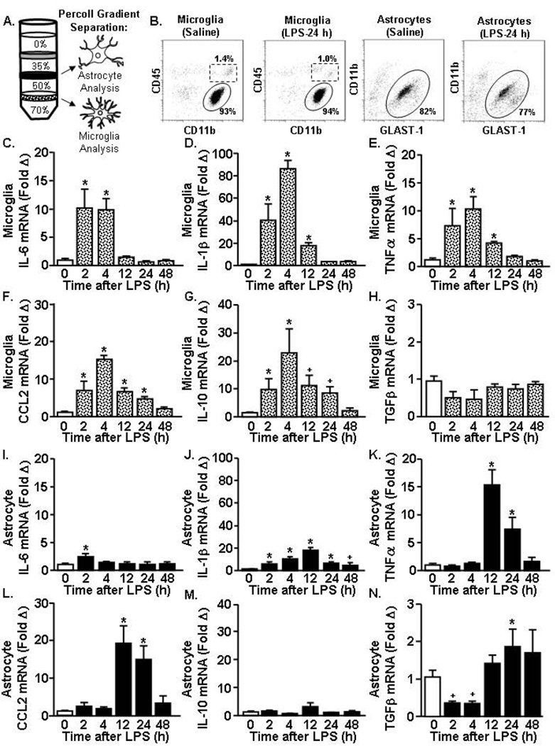
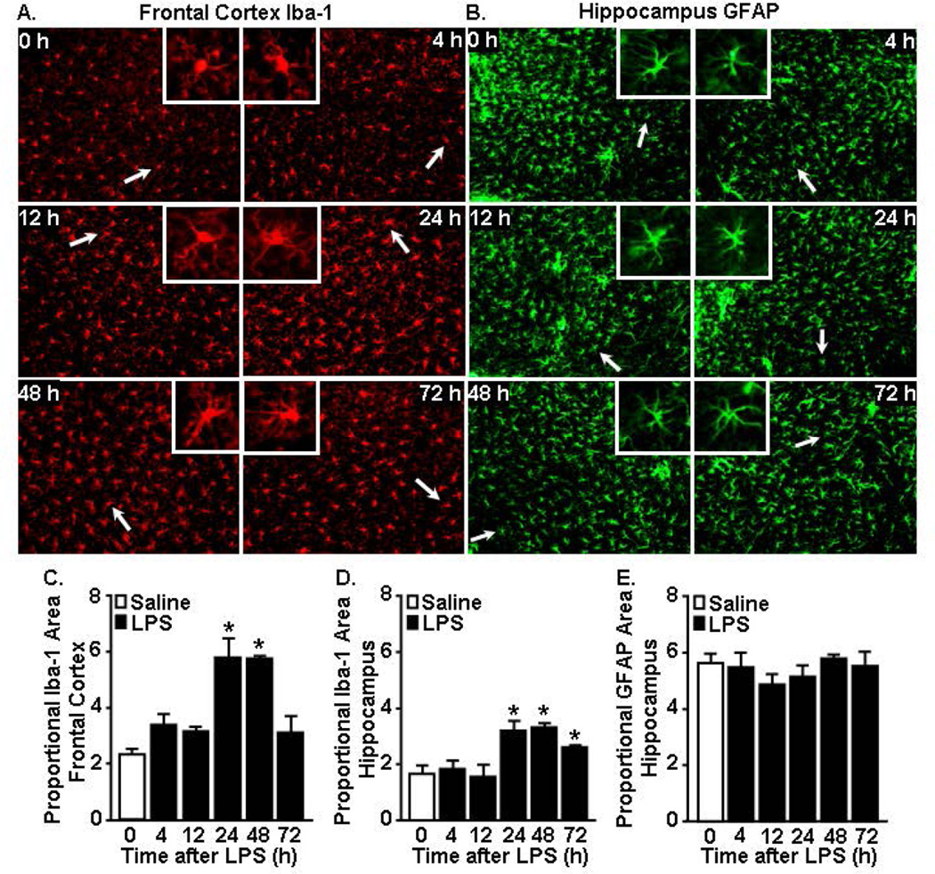
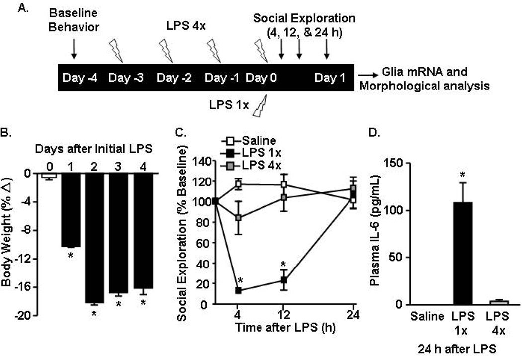
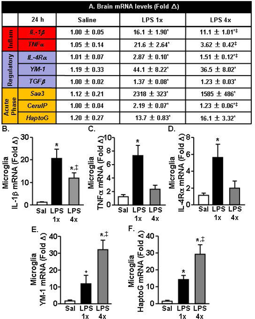
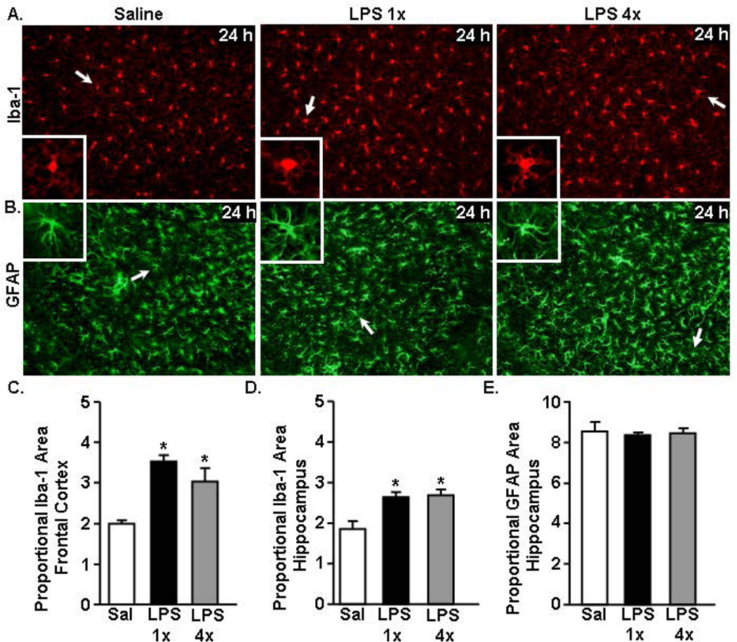
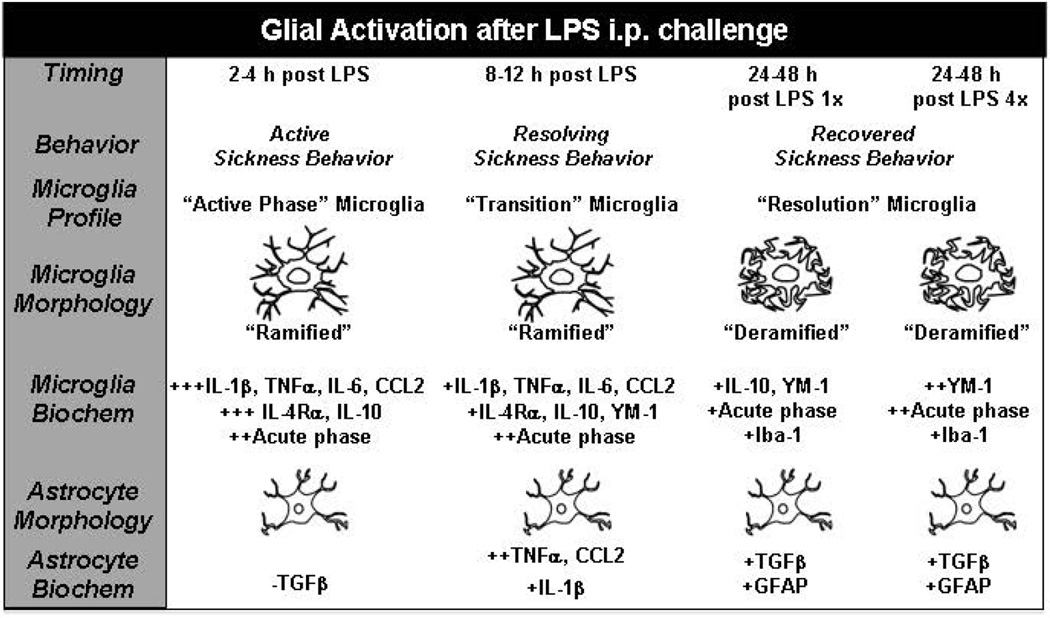
Activation of the peripheral immune system elicits a coordinated response from the central nervous system. Key to this immune to brain communication is that glia, microglia, and astrocytes, interpret and propagate inflammatory signals in the brain that influence physiological and behavioral responses. One issue in glial biology is that morphological analysis alone is used to report on glial activation state. Therefore, our objective was to compare behavioral responses after in vivo immune (lipopolysaccharide, LPS) challenge to glial specific mRNA and morphological profiles. Here, LPS challenge induced an immediate but transient sickness response with decreased locomotion and social interaction. Corresponding with active sickness behavior (2-12 h), inflammatory cytokine mRNA expression was elevated in enriched microglia and astrocytes. Although proinflammatory cytokine expression in microglia peaked 2-4 h after LPS, astrocyte cytokine, and chemokine induction was delayed and peaked at 12 h. Morphological alterations in microglia (Iba-1(+)) and astrocytes (GFAP(+)), however, were undetected during this 2-12 h timeframe. Increased Iba-1 immunoreactivity and de-ramified microglia were evident 24 and 48 h after LPS but corresponded to the resolution phase of activation. Morphological alterations in astrocytes were undetected after LPS. Additionally, glial cytokine expression did not correlate with morphology after four repeated LPS injections. In fact, repeated LPS challenge was associated with immune and behavioral tolerance and a less inflammatory microglial profile compared with acute LPS challenge. Overall, induction of glial cytokine expression was sequential, aligned with active sickness behavior, and preceded increased Iba-1 or GFAP immunoreactivity after LPS challenge.

Keywords: astrocytes; lipopolysaccharide; microglia; neuroinflammation; sickness behavior.


Figures

Figure 1
Figure 1. Acute LPS challenge promoted a transient sickness response that was resolved within 48 h
Adult BALB/c mice were injected (i.p.) with saline or LPS (10 µg). Over a 72 h time course, A) body weight, B) locomotor activity, and C) social exploratory behavior were determined. D) IL-6 protein levels were determined in plasma collected 2, 4, 12, 24, and 48 h later. Data expressed as percent change from baseline. Means with (*) are different from saline controls (p<0.05) and means with (+) tend to be different from saline controls (p=0.06–0.10).
Figure 2
Figure 2. Rapid microglial cytokine induction after acute LPS challenge preceded astrocyte cytokine expression
Adult mice received (i.p.) saline or LPS (10 µg) and A) astrocytes and microglia were isolated and Percoll-enriched 2, 4, 12, 24, and 48 h later. B) Representative bivariate dot plots of CD11b and CD45 labeling of microglia and CD11b and Glast-1 labeling of astrocytes collected after Percoll enrichment. Levels of C) IL-6, D) IL-1β, E) TNFα, F) CCL2, G) IL-10, and H) TGFβ mRNA were determined in enriched microglia. The expression levels for I) IL-6, J) IL-1β, K) TNFα, L) CCL2, M) IL-10, and N) TGFβ were determined in enriched astrocytes. Means with (*) are different from saline controls (p<0.05) and means with (+) tend to be different from saline controls (p=0.06–0.10).
Figure 3
Figure 3. Iba-1 immunoreactivity of microglia increased 24 and 48 h after acute LPS challenge while GFAP immunoreactivity of astrocytes was unaffected
Brains were collected and Iba-1 and GFAP immunoreactivity was determined 4, 12, 24, 48, and 72 h after saline or LPS (10 µg) injection. Representative images of labeling for A) Iba-1 (cortex) and B) GFAP (hippocampus) are shown. White arrows indicate the enlarged insert of a representative cell. Proportional area of Iba-1 in the C) frontal cortex and D) hippocampus. E) Proportional area of GFAP in the hippocampus. *p<0.05 from saline.
Figure 4
Figure 4. Acute LPS challenge corresponded to a more pronounced active sickness response over 24 h compared to the behavioral response 24 h after the last injection of repeated LPS
A) Experiment schematic injection paradigm for the single- and four repeated-injections of LPS. Adult BALB/c mice were injected i.p. with saline, 1× LPS (20 µg), or 4× LPS (20 µg). In LPS 4× mice, the four repeated LPS injections were provided in 24 h increments. B) Over the LPS 4× injection time course, body weight was determined daily. C) Social exploratory behavior was determined prior to injections and 4–24 h after the last injection of LPS. D) Plasma IL-6 levels were determined at 24 h after the last LPS injection. *p<0.05 from saline.
Figure 5
Figure 5. A more pronounced active inflammatory response was evident in the brain 24 h after acute LPS challenge compared to repeated LPS challenge
Adult BALB/c mice were injected (i.p.) with saline, LPS 1× (20 µg), or LPS 4× (20 µg), and a 1 mm coronal brain section was collected 24 h after the last injection. A) mRNA expression of several inflammatory (“Inflam”), “Regulatory,” and “Acute Phase” markers were determined. In the same mice, mRNA levels of B) IL-1β, C) TNFα, D) IL-4Rα, E) YM-1, and F) HaptoG were determined in enriched microglia. Means with (*) are different from saline controls (p<0.05). Means with (‡) are different from LPS 1× mice (p<0.05).
Figure 6
Figure 6. Increased Iba-1 immunoreactivity of microglia 24 h after acute and repeated LPS challenge with no change of GFAP immunoreactivity
Adult BALB/c mice were injected i.p. with saline, LPS 1× (20 µg), or LPS 4× (20 µg). After 24 h, brains were collected and Iba-1 and GFAP immunoreactivity was determined. Representative images of labeling for A) Iba-1 (cortex) and B) GFAP (hippocampus) are shown. White arrows indicate the cell represented in the inset. Proportional area of Iba-1 in the C) frontal cortex and D) hippocampus. E) Proportional area of hippocampal GFAP labeling. *p<0.05 from saline.
Figure 7
Figure 7. Time course of biochemical and morphological alterations of microglial and astrocyte activation in the context of LPS-induced sickness behavior
This illustration summarizes the biochemical and morphological alterations of microglia and astrocytes after peripheral LPS challenge. Activation: Microglia were rapidly and robustly activated at 2–4 h after LPS to express cytokines, chemokines and acute phase markers. This microglial activation was associated with a ramified Iba-1 morphology and an active sickness behavioral response. At 2–4 h astrocytes had a down regulation of TGFβ, limited cytokine induction and no alteration in GFAP morphology. Transition: By 12 h after LPS, the profile of microglia was attenuated. This change was associated with a ramified Iba-1 morphology and a resolving sickness behavioral response. Astrocytes had a peak activation of cytokine and chemokine expression 12 h after LPS but no alteration in GFAP morphology. Resolution: By 24–48 h after LPS microglia had baseline expression of cytokines and chemokines, but also had some level of maintenance of IL-10, YM-1 and acute phase genes in acute and repeated LPS injection. This regulatory profile, however, was augmented by repeated LPS injection. In both cases, microglia resolution was associated with de-ramified Iba-1 morphology and resolved sickness behavior. Astrocytes had an altered gene expression profile after LPS with increased TGFβ, but no changes were detected in their GFAP morphology.

Similar articles

Cited by

References

    1. Berg BM, Godbout JP, Kelley KW, Johnson RW. Alpha-tocopherol attenuates lipopolysaccharide-induced sickness behavior in mice. Brain, behavior, and immunity. 2004;18:149–157. - PubMed
    1. Biber K, Neumann H, Inoue K, Boddeke HW. Neuronal 'On' and 'Off' signals control microglia. Trends in neurosciences. 2007;30:596–602. - PubMed
    1. Biswas SK, Lopez-Collazo E. Endotoxin tolerance: new mechanisms, molecules and clinical significance. Trends in immunology. 2009;30:475–487. - PubMed
    1. Bluthe RM, Laye S, Michaud B, Combe C, Dantzer R, Parnet P. Role of interleukin-1beta and tumour necrosis factor-alpha in lipopolysaccharide-induced sickness behaviour: a study with interleukin-1 type I receptor-deficient mice. The European journal of neuroscience. 2000a;12:4447–4456. - PubMed
    1. Bluthe RM, Michaud B, Poli V, Dantzer R. Role of IL-6 in cytokine-induced sickness behavior: a study with IL-6 deficient mice. Physiology & behavior. 2000b;70:367–373. - PubMed

Publication types