Case Reports
. 2016 Jun;27(6):1665-77.
doi: 10.1681/ASN.2015040348. Epub 2015 Oct 15.

A Familial C3GN Secondary to Defective C3 Regulation by Complement Receptor 1 and Complement Factor H

Affiliations
Case Reports

A Familial C3GN Secondary to Defective C3 Regulation by Complement Receptor 1 and Complement Factor H

Sophie Chauvet et al. J Am Soc Nephrol. 2016 Jun.

Abstract

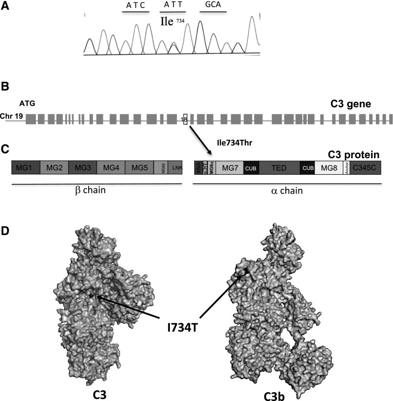
C3 glomerulopathy is a recently described form of CKD. C3GN is a subtype of C3 glomerulopathy characterized by predominant C3 deposits in the glomeruli and is commonly the result of acquired or genetic abnormalities in the alternative pathway (AP) of the complement system. We identified and characterized the first mutation of the C3 gene (p. I734T) in two related individuals diagnosed with C3GN. Immunofluorescence and electron microscopy studies showed C3 deposits in the subendothelial space, associated with unusual deposits located near the complement receptor 1 (CR1)-expressing podocytes. In vitro, this C3 mutation exhibited decreased binding to CR1, resulting in less CR1-dependent cleavage of C3b by factor 1. Both patients had normal plasma C3 levels, and the mutant C3 interacted with factor B comparably to wild-type (WT) C3 to form a C3 convertase. Binding of mutant C3 to factor H was normal, but mutant C3 was less efficiently cleaved by factor I in the presence of factor H, leading to enhanced C3 fragment deposition on glomerular cells. In conclusion, our results reveal that a CR1 functional deficiency is a mechanism of intraglomerular AP dysregulation and could influence the localization of the glomerular C3 deposits.

Keywords: C; glomerular disease; immunology and pathology.


Figures

Figure 1.
Figure 1.
Pedigree with familial C3G and microscopy of kidney biopsies of patient II-1. (A) Pedigree demonstrating that two members of generation II carry the C3 mutation I734T (II-1 and II-2, black squares). (B) By light microscopy, patient II-1 presented with a membranoproliferative GN pattern characterized by hypertrophy of the mesangial matrix with hypercellularity secondary to infiltration by neutrophils and macrophages, and mesangial and subendothelial deposits. (C) Immunofluorescence staining was positive for C3. Staining for Ig was negative (not shown). Deposits were granular and localized in the mesangium and along the GBM, as well as in the subepithelial or subendothelial spaces. (D) Electron microscopy (original magnification, ×3000) showed subendothelial and voluminous extramembranous deposits (humps) (*) (E) Electron microscopy (original magnification, ×10,000) showed microvesicular structures (single arrow) and laminated thread-like structures (double arrow). (F, G) Recurrence of C3GN with a membranoproliferative GN pattern was observed after kidney transplantation in patient II-1 by light microscopy. (H) Immunofluorescence staining was positive only for C3. (I) Deposits were predominantly localized in the mesangium, as detected by electron microscopy.
Figure 2.
Figure 2.
Localization of C3 I734T mutation. (A) A chromatogram corresponding to the DNA sequence surrounding the mutated nucleotides in C3 of a patient carrying I734T. (B) Position within the C3 gene and (C) the primary structure of C3. (D) Position of the residue on the protein surface. Mapping of I734T on the surface of C3 and C3b was performed using Pymol software.
Figure 3.
Figure 3.
Complement AP activation on resting and apoptotic-necrotic glomerular endothelial cells. Cells were incubated with sera from C3 I734T patients. (A) C3 fragment deposition on resting GEnC in the presence of sera from NHS, serum depleted in FH (FH-dpl), and C3 I734T patient II-2. (B) C3 fragment deposition after incubation of apoptotic-necrotic GEnC with NHS or patient sera. (C) C5b-9 deposition on apoptotic-necrotic GEnC in the presence of NHS and patient sera. These analyses were carried out three times using patient II-1 serum. In every experiment the deposition was compared with a control NHS as well as a total of four NHS, which were very similar to the control NHS (not shown). A representative flow cytometry histogram is shown. C3 deposition obtained with patient II-2 and FH-dpl were normalized to the C3 deposition from the reference NHS on cells to compare the fold increase. (D) C3 deposition on apoptotic-necrotic GEnC in the presence of patients’ sera, FH-dpl serum, and serum of a patient with C-terminal mutation in FH (W1183R), supplemented with increasing concentrations of purified FH. The C3 fragments deposition in absence of FH was taken as 100% and the decrease at any dose of FH was calculated as a percentage of this basal level. Iso, isotope; RFI, relative fluorescent intensity.
Figure 4.
Figure 4.
Complement AP activation on podocytes. (A) Podocytes express CR1 on their surface. Labeling of CR1 with anti–CR1-FITC was positive compared with the isotype control. (B, C) The deposition of C3 activation fragments on podocytes was compared after incubation with NHS and serum from patient II-1. (B) C3c staining (recognizing C3b/iC3b deposition). Stronger staining on the cell surface and in the extracellular matrix was observed with the patient serum (arrow). (C) C3d staining (recognizing primarily C3dg/C3d and to a smaller extent C3b/iC3b). Similar staining was detected on the cell surface but, in the presence of patient serum, deposits in the extracellular matrix were also observed (double arrow). Experiments were performed two times with same results.
Figure 5.
Figure 5.
AP C3 convertase formation by C3 I734T and cleavage of C3 I734T by the C3 convertase. (A) I734 position on the structures of C3b with FB in closed (refractory to cleavage by FD) and open (prone to cleavage by FD) conformations and the active convertase C3bBb. C3b is colored light gray and FB is dark gray. (B) The binding of FB to recombinant WT or C3 I734T protein bound to an anti-C3d mAb on the biosensor chip studied by SPR. Binding of FB was analyzed by flowing recombinant C3 WT (gray line) or C3 I734T (black line) over an anti-C3d antibody coated chip, followed by FB. Binding of FB to the mutant protein was similar to WT. (C) Formation of C3 convertase by injection of recombinant C3 WT (gray line) or C3 I734T (black line) on anti-C3d antibody coated chip surface, followed by injection of FB plus FD. Protein deposition on the chip was followed over time. No difference was observed in the two cases. (D) Hemolytic assay for the C3 convertase forming capacity of normal plasma and patient II-1 plasma on rabbit erythrocytes. Lysis was similar in the three different experiments. (E) Recombinant C3 WT and C3 I734T were tested for their capacity to be cleaved to C3b in the presence of a preformed solid-phase C3 convertase. Both α-chains of C3 WT and C3 I734T were cleaved to α′. The experiment was repeated twice with identical results.
Figure 6.
Figure 6.
Interaction of C3 I734T with FH was normal with normal regulation of C3 convertase on sheep erythrocytes, but was associated with decreased inactivation by FI. (A) The location of I734 on the structure of C3b in a complex with FH CCP1–4. I734 is close to the binding site of FH-CCP1. The arrow shows the position of the mutated residue, which is hidden by FH. C3b is shown in light gray and FH in dark gray. (B) The real-time binding of purified recombinant WT or C3 I734T to immobilized FH was studied by SPR. FH was coupled to the biosensor chip followed by injection of recombinant C3 WT (gray line) or C3 I734T (black line) over the chip. The affinity for FH was determined by fitting the sensorgrams with the ProteON Manager software. The affinity of recombinant C3 I734T and C3 WT for FH were similar. Results were also compared with those obtained with the mutant R139W, which has known functional consequences. (C) Hemolytic assays with sheep erythrocytes showed similar lysis of cells in the presence of plasma of patient II-1 compared with normal plasma (n=3). (D) Inactivation of C3(H2O) I734T to iC3 I734T by FI was decreased compared with C3(H2O) WT. Recombinant C3(H2O) I734T and C3(H2O) WT were incubated with FI and FH. Cleavage of C3b to iC3 was indicated by generation of α43 product and decrease of the α-chain. Commercially available plasma-derived C3, treated identically to the recombinant proteins to obtain C3(H2O) and incubated in presence of FH and FI, served as a positive control for these experiments. The experiment was repeated three times with statistically significant results.
Figure 7.
Figure 7.
Interactions of C3 WT and C3 I734T was normally inactivated by FI in the presence of MCP. (A) Position of I734T on the structure of C3b in a complex with MCP. I734 is remote from the binding site for MCP. C3b is colored in light gray and MCP in dark gray. (B) The real-time binding of recombinant C3 WT or mutant C3 I734T to MCP was studied by SPR. MCP was coupled to the biosensor chip followed by injection of recombinant C3 WT (gray line) or C3 I734T (black line). The affinity for MCP was determined by fitting the sensorgrams with Langmuir 1:1 model by ProteON Manager software. The affinity of recombinant C3 I734T for MCP was similar compared with the C3 WT. (C) Cofactor activity of MCP for the inactivation of recombinant C3(H2O) to iC3 by FI. Inactivation of C3 I734T to iC3 I734T by FI and MCP was similar to that of C3 WT. Recombinant C3(H2O) I734T or C3(H2O) WT was incubated with FI and MCP. Cleavage of C3(H2O) to iC3 was measured by the generation of α43 fragment and decrease of the α-chain. The experiment was repeated three times with similar results.
Figure 8.
Figure 8.
C3 binding to CR1 and cofactor activity of CR1 were decreased. (A) I734 position on the structure of C3b in a complex with CR1 (putative binding site in dark gray). I734 is close to the binding site of CR1. C3b is colored in light gray. (B) The real-time binding of purified recombinant C3WT or C3 I734T to CR1 was studied by SPR. CR1 was coupled to the biosensor chip and C3WT (gray line) or C3 I734T (black line) was flowed over the surface. The affinity for CR1 was determined by fitting the sensorgrams. The affinity of recombinant C3 I734T for CR1 was significantly decreased compared with C3 WT. (C) Cofactor activity of CR1 for the inactivation of recombinant C3(H2O) to iC3 by FI. Inactivation of C3(H20) I734T by FI in the presence of CR1 was decreased compared with inactivation of C3 WT. Recombinant C3 I734T and C3 WT were incubated with FI and FH. Cleavage of C3(H2O) to iC3 was indicated by generation of α43 fragment and decrease of the α-chain. The experiment was repeated four times with similar results.
Figure 9.
Figure 9.
Schematic diagram of AP dysregulation caused by the C3 I734T mutation leading to glomerular C3 deposits. Generated C3b and its cleavage fragments from the capillary lumen (CL) cross the fenestrated glomerular endothelium (GEc) and reach the subendothelial space and the mesangium (M) in the vicinity of mesangial cells (Mc), where C3b is only partially regulated by FH, leading to the accumulation of C3b and iC3b in these areas. C3b and iC3b fragments cross the GBM toward the subepithelial space. They cannot be appropriately regulated by CR1 expressed on the foot podocytes, resulting in iC3b/C3d accumulation (humps). US, urinary space.

References

    1. Fakhouri F, Frémeaux-Bacchi V, Noël LH, Cook HT, Pickering MC: C3 glomerulopathy: a new classification. Nat Rev Nephrol 6: 494–499, 2010 - PubMed
    1. Hou J, Markowitz GS, Bomback AS, Appel GB, Herlitz LC, Barry Stokes M, D’Agati VD: Toward a working definition of C3 glomerulopathy by immunofluorescence. Kidney Int 85: 450–456, 2014 - PubMed
    1. Pickering MC, D’Agati VD, Nester CM, Smith RJ, Haas M, Appel GB, Alpers CE, Bajema IM, Bedrosian C, Braun M, Doyle M, Fakhouri F, Fervenza FC, Fogo AB, Frémeaux-Bacchi V, Gale DP, Goicoechea de Jorge E, Griffin G, Harris CL, Holers VM, Johnson S, Lavin PJ, Medjeral-Thomas N, Paul Morgan B, Nast CC, Noel LH, Peters DK, Rodríguez de Córdoba S, Servais A, Sethi S, Song WC, Tamburini P, Thurman JM, Zavros M, Cook HT: C3 glomerulopathy: consensus report. Kidney Int 84: 1079–1089, 2013 - PMC - PubMed
    1. Sethi S, Fervenza FC: Membranoproliferative glomerulonephritis--a new look at an old entity. N Engl J Med 366: 1119–1131, 2012 - PubMed
    1. Sethi S, Fervenza FC, Zhang Y, Nasr SH, Leung N, Vrana J, Cramer C, Nester CM, Smith RJ: Proliferative glomerulonephritis secondary to dysfunction of the alternative pathway of complement. Clin J Am Soc Nephrol 6: 1009–1017, 2011 - PMC - PubMed

Publication types