. 2016 May;27(5):1379-88.
doi: 10.1681/ASN.2015040460. Epub 2015 Oct 15.

Endothelial Krüppel-Like Factor 4 Mediates the Protective Effect of Statins against Ischemic AKI

Affiliations

Endothelial Krüppel-Like Factor 4 Mediates the Protective Effect of Statins against Ischemic AKI

Tadashi Yoshida et al. J Am Soc Nephrol. 2016 May.

Abstract

Endothelial cells participate in the pathophysiology of ischemic AKI by increasing the expression of cell adhesion molecules and by recruiting inflammatory cells. We previously showed that endothelial Krüppel-like factor 4 (Klf4) regulates vascular cell adhesion molecule 1 (Vcam1) expression and neointimal formation after carotid injury. In this study, we determined whether endothelial Klf4 is involved in ischemic AKI using endothelial Klf4 conditional knockout (Klf4 cKO) mice generated by breeding Tek-Cre mice and Klf4 floxed mice. Klf4 cKO mice were phenotypically normal before surgery. However, after renal ischemia-reperfusion injury, Klf4 cKO mice exhibited elevated serum levels of urea nitrogen and creatinine and aggravated renal histology compared with those of Klf4 floxed controls. Moreover, Klf4 cKO mice exhibited enhanced accumulation of neutrophils and lymphocytes and elevated expression of cell adhesion molecules, including Vcam1 and Icam1, in injured kidneys. Notably, statins ameliorated renal ischemia-reperfusion injury in control mice but not in Klf4 cKO mice. Mechanistic analyses in cultured endothelial cells revealed that statins increased KLF4 expression and that KLF4 mediated the suppressive effect of statins on TNF-α-induced VCAM1 expression by reducing NF-κB binding to the VCAM1 promoter. These results provide evidence that endothelial Klf4 is renoprotective and mediates statin-induced protection against ischemic AKI by regulating the expression of cell adhesion molecules and concomitant recruitment of inflammatory cells.

Keywords: acute renal failure; adhesion molecule; endothelial cells; ischemia-reperfusion; transcription factors.


Figures

Figure 1.
Figure 1.
Generation of Klf4 cKO mice. (A) Schematic representation of endothelial deletion of the Klf4 gene is shown. The numbers shown represent Klf4 exons. The triangles represent the loxP sites. X represents breeding. (B) Recombination of the Klf4loxP allele was examined in the kidneys of Klf4 cKO mice and control mice, as determined by PCR. (C) Klf4 expression was examined by immunohistochemistry in the kidneys of Klf4 cKO mice and control mice. Klf4 expression was visualized by diaminobenzidine, and sections were counterstained with hematoxylin. Representative pictures are shown from six mice analyzed per each genotype. Magnification ×20. Bar: 100 μm. Arrowheads indicate the glomeruli, and arrows indicate the vessels. Inset: Dotted area is enlarged.
Figure 2.
Figure 2.
Endothelial Klf4 deletion exacerbated renal I/R injury. Klf4 cKO mice and control mice were subjected to bilateral renal ischemia for 35 minutes (I/R) or sham operation, and serum levels of urea nitrogen (A) and creatinine (B) were measured 24 hours after reperfusion. n=5–6 per each group. *P<0.05 compared with sham-operated mice. #P<0.05 compared with I/R-injured control mice.
Figure 3.
Figure 3.
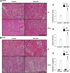
Endothelial Klf4 deletion exacerbated renal histologic damages after I/R injury. Klf4 cKO mice and control mice were subjected to bilateral renal ischemia for 35 minutes (I/R) or sham operation, and renal histology was examined 24 hours after reperfusion. (A and B) Representative hematoxylin-eosin staining for the outer stripe of outer medulla (OSOM) and the inner stripe of outer medulla (ISOM) are shown. Bars: 200 μm. (C–E) Levels of the formation of proteinaceous casts (C), tubular necrosis (D), and medullary congestion (E) were scored semiquantitatively. n=5–6 per each group. Magnification ×10. *P<0.05 compared with sham-operated mice. #P<0.05 compared with I/R-injured control mice.
Figure 4.
Figure 4.
Accumulation of inflammatory cells was enhanced in Klf4 cKO mice after renal I/R injury. Klf4 cKO mice and control mice were subjected to bilateral renal ischemia for 35 minutes (I/R) or sham operation, and the accumulation of neutrophils (A and B) and lymphocytes (C and D) was examined 24 hours after reperfusion. (A and C) Representative pictures for immunohistochemical staining for neutrophils (A) and lymphocytes (C) are shown. Neutrophils (A) and lymphocytes (C) were visualized by diaminobenzidine, and sections were counterstained with hematoxylin. Bars: 100 μm. Red arrowheads indicate the positive cells. Insets: Dotted areas are enlarged. (B and D) The numbers of neutrophils (B) and lymphocytes (D) per five random fields in the kidneys were quantified. Magnification ×10. n=5–6 per each group. *P<0.05 compared with sham-operated mice. #P<0.05 compared with I/R-injured control mice.
Figure 5.
Figure 5.
Induction of cell adhesion molecules was augmented in Klf4 cKO mice after renal I/R injury. Klf4 cKO mice and control mice were subjected to bilateral renal ischemia for 35 minutes (I/R) or sham operation, and the expression of cell adhesion molecules was examined 24 hours after reperfusion. Expression of Vcam1 (A) and Icam1 (B) and Cxcl1, Cxcl2, Tnf, Il6, and Il10 (C) in the kidneys was determined by real-time RT-PCR. n=5–6 per each group. *P<0.05 compared with sham-operated mice. #P<0.05 compared with I/R-injured control mice.
Figure 6.
Figure 6.
Fluvastatin ameliorated renal I/R injury in control mice, but not in Klf4 cKO mice. Klf4 cKO mice and control mice were pretreated with fluvastatin (Fluva) orally for 3 days, and then they were subjected to bilateral renal ischemia for 35 minutes (I/R) or sham operation. Renal damages were evaluated 24 hours after reperfusion. (A and B) Serum levels of urea nitrogen (A) and creatinine (B) were measured. (C and D) Levels of the formation of proteinaceous casts (C) and tubular necrosis (D) were scored semiquantitatively. (E and F) Expression of Vcam1 (E) and Icam1 (F) in the kidneys was determined by real-time RT-PCR. n=5–6 per each group. Data for mice that were not treated with fluvastatin are reproduced from Figures 2, A and B, 3, C and D, and 5, A and B. *P<0.05 compared with sham-operated mice. #P<0.05 compared with control mice receiving the same treatment. $P<0.05 compared with I/R-injured mice without fluvastatin treatment.
Figure 7.
Figure 7.
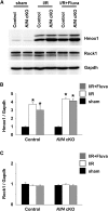
Hmox1, but not Rock1, was increased in the kidneys after I/R injury. Klf4 cKO mice and control mice were pretreated with fluvastatin (Fluva) orally for 3 days, and then they were subjected to bilateral renal ischemia for 35 minutes (I/R) or sham operation. Expression of Hmox1, Rock1, and Gapdh (glyceraldehyde 3-phosphate dehydrogenase) was determined by Western blotting. (A) Representative pictures are shown. (B and C) Expression of Hmox1, Rock, and Gapdh was determined by densitometry. n=5–6 per each group. *P<0.05 compared with sham-operated mice.
Figure 8.
Figure 8.
KLF4 mediated the suppressive effect of statins on TNF-induced VCAM1 expression by reducing the association of NF-κB with the VCAM1 promoter in HUVECs. HUVECs were transfected with siRNAs for KLF4 (siKLF4) or a scrambled sequence (siScramble), and then they were treated with fluvastatin, simvastatin, or TNF for 24 hours. (A and B) Expression of KLF4 (A) and VCAM1 (B) was determined by real-time RT-PCR. (C) Expression of VCAM1, phosphorylated p65 (p-p65), p65, and GAPDH (glyceraldehyde 3-phosphate dehydrogenase) was examined by Western blotting. (D) Association of p65 with the VCAM1 promoter was determined by chromatin immunoprecipitation assays. n=4. *P<0.05 compared with cells without TNF treatment. #P<0.05 compared with cells transfected with siScramble. $P<0.05 compared with cells untreated with statins.

References

    1. Kinsey GR, Li L, Okusa MD: Inflammation in acute kidney injury. Nephron, Exp Nephrol 109: e102–e107, 2008 - PMC - PubMed
    1. Molitoris BA: Therapeutic translation in acute kidney injury: The epithelial/endothelial axis. J Clin Invest 124: 2355–2363, 2014 - PMC - PubMed
    1. Kelly KJ, Williams WW Jr, Colvin RB, Bonventre JV: Antibody to intercellular adhesion molecule 1 protects the kidney against ischemic injury. Proc Natl Acad Sci U S A 91: 812–816, 1994 - PMC - PubMed
    1. Rabb H, Mendiola CC, Saba SR, Dietz JR, Smith CW, Bonventre JV, Ramirez G: Antibodies to ICAM-1 protect kidneys in severe ischemic reperfusion injury. Biochem Biophys Res Commun 211: 67–73, 1995 - PubMed
    1. Kapitsinou PP, Sano H, Michael M, Kobayashi H, Davidoff O, Bian A, Yao B, Zhang MZ, Harris RC, Duffy KJ, Erickson-Miller CL, Sutton TA, Haase VH: Endothelial HIF-2 mediates protection and recovery from ischemic kidney injury. J Clin Invest 124: 2396–2409, 2014 - PMC - PubMed

MeSH terms

Substances