. 2015 Oct 15:15:706.
doi: 10.1186/s12885-015-1738-3.

MiR-449a suppresses the epithelial-mesenchymal transition and metastasis of hepatocellular carcinoma by multiple targets

Affiliations

MiR-449a suppresses the epithelial-mesenchymal transition and metastasis of hepatocellular carcinoma by multiple targets

Shu-Peng Chen et al. BMC Cancer. .

Abstract

Background: Increasing evidence indicates that Epithelial-mesenchymal transition (EMT) can be regulated by microRNAs (miRNAs). MiR-449a is a liver abundant miRNA. However, the role of miR-449a in the metastasis of hepatocellular carcinoma (HCC) remains largely unknown.

Methods: The expression levels of miR-449a were first examined in HCC cell lines and tumour tissues by real-time PCR. The in vitro and in vivo functional effect and underlying molecular mechanisms of miR-449a were examined further.

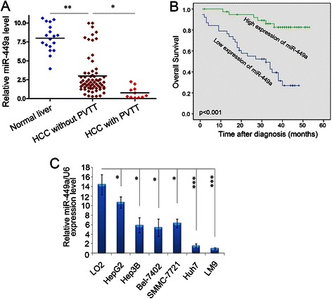
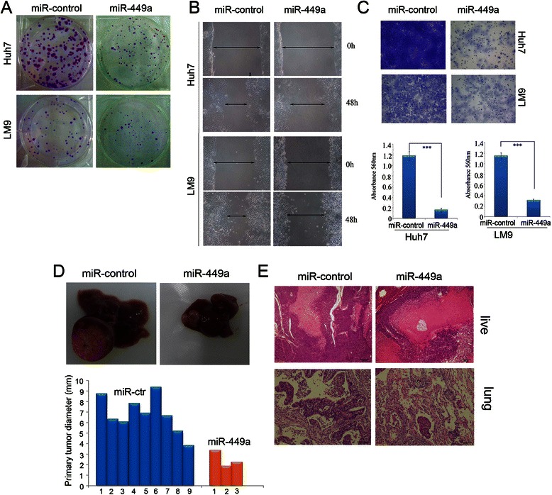
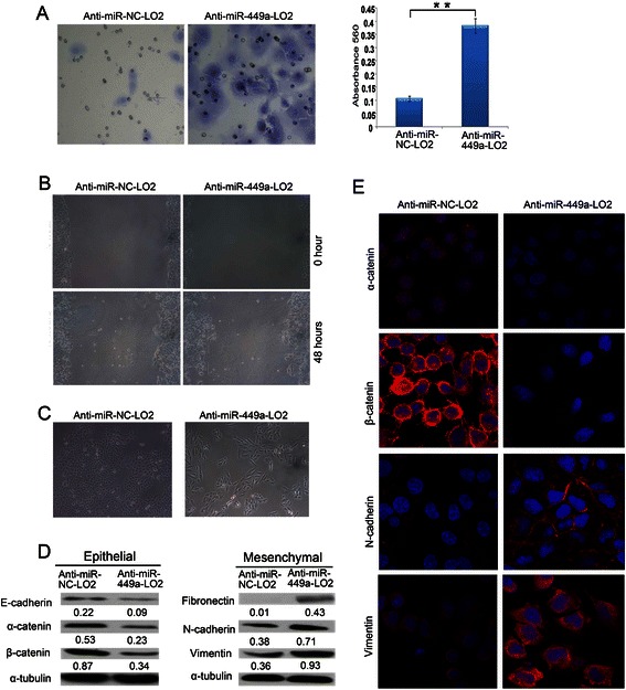
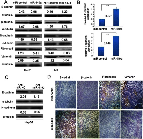
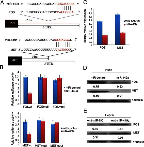
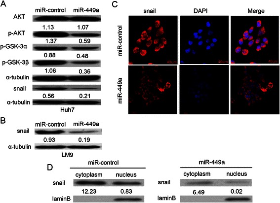
Results: In the present study, we found that miR-449a was significantly decreased in HCC cells and tissues, especially in those with the portal vein tumor thrombus. In HCC cell lines, stable overexpression of miR-449a was sufficient to inhibit cell motility in vitro, and pulmonary metastasis in vivo. In addition, ectopic overexpression of miR-449a in HCC cells promoted the expression of epithelial markers and reduced the levels of mesenchymal markers. Further studies revealed that the reintroduction of miR-449a attenuated the downstream signaling of Met, and consequently reduced the accumulation of Snail in cell nucleus by targeting the 3'-untranslated regions (3'-UTR) of FOS and Met.

Conclusions: Our data highlight an important role of miR-449a in the molecular etiology of HCC, and implicate the potential application of miR-449a in cancer therapy.


Figures

Fig. 1
Fig. 1
The levels of mature miR-449a expression in human HCC cell lines and tissues by real-time PCR. a miR-449a expression was reduced in HCC tissues with the PVTT. The levels of miR-449a in normal livers (n = 18), HCC tissues without the PVTT (n = 66) and with the PVTT (n = 11) were detected by qPCR and were then normalized to the expression of RNU6B to yield the relative miR-449a levels for individual sample. The median level of miR-449a in all HCC tissues was set as cut-off. Mann–Whitney U-test was applied to compare the differences in the miR-449a expression between the indicated cohorts. *P < 0.05, ***P < 0.001. b KaplaneMeier analysis for survival of patients with HCC as a function of miR-449a levels. Probability of patient survival, high-expression of miR-449a, n = 39; low-expression of miR-449a, n = 38 (p < 0.001). c Expression levels of miR-449a were examined by real-time PCR in LO2 cells and six HCC cell lines. Experiments were performed three times. Data are presented as means ± SE (p < 0.0001, independent t test)
Fig. 2
Fig. 2
Exogenetic expression of miR-449a suppresses hepatocellular carcinoma cell invasion in vitro and reduces metastasis in vivo. a Effect of miR-449a on colony formation of the HCC cell line. Two hundred or 500 miR-124-infected Huh7 and LM9 cells were plated and a colony formation assay carried out. Representative results of colony formation of mock, miR-control-lentivirus-infected, miR-449a-lentivirus-infected Huh7 and LM9 cells. The results were reproducible in three independent experiments. b The wound-healing assay showed different cell motilities in miR-control-LM9, miR-449a-LM9, miR-control-Huh7 and miR-449a-Huh7 cells. The ectopic expression of miR-449a obviously inhibited the migration of LM9 and Huh7 cells. c Cell invasion was evaluated using a Matrigel invasion chamber. LM9 and Huh7 cells were infected by miR-control-lentivirus and miR-449a-lentivirus, respectively. All cells were subjected to a Matrigel invasion assay with fetal bovine serum as chemoattractant. Invasive cells were fixed and stained with crystal violet. The inserts were treated with 10 % acetic acid and the absorbance was measured. Both overexpression of miR-449a clearly inhibited the invasion of LM9 and Huh7 cells. Data are the means ± SD of three independent experiments. *p < 0.05, **p < 0.01. Scale bar: 100 mm. d Up, representative liver, treatments indicated. Down, the sizes of primary tumours in livers of mice, thirty-eight days after implantation of miR-control-Huh7 cells (average size, ± SE, 6.199 ± 1.63 mm) and miR-449a-Huh7 cells (average size, ± SE, 2.50 ± 0.79 mm). e The restoration of miR-449a inhibited the pulmonary metastases of HCC cells in vivo. miR-449a-Huh7 (Huh7 cells with stable expression of miR-449a) and miR-control-Huh7(control cells) were inoculated under the capsules of the left hepatic lobes of nude mice. Hematoxylin-eosin (HE) staining was performed on the serial sections of paraffin-embedded lung tissues (left and middle panels). Scale bar, ×100
Fig. 3
Fig. 3
Silencing endogenous miR-449a promotes cell motility and induces the EMT phenotype. a The invasive properties of the LO2 cells transfected with between anti-miR-NC and anti-miR-449a were analyzed by an invasion assay using a Matrigel Invasion Chamber. Migrated cells were plotted as the average number of cells per field of view from 3 different experiments, as described in Methods. b Anti-miR-449a- LO2-transfected cells showed higher motility in a wound-healing assay. 48 hours posttreatment. c Cell morphyology of anti-miR-NC- LO2 and anti-miR-449a- LO2 cells. d Expressions of epithelial markers α-catenin, β-catenin and mesenchymal markers fibronectin, N-cadherin and vimentin were compared by western blot analysis between anti-miR-NC-LO2 and anti-miR-449a- LO2 cells. α-tubulin was used as a loading control. e IF was used to compare expression level/pattern of epithelial markers and mesenchymal markers between anti-miR-NC-LO2 and anti-miR-449a-LO2 cells. Epithelial markers α-catenin, β-catenin (red signal) were downregulated in anti-miR-449a cells; mesenchymal markers fibronectin, N-cadherin and vimentin (red cytoplastic signal) were upregulated in anti-miR-449a cells
Fig. 4
Fig. 4
Overexpression of miR-449a in Huh7 and LM9 cells reverses epithelial mesenchymal transfer. a Expression of epithelial markers and mesenchymal markers were compared by western blot analysis between miR-control-Huh7, miR-control-LM9 and miR-449a-Huh7, miR-449a-LM9 cells. α-tubulin was used as a loading control. b miR-449a overexpression enhanced the mRNA level of E-cadherin in hepatoma cells. **P < 0.01. c The antagonism of endogenous miR-449a promoted the EMT of hepatoma cells. HepG2 cells were transfected with anti-miR-449a or its control (anti-miR-NC) for 72 h and analyzed by immunoblotting. α-tubulin was used as a loading control. d Immunohistochemistry staining showed a decreased expression of fibronectin and vimentin and an increased expression of E-cadherin and β-catenin in tumour tissues originating from miR-449a-Huh7 cells, compared with that originating from miR-control-Huh7 cells. Scale bar: 50 mm
Fig. 5
Fig. 5
FOS and MET are targets of miR-449a. a Schematic illustration of the predicted miR-449a-binding sites in FOS and MET 3’-UTR. b FOS and MET were targets of miR-449a. MiR report constructs, containing a wildtype and two mutated FOS and MET 3’-UTR, were co-transfected into LM9 cells which were infected by miRcontrol-lentivirus or miR-449a-lentivirus. Relative repression of firefly luciferase expression was standardised to a transfection control. Data of the reporter assays are the means ± SE of three independent experiments. c mRNA levels of FOS and MET after miR-449a-induced expression in LM9 cells examined by real-time PCR. d Ectopic expression of miR-449a decreased endogenous levels of FOS and MET protein in LM9 cells. LM9 cells were infected with either lentivirus-miR-control or lentivirus-miR-449a for 72 h. FOS and MET expression was assessed by western blot. e The antagonism of endogenous miR-449a increased FOS and MET expression in the HepG2 cell. Anti-miR-NC or anti-miR- 449a was transfected into HepG2 cells for 72 h and analyzed by immunoblotting. α-tubulin was used as a loading control
Fig. 6
Fig. 6
MiR-449a regulates AKT/GSK-3α/GSK-3β/Snail signaling and subcellular location of Snail. a miR-449a inhibited AKT/GSK-3α/GSK-3β/Snail signaling in Huh7 cells. b miR-449a repressed the expression of Snail in LM9 cells. c miR-449a reduced the nuclear accumulation of Snail in LM9 cells. miR-control-Huh7, miR-control-LM9 and miR-449a-Huh7, miR-449a-LM9 cells were analyzed by immunoblotting and immunofluorescent staining. Scale bar, 40 μm. d Expression of snail was compared by western blot analysis nuclear and cytoplasmic extraction of miR-control-Huh7 and miR-449a-Huh7 cells. LaminB was used as a loading control

Similar articles

Cited by

References

    1. Polyak K, Weinberg RA. Transitions between epithelial and mesenchymal states: acquisition of malignant and stem cell traits. Nat Rev Cancer. 2009;9(4):265–273. doi: 10.1038/nrc2620. - DOI - PubMed
    1. Kato M, Slack FJ. MicroRNAs: small molecules with big roles - C. elegans to human cancer. Biology of the cell / under the auspices of the European Cell Biology Organization. 2008;100(2):71–81. doi: 10.1042/BC20070078. - DOI - PubMed
    1. Szabo G, Bala S. MicroRNAs in liver disease. Nat Rev Gastroenterol Hepatol. 2013;10(9):542–552. doi: 10.1038/nrgastro.2013.87. - DOI - PMC - PubMed
    1. Iorio MV, Croce CM. MicroRNA dysregulation in cancer: diagnostics, monitoring and therapeutics. A comprehensive review. EMBO Mol Med. 2012;4(3):143–159. doi: 10.1002/emmm.201100209. - DOI - PMC - PubMed
    1. Oishi N, Kumar MR, Roessler S, Ji J, Forgues M, Budhu A, et al. Transcriptomic profiling reveals hepatic stem-like gene signatures and interplay of miR-200c and epithelial-mesenchymal transition in intrahepatic cholangiocarcinoma. Hepatology. 2012;56(5):1792–1803. doi: 10.1002/hep.25890. - DOI - PMC - PubMed

Publication types