. 2016 Mar;94(3):285-92.
doi: 10.1038/icb.2015.91. Epub 2015 Oct 16.

Calcineurin B stimulates cytokine production through a CD14-independent Toll-like receptor 4 pathway

Affiliations

Calcineurin B stimulates cytokine production through a CD14-independent Toll-like receptor 4 pathway

Wu Wu et al. Immunol Cell Biol. 2016 Mar.

Abstract

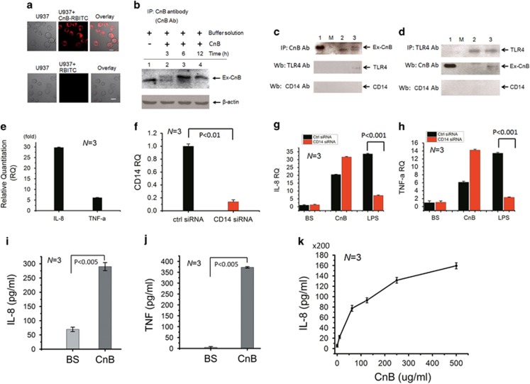
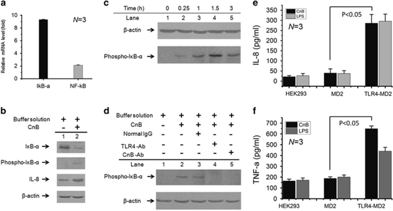
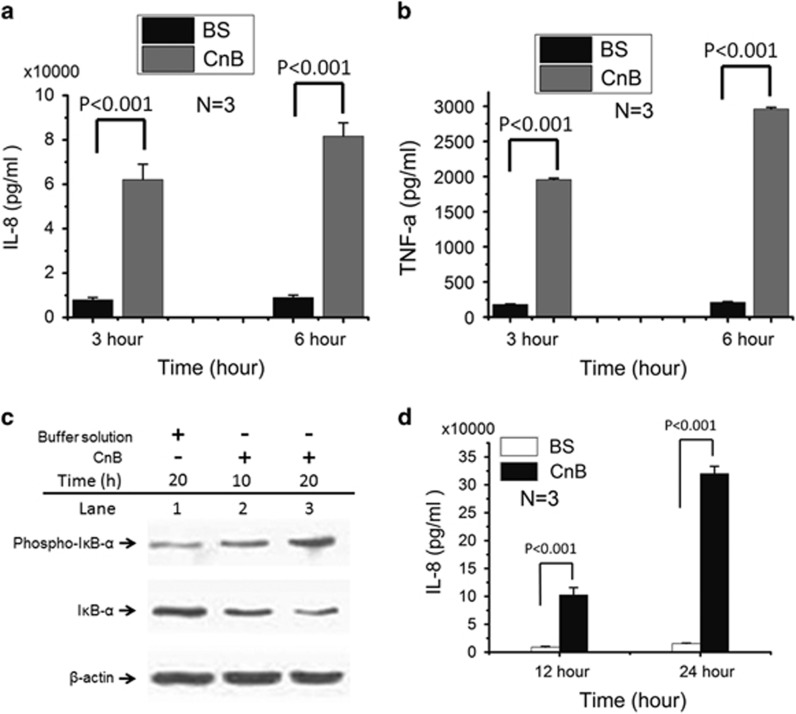
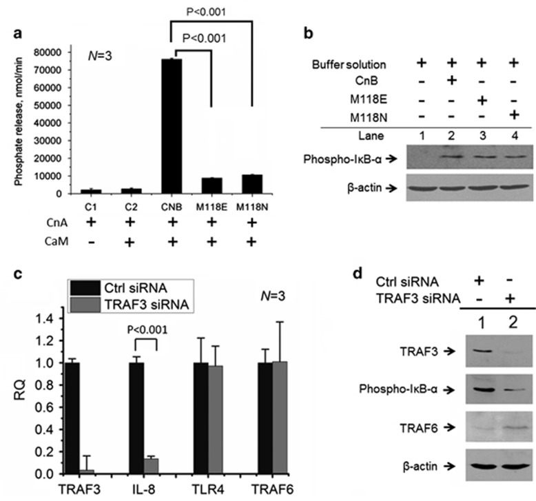
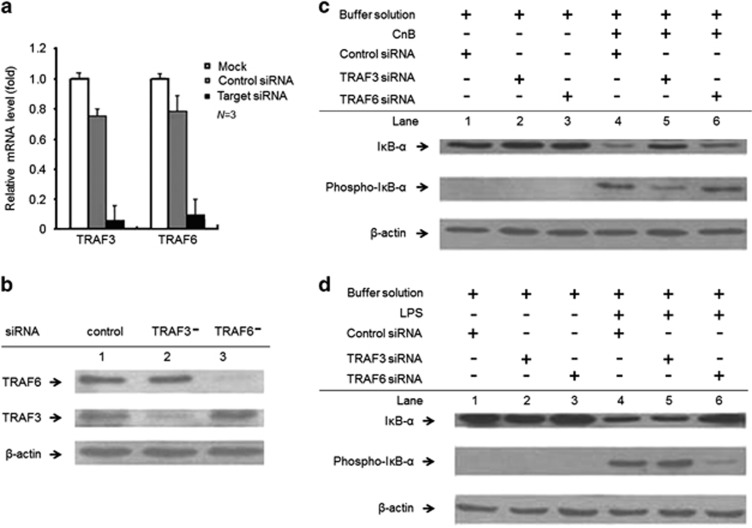
The calcineurin B subunit (CnB) is the regulatory subunit of Cn, a Ca(2+)/calmodulin-dependent serine/threonine protein phosphatase. In this study, we demonstrate that extracellular CnB was effectively internalized through a CD14-independent Toll-like receptor 4 (TLR4) pathway, which led to the phosphorylation of nuclear factor (NF)-kappa-B inhibitor alpha (IκB-α) and upregulation of pro-inflammatory cytokines in human monocytes. CnB-induced IκB-α phosphorylation is completely dependent on TNF receptor-associated factor 3 (TRAF3) but not TRAF6, which is indispensable for IκB-α phosphorylation in response to lipopolysaccharide. The loss-of-function CnB mutants were able to induce IκB-α phosphorylation, further indicating that this novel role of CnB is completely independent of the phosphatase function of Cn. Taken together, these findings demonstrate that CnB is a novel host-derived immunostimulatory factor, having a role as an agonist in monocytes, and specificity in TLR4 signaling through TRAF3 and TRAF6, in response to various agonists.


Figures

Figure 1
Figure 1
CnB-induced endocytosis of TLR4 and pro-inflammatory cytokine production are independent of CD14. (a) Confocal microscopy images showing CnB-RBITC and RBITC internalization in U937 cells. (b) Immunoprecipitation analysis of exogenous CnB (Ex-CnB) ingested into U937 cells. Lane 1: U937 cell lysate, the band is the endogenous CnB protein; lanes 2–4: CnB-treated U937 cells at various time points. (c and d) Co-immunoprecipitation (repeated for three times) of exogenous CnB (Ex-CnB) and endogenous TLR4 using exogenous CnB (c) and endogenous TLR4 (d), respectively, as the bait protein, followed by western blotting for CnB, TLR4 and CD14. Lane 1: purified CnB was used as a control added directly to the SDS–PAGE gel; lane M: protein marker; lane 2: buffer solution-treated U937 cells; lane 3: CnB-treated U937 cells. (e) Quantitative PCR of IL-8 and TNF-α, which were normalized to the mRNA transcripts from the buffer solution-treated cells (as a control). (f) Quantitative PCR for CD14 expression in U937 cells transfected with a control siRNA or CD14 siRNA, which were normalized to the β-actin mRNA. CD14 expression was knocked down to 13.9%. (g and h) Quantitative PCR for IL-8 and TNF-α in U937 cells transfected with a CD14 siRNA or control siRNA and treated with CnB, buffer solution (BS) or LPS for 3 h. (i and j) ELISA for IL-8 and TNF-α in U937 cells treated with CnB for 3 h. (k) ELISA for IL-8 in U937 cells treated with different concentrations of CnB (0, 10, 62.5, 125, 250 and 500 μg) for 24 h. The scale bar represents 10 μm in a, and the error bars represent the mean±s.d.
Figure 2
Figure 2
The CnB–TLR4 interaction results in cytokine production through IκB-α phosphorylation. (a), Quantitative PCR for NF-κB (2.1-fold) and IκB-α (9.3-fold). P<0.001, N=3. (b) Western blot to detect IκB-α (with or without phosphorylation) and IL-8 in CnB-treated (lane 2) or buffer solution-treated (lane 1) U937 cells; the cells were stimulated with CnB or its buffer solution for 3 h. (c) Western blot to detect IκB-α phosphorylation at various time points in CnB-treated U937 cells. (d) Western blot to detect IκB-α phosphorylation in U937 cells treated with different antibodies. (e and f) ELISA for IL-8 and TNF-α in HEK293 cells transfected with TLR4 and/or MD2 and treated with CnB and LPS for 3 h. The error bars represent the mean±s.d.
Figure 3
Figure 3
Effect of CnB on human monocytes from peripheral blood mononuclear cells (PBMCs). (a and b) ELISA for IL-8 and TNF-α in human monocytes from PBMCs that were treated with CnB or its buffer solution (BS) for 3 or 6 h. (c) Western blot analysis of IκB-α and its phosphorylation in human monocytes treated with CnB or its buffer solution (BS). (d) Human monocytes were incubated with CnB or its buffer solution alone as negative control for 12 or 24 h, and analyzed using a human IL-8 ELISA kit. IL-8 was translated and released in a time-dependent manner and showed a 10-fold increase after 12 h and a 20-fold increase after 24 h. The error bars represent the mean±s.d.
Figure 4
Figure 4
The role of exogenous CnB as an agonist independent of Cn activity, but dependent on TRAF3. (a) Dephosphorylation activity of CnA (CnA subunit) toward [32P] RII regulated by wild-type CnB and its mutants. Controls 1 and 2 (C1 and C2) did not include CnB or its mutants. CaM, calmodulin; N=3. (b) Western blot analysis of IκB-α phosphorylation induced by the buffer solution, CnB or its mutants in U937 cells; the cells were stimulated with buffer solution, CnB and its mutants for 3 h. (c) Quantitative PCR for TRAF3 IL-8, TLR4 and TRAF6 in U937 cells transfected with TRAF3 siRNA or its control siRNA, respectively, and then induced by CnB. (d) Western blot analysis to detect the expression of TRAF3 and TRAF6 and IκB-α phosphorylation; lane 1: control siRNA and lane 2: TRAF3 siRNA. The error bars represent the mean±s.d.
Figure 5
Figure 5
TLR4 signaling through TRAF3 and TRAF6 in response to different agonists. (a and b) Quantitative PCR (a) and western blot (b) analyses of TRAF3 and TRAF6 expression. The TRAF3 and TRAF6 mRNA levels were decreased by ~6% and ~9% (shown by qRT–PCR), respectively, compared with the levels in the mock transfected cells (without siRNA), or by ~7 and ~12%, respectively, compared with the levels in the cells transfected with the negative control siRNA. P<0.001. (c and d) Western blot analysis of CnB-induced or LPS-induced IκB-α phosphorylation in the siRNA-transfected cells; the cells were stimulated with LPS, CnB or its buffer solution for 3 h. The LPS-treated, TRAF6-knockdown cells remarkably decreased the phosphorylation of IκB-α compared with the negative control and TRAF3-knockdown cells. Furthermore, the TRAF3-knockdown cells slightly increased LPS-induced IκB-α phosphorylation, but considerably impaired CnB-induced IκB-α phosphorylation. The experiments were repeated three times. The error bars represent the mean±s.d.

References

    1. Seong SY, Matzinger P. Hydrophobicity: an ancient damage-associated molecular pattern that initiates innate immune responses. Nat Rev Immunol 2004; 4: 469–478. - PubMed
    1. Zhong B, Tien P, Shu HB. Innate immune responses: crosstalk of signaling and regulation of gene transcription. Virology 2006; 352: 14–21. - PubMed
    1. Häcker H, Redecke V, Blagoev B, Kratchmarova I, Hsu LC, Wang GG et al. Specificity in Toll-like receptor signalling through distinct effector functions of TRAF3 and TRAF6. Nature 2006; 439: 204–207. - PubMed
    1. Griffith JP, Kim JL, Kim EE, Sintchak MD, Thomson JA, Fitzgibbon MJ et al. X-ray structure of calcineurin inhibited by the immunophilin-immunosuppressant FKBP12-FK506 complex. Cell 1995; 82: 507–522. - PubMed
    1. Milan D, Griffith J, Su M, Price ER, McKeon F. The latch region of calcineurin B is involved in both immunosuppressant-immunophilin complex docking and phosphatase activation. Cell 1994; 79: 437–447. - PubMed

Publication types

MeSH terms