. 2015 Oct 16:17:291.
doi: 10.1186/s13075-015-0794-z.

Conditional deletion of caspase-8 in macrophages alters macrophage activation in a RIPK-dependent manner

Affiliations

Conditional deletion of caspase-8 in macrophages alters macrophage activation in a RIPK-dependent manner

Carla M Cuda et al. Arthritis Res Ther. .

Abstract

Introduction: Although caspase-8 is a well-established initiator of apoptosis and suppressor of necroptosis, recent evidence suggests that this enzyme maintains functions beyond its role in cell death. As cells of the innate immune system, and in particular macrophages, are now at the forefront of autoimmune disease pathogenesis, we examined the potential involvement of caspase-8 within this population.

Methods: Cre (LysM) Casp8 (fl/fl) mice were bred via a cross between Casp8 (fl/fl) mice and Cre (LysM) mice, and RIPK3 (-/-) Cre (LysM) Casp8 (fl/fl) mice were generated to assess the contribution of receptor-interacting serine-threonine kinase (RIPK)3. Immunohistochemical and immunofluorescence analyses were used to examine renal damage. Flow cytometric analysis was employed to characterize splenocyte distribution and activation. Cre (LysM) Casp8 (fl/fl) mice were treated with either Toll-like receptor (TLR) agonists or oral antibiotics to assess their response to TLR activation or TLR agonist removal. Luminex-based assays and enzyme-linked immunosorbent assays were used to measure cytokine/chemokine and immunoglobulin levels in serum and cytokine levels in cell culture studies. In vitro cell culture was used to assess macrophage response to cell death stimuli, TLR activation, and M1/M2 polarization. Data were compared using the Mann-Whitney U test.

Results: Loss of caspase-8 expression in macrophages promotes onset of a mild systemic inflammatory disease, which is preventable by the deletion of RIPK3. In vitro cell culture studies reveal that caspase-8-deficient macrophages are prone to a caspase-independent death in response to death receptor ligation; yet, caspase-8-deficient macrophages are not predisposed to unchecked survival, as analysis of mixed bone marrow chimeric mice demonstrates that caspase-8 deficiency does not confer preferential expansion of myeloid populations. Loss of caspase-8 in macrophages dictates the response to TLR activation, as injection of TLR ligands upregulates expression of costimulatory CD86 on the Ly6C(high)CD11b(+)F4/80(+) splenic cells, and oral antibiotic treatment to remove microbiota prevents splenomegaly and lymphadenopathy in Cre (LysM) Casp8 (fl/fl) mice. Further, caspase-8-deficient macrophages are hyperresponsive to TLR activation and exhibit aberrant M1 macrophage polarization due to RIPK activity.

Conclusions: These data demonstrate that caspase-8 functions uniquely in macrophages by controlling the response to TLR activation and macrophage polarization in an RIPK-dependent manner.


Figures
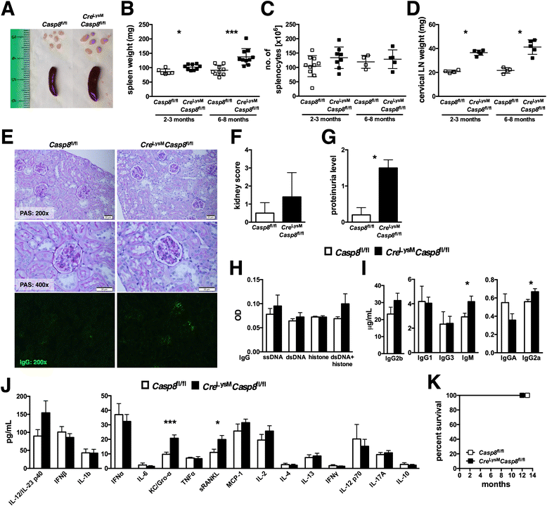
Fig. 1
Fig. 1
Mice with conditionally deleted caspase-8 exhibit mild systemic inflammation. We evaluated 2–3-month old (young) and 6–8-month-old (aged) female Casp8 fl/fl (control) and Cre LysM Casp8 fl/fl mice (n ≥ 4) for systemic autoimmune disease phenotypes. a Representative spleens and cervical lymph nodes from aged mice. b Spleen weights of young and aged mice. c Total numbers of live splenocytes from young and aged mice. d Cervical lymph node weights of young and aged mice. e Formalin-fixed kidney sections (5 μm) of aged mice stained with periodic acid–Schiff (PAS) and frozen kidney sections (10 μm) stained with IgGα-fluorescein isothiocyanate. f Kidney score of aged mice. g Proteinuria of aged mice assessed using Uristix reagent strips. Serum from aged mice was evaluated for levels of h ssDNA-, dsDNA-, histone-, and chromatin-reactive IgG antibodies; i total IgM and IgG isotypes; and j cytokines and chemokines. k Survival study. Data are represented as mean ± SD and were compared by Mann–Whitney U test: *p < 0.05; ***p < 0.0005. Casp8 caspase-8, dsDNA double-stranded DNA, Gro-α growth-regulated oncogene-α, IFN interferon, Ig immunoglobulin, IL interleukin, KC keratinocyte chemoattractant, LN lymph node, MCP monocyte chemoattractant protein, OD optical density, sRANKL soluble receptor activator of nuclear factor κB ligand, ssDNA single-stranded DNA, TNF tumor necrosis factor
Fig. 2
Fig. 2
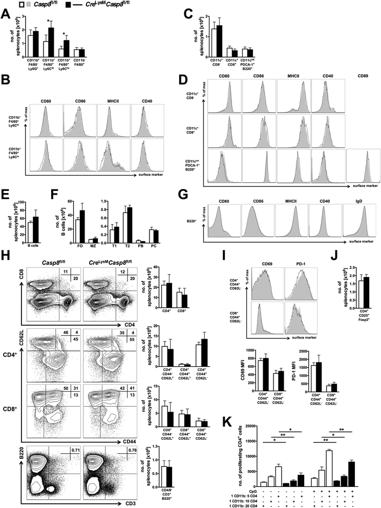
Splenic cellularity and activation profiles of Cre LysM Casp8 fl/fl mice are not drastically altered. Splenocytes from 6–8-month-old (aged) female Casp8 fl/fl (control) and Cre LysM Casp8 fl/fl mice (n ≥ 4) were analyzed by flow cytometry. a Number of CD11b+F4/80Ly6G+ neutrophils, Ly6Chigh and Ly6Clow CD11b+F4/80+ cells, and CD11bF4/80+ red pulp macrophages. b Representative fluorescence-activated cell sorting (FACS) plots of CD11b+F4/80+Ly6Chigh and CD11b+F4/80+Ly6Clow splenocytes displaying levels of surface activation marker expression. c Number of CD11c+CD8 and CD11c+CD8 conventional DCs and CD11cintermediatePDCA-1+B220+ plasmacytoid DCs. d Representative FACS plots of CD11c+CD8 and CD11c+CD8 conventional DCs and CD11cintermediatePDCA-1+B220+ plasmacytoid DCs displaying levels of surface activation marker expression. e Total B-cell (CD11cB220+) numbers. f B-cell subsets: follicular (FO; CD19+CD21/35+CD23+), marginal zone (MZ; CD19+CD21/35+CD23low), transitional 1 (T1; B220+AA4.1+CD23), transitional 2 (T2; B220+AA4.1+CD23+), plasmablasts (PB; CD19+B220lowCD138+CD21/35CD23), and plasma cells (PC; CD19+B220+CD138+CD21/35CD23). g Representative FACS plots from B cells displaying levels of surface activation and maturation marker expression. h Representative FACS plots and quantitative graphs of total CD4+ and CD8+ T-cell numbers, naïve (CD44CD62L+), central memory (CD44+CD62L+) and activated (CD44+CD62L) CD4+ and CD8+ T-cell numbers, and CD4CD8CD3+B220+ double-negative T-cell numbers. i Representative FACS plots and quantitative graphs depicting activated CD4+ and CD8+ T cells displaying levels of surface activation marker expression. j CD4+CD25+Foxp3+ regulatory T-cell numbers. Data are represented as mean ± SD and were compared by Mann–Whitney U test: **p < 0.005, ***p < 0.0005. k Bead-separated CD11b+ cells incubated with ovalbumin were cocultured with B6.CD45.1/OT-II/RAG −/− CD4+ T cells at various ratios with or without CpG. Data are represented as mean ± SD of biological triplicates, and experiments were repeated twice. Data were compared by Mann–Whitney U test: *p < 0.05; **p < 0.005. PDCA-1 plasmacytoid dendritic cell antigen 1
Fig. 3
Fig. 3
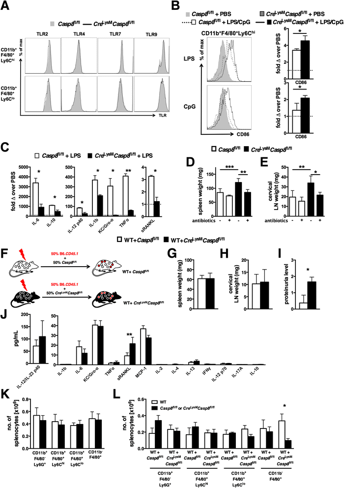
Caspase-8 deficiency alters the macrophage TLR response in vivo but does not affect cell survival. a Splenocytes from 6–8-month-old (aged) female Casp8 fl/fl (control) and Cre LysM Casp8 fl/fl mice (n ≥ 7) were analyzed by flow cytometry. Shown are representative fluorescence-activated cell sorting (FACS) plots of splenic CD11b+F4/80+Ly6Chigh and CD11b+F4/80+Ly6Clow populations displaying relative levels of TLR expression. b and c Representative FACS plots and quantitative graphs of results representing the fold change in CD86 expression over PBS injection alone. b 3-month-old control and Cre LysM Casp8 fl/fl mice (n = 4) that received LPS or CpG injection (200 μg/mouse) were evaluated 4 h later for splenic CD11b+F4/80+Ly6Chigh cell expression of CD86. c Serum levels of cytokines and chemokines from TLR agonist–injected mice. d and e 3-week-old control and Cre LysM Casp8 fl/fl mice (n = 4) treated with oral antibiotics (ampicillin, vancomycin, neomycin sulfate, metronidazole) for 8 weeks were evaluated for d spleen weight and e cervical lymph node weight. fl Mice reconstituted with equal portions of B6.CD45.1 (wild-type [WT]) and either control or Cre LysM Casp8 fl/fl FACS-sorted LSK populations (n = 5) were maintained on low-dose oral antibiotics. f Representation of chimera generation. Chimeric mice were evaluated 8 months posttransfer for g splenomegaly, h lymphadenopathy, i proteinuria, j serum cytokine and chemokine levels, k myeloid cell subset numbers, and l distribution of WT (45.1)-derived and control or Cre LysM Casp8 fl/fl (45.2)-derived myeloid populations. Data are represented as mean ± SD and were compared by Mann–Whitney U test: *p < 0.05; **p < 0.005; ***p < 0.0005. TLR Toll-like receptor, Casp8 caspase-8, LPS lipopolysaccharide, PBS phosphate-buffered saline, IL interleukin, Gro-α growth-regulated oncogene-α, IFN interferon, Ig immunoglobulin, KC keratinocyte chemoattractant, TNF tumor necrosis factor, sRANKL soluble receptor activator of nuclear factor κB ligand, LN lymph node, WT wild type, MCP monocyte chemoattractant protein, LSK lineage-negative, Sca-1+, c-kit+
Fig. 4
Fig. 4
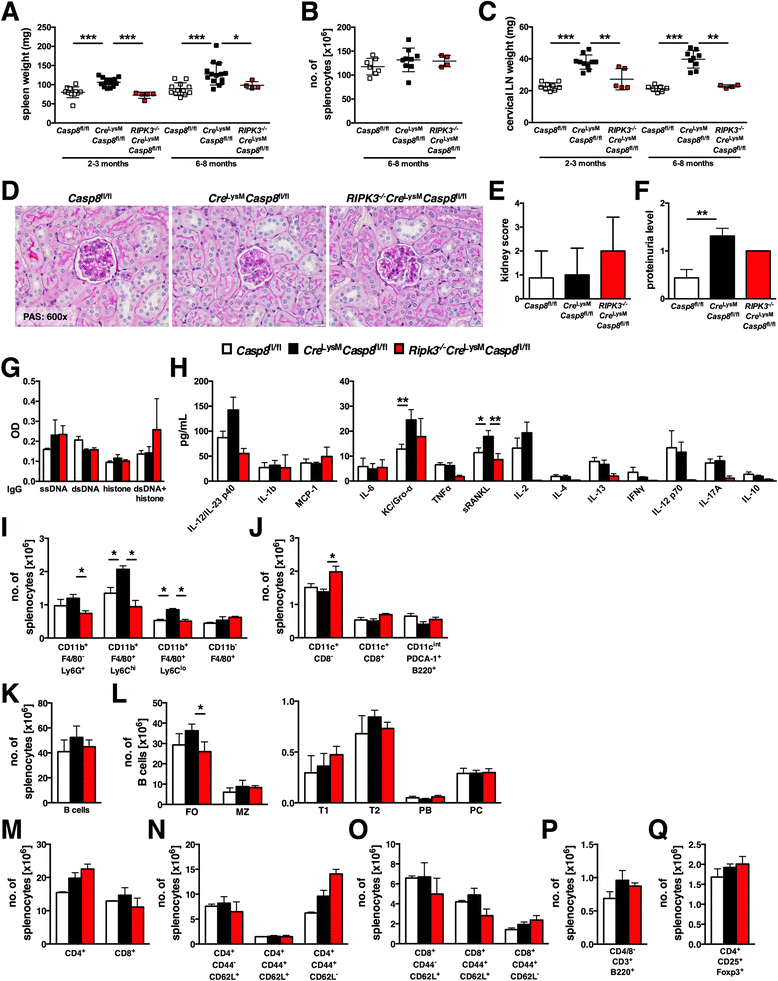
RIPK3 deletion prevents inflammation in Cre LysM Casp8 fl/fl mice. 2–3-month-old (young) and 6–8-month-old (aged) female Casp8 fl/fl (control), Cre LysM Casp8 fl/fl, and RIPK3 −/− Cre LysM Casp8 fl/fl mice (n ≥ 4) were evaluated for systemic autoimmune disease phenotypes, and splenocytes from aged mice were analyzed by flow cytometry. a Spleen weights of young and aged mice. b Total numbers of live splenocytes from aged mice. c Cervical lymph node weights of young and aged mice. d Formalin-fixed kidney sections (5 μm) from aged mice stained with periodic acid–Schiff (PAS). e Kidney scores of aged mice. f Proteinuria of aged mice assessed using Uristix reagent strips. Serum from aged mice was evaluated for levels of g ssDNA-, dsDNA-, histone-, and chromatin-reactive IgG antibodies and h cytokines and chemokines. i Number of CD11b+F4/80Ly6G+ neutrophils, Ly6Chigh and Ly6Clow CD11b+F4/80+ cells and CD11bF4/80+ red pulp macrophages. j Number of CD11c+CD8 and CD11c+CD8 conventional DCs and CD11cintermediatePDCA-1+B220+ plasmacytoid DCs. k Total B-cell (CD11cB220+) numbers. l B-cell subsets: follicular (FO; CD19+CD21/35+CD23+), marginal zone (MZ; CD19+CD21/35+CD23low), transitional 1 (T1; B220+AA4.1+CD23), transitional 2 (T2; B220+AA4.1+CD23+), plasmablasts (PB; CD19+B220lowCD138+CD21/35CD23), and plasma cells (PC; CD19+B220+CD138+CD21/35CD23). m Total CD4+ and CD8+ T-cell numbers. Naïve (CD44CD62L+), central memory (CD44+CD62L+), and activated (CD44+CD62L) n CD4+ and o CD8+ T-cell numbers. p CD4CD8CD3+B220+ double-negative T-cell numbers. q CD4+CD25+Foxp3+ regulatory T-cell numbers. ah represent analyses that included data from control and Cre LysM Casp8 fl/fl mice shown in Figs. 1b–d, f–h, and j. Data are represented as mean ± SD and were compared by Mann–Whitney U test: *p < 0.05; **p < 0.005; ***p < 0.0005. Casp8 caspase-8, LPS lipopolysaccharide, IL interleukin, Gro-α growth-regulated oncogene-α, IFN interferon, Ig immunoglobulin, KC keratinocyte chemoattractant, TNF tumor necrosis factor, MCP monocyte chemoattractant protein, LN, lymph node, OD optical density, RIPK receptor-interacting serine/threonine protein kinase, PDCA-1 plasmacytoid dendritic cell antigen 1, sRANKL soluble receptor activator of nuclear factor κB ligand
Fig. 5
Fig. 5
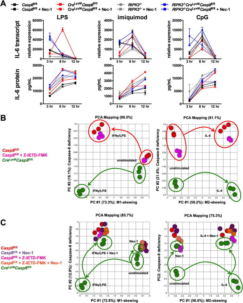
Caspase-8 deficiency in macrophages alters the response to Toll-like receptor activation and macrophage polarization in vitro. a Casp8 fl/fl (control), Cre LysM Casp8 fl/fl, RIPK3 −/−, and RIPK3 −/− Cre LysM Casp8 fl/fl BMDMs were stimulated with LPS (10 ng/ml), imiquimod (5 μg/ml), and CpG (5 μg/ml) with or without Nec-1 (30 μM) for 3, 6, and 12 h and evaluated for transcript and supernatant levels of IL-6. Data are represented as mean ± SD of biological triplicates, and experiments were repeated twice. b and c Control, control + Z-IETD-FMK (20 μM), and Cre LysM Casp8 fl/fl BMDMs were cultured with M1-polarizing conditions (primed overnight with IFN-γ 100 ng/ml and stimulated for 3 h with LPS 10 ng/ml) and M2-polarizing conditions (stimulated for 24 h with IL-4 40 ng/ml) with or without Nec-1. Principal component analysis of gene expression was performed in BMDMs under (b) M1- and M2-polarizing conditions and (c) M1- and M2-polarizing conditions + Nec-1. Data are represented as biological triplicates. FMK fluoromethylketone, IFN interferon, IL interleukin, LPS lipopolysaccharide, Nec-1 necrostatin-1, PCA principal component analysis, Z-IETD Z-Ile-Glu-(O-ME)-Thr-Asp-(O-Me)

Similar articles

Cited by

  • The inflammatory role of phagocyte apoptotic pathways in rheumatic diseases.
    Cuda CM, Pope RM, Perlman H. Cuda CM, et al. Nat Rev Rheumatol. 2016 Aug 23;12(9):543-58. doi: 10.1038/nrrheum.2016.132. Nat Rev Rheumatol. 2016. PMID: 27549026 Free PMC article. Review.
  • Targeting caspase-8: a new strategy for combating hepatocellular carcinoma.
    Chen H, Lin Y, Chen J, Luo X, Kan Y, He Y, Zhu R, Jin J, Li D, Wang Y, Han Z. Chen H, et al. Front Immunol. 2024 Dec 12;15:1501659. doi: 10.3389/fimmu.2024.1501659. eCollection 2024. Front Immunol. 2024. PMID: 39726605 Free PMC article. Review.
  • Apoptotic cell death in disease-Current understanding of the NCCD 2023.
    Vitale I, Pietrocola F, Guilbaud E, Aaronson SA, Abrams JM, Adam D, Agostini M, Agostinis P, Alnemri ES, Altucci L, Amelio I, Andrews DW, Aqeilan RI, Arama E, Baehrecke EH, Balachandran S, Bano D, Barlev NA, Bartek J, Bazan NG, Becker C, Bernassola F, Bertrand MJM, Bianchi ME, Blagosklonny MV, Blander JM, Blandino G, Blomgren K, Borner C, Bortner CD, Bove P, Boya P, Brenner C, Broz P, Brunner T, Damgaard RB, Calin GA, Campanella M, Candi E, Carbone M, Carmona-Gutierrez D, Cecconi F, Chan FK, Chen GQ, Chen Q, Chen YH, Cheng EH, Chipuk JE, Cidlowski JA, Ciechanover A, Ciliberto G, Conrad M, Cubillos-Ruiz JR, Czabotar PE, D'Angiolella V, Daugaard M, Dawson TM, Dawson VL, De Maria R, De Strooper B, Debatin KM, Deberardinis RJ, Degterev A, Del Sal G, Deshmukh M, Di Virgilio F, Diederich M, Dixon SJ, Dynlacht BD, El-Deiry WS, Elrod JW, Engeland K, Fimia GM, Galassi C, Ganini C, Garcia-Saez AJ, Garg AD, Garrido C, Gavathiotis E, Gerlic M, Ghosh S, Green DR, Greene LA, Gronemeyer H, Häcker G, Hajnóczky G, Hardwick JM, Haupt Y, He S, Heery DM, Hengartner MO, Hetz C, Hildeman DA, Ichijo H, Inoue S, Jäättelä M, Janic A, Joseph B, Jost PJ, Kanneganti TD, Karin M, Kashkar H, Kaufmann T, Kelly … See abstract for full author list ➔ Vitale I, et al. Cell Death Differ. 2023 May;30(5):1097-1154. doi: 10.1038/s41418-023-01153-w. Epub 2023 Apr 26. Cell Death Differ. 2023. PMID: 37100955 Free PMC article. Review.
  • The intersection of cell death and inflammasome activation.
    Vince JE, Silke J. Vince JE, et al. Cell Mol Life Sci. 2016 Jun;73(11-12):2349-67. doi: 10.1007/s00018-016-2205-2. Epub 2016 Apr 11. Cell Mol Life Sci. 2016. PMID: 27066895 Free PMC article. Review.
  • Bim suppresses the development of SLE by limiting myeloid inflammatory responses.
    Tsai F, Homan PJ, Agrawal H, Misharin AV, Abdala-Valencia H, Haines GK 3rd, Dominguez S, Bloomfield CL, Saber R, Chang A, Mohan C, Hutcheson J, Davidson A, Budinger GRS, Bouillet P, Dorfleutner A, Stehlik C, Winter DR, Cuda CM, Perlman H. Tsai F, et al. J Exp Med. 2017 Dec 4;214(12):3753-3773. doi: 10.1084/jem.20170479. Epub 2017 Nov 7. J Exp Med. 2017. PMID: 29114065 Free PMC article.

References

    1. Hutcheson J, Perlman H. BH3-only proteins in rheumatoid arthritis: potential targets for therapeutic intervention. Oncogene. 2008;27(Suppl 1):S168–75. doi: 10.1038/onc.2009.54. - DOI - PMC - PubMed
    1. Oberst A, Dillon CP, Weinlich R, McCormick LL, Fitzgerald P, Pop C, et al. Catalytic activity of the caspase-8–FLIPL complex inhibits RIPK3-dependent necrosis. Nature. 2011;471:363–7. doi: 10.1038/nature09852. - DOI - PMC - PubMed
    1. Rajput A, Kovalenko A, Bogdanov K, Yang SH, Kang TB, Kim JC, et al. RIG-I RNA helicase activation of IRF3 transcription factor is negatively regulated by caspase-8-mediated cleavage of the RIP1 protein. Immunity. 2011;34:340–51. doi: 10.1016/j.immuni.2010.12.018. - DOI - PubMed
    1. Sears N, Sen GC, Stark GR, Chattopadhyay S. Caspase-8-mediated cleavage inhibits IRF-3 protein by facilitating its proteasome-mediated degradation. J Biol Chem. 2011;286:33037–44. doi: 10.1074/jbc.M111.257022. - DOI - PMC - PubMed
    1. Kang TB, Yang SH, Toth B, Kovalenko A, Wallach D. Caspase-8 blocks kinase RIPK3-mediated activation of the NLRP3 inflammasome. Immunity. 2013;38:27–40. doi: 10.1016/j.immuni.2012.09.015. - DOI - PubMed

Publication types

MeSH terms

LinkOut - more resources